. 2021 Aug 10;95(17):e0069321.
doi: 10.1128/JVI.00693-21. Epub 2021 Aug 10.

Murine Cytomegalovirus MCK-2 Facilitates In Vivo Infection Transfer from Dendritic Cells to Salivary Gland Acinar Cells

Affiliations

Murine Cytomegalovirus MCK-2 Facilitates In Vivo Infection Transfer from Dendritic Cells to Salivary Gland Acinar Cells

Jiawei Ma et al. J Virol. .

Abstract

The cytomegaloviruses (CMVs) spread systemically via myeloid cells and demonstrate broad tissue tropism. Human CMV (HCMV) UL128 encodes a component of the virion pentameric complex (PC) that is important for entry into epithelial cells and cell-cell spread in vitro. It possesses N-terminal amino acid sequences similar to those of CC chemokines. While the species specificity of HCMV precludes confirmation of UL128 function in vivo, UL128-like counterparts in experimental animals have demonstrated a role in salivary gland infection. How they achieve this has not been defined, although effects on monocyte tropism and immune evasion have been proposed. By tracking infected cells following lung infection, we show that although the UL128-like protein in mouse CMV (MCMV) (designated MCK-2) facilitated entry into lung macrophages, it was dispensable for subsequent viremia mediated by CD11c+ dendritic cells (DCs) and extravasation to the salivary glands. Notably, MCK-2 was important for the transfer of MCMV infection from DCs to salivary gland acinar epithelial cells. Acinar cell infection of MCMVs deleted of MCK-2 was not rescued by T-cell depletion, arguing against an immune evasion mechanism for MCK-2 in the salivary glands. In contrast to lung infection, peritoneal MCMV inoculation yields mixed monocyte/DC viremia. In this setting, MCK-2 again promoted DC-dependent infection of salivary gland acinar cells, but it was not required for monocyte-dependent spread to the lung. Thus, the action of MCK-2 in MCMV spread was specific to DC-acinar cell interactions. IMPORTANCE Cytomegaloviruses (CMVs) establish myeloid cell-associated viremias and persistent shedding from the salivary glands. In vitro studies with human CMV (HCMV) have implicated HCMV UL128 in epithelial tropism, but its role in vivo is unknown. Here, we analyzed how a murine CMV (MCMV) protein with similar physical properties, designated MCK-2, contributes to host colonization. We demonstrate that MCK-2 is dispensable for initial systemic spread from primary infection sites but within the salivary gland facilitates the transfer of infection from dendritic cells (DCs) to epithelial acinar cells. Virus transfer from extravasated monocytes to the lungs did not require MCK-2, indicating a tissue-specific effect. These results provide new information about how persistent viral tropism determinants operate in vivo.

Keywords: chemokines; dendritic cells; mouse cytomegalovirus.


Figures
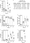
FIG 1
FIG 1
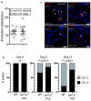
MCMV salivary gland tropism afforded by MCK-2 is mediated downstream of DC-dependent systemic spread from the lung mucosa. (A) BALB/c mice were nose inoculated (105 PFU) with either wild-type (wt) K181 (open circles) or Δm131-stop (open squares). Infectious virus titers in the nose and the salivary glands (SG) were determined by plaque assays. (B) BALB/c mice were inoculated via the lung (106 PFU) with either wt K181 MCMV or Δm131-stop, and lungs were harvested 1 day later to quantify in situ the colocalization of infected cells with the following cell surface markers: CD206 and CD68 (monocytes/macrophages), surfactant protein C (SPC-1; alveolar type II epithelial cells [AEC2]), and CD11c (DCs). For each virus, the percentages of marker-positive cells that were also MCMV+ were counted across 5 sections each for 3 mice per group. Experimental groups were compared by Fisher’s exact test. (C to F) BALB/c mice were inoculated via the lung (106 PFU) with either wt K181 or Δm131-stop. Infectious virus titers in the lungs (C) and the salivary glands (E) and genomic loads quantified by qPCR in the blood (D) and salivary glands (F) were determined on the days indicated. Individual titers/viral copies are depicted by symbols; bars show mean values for groups of mice (n = 4/group). The dotted horizontal line shows the assay sensitivity limit. Significant differences are indicated (**, P < 0.01; ***, P < 0.001).
FIG 2
FIG 2
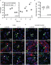
MCK-2 confers spread from CD11c+ to CD11c cells in the salivary gland. Mice were lung inoculated with wild-type K181 (wt) or Δm131-stop (106 PFU/mouse; n = 5 per group). (A) Frequency of MCMV-infected cells in sections of salivary glands at day 4 p.i. for wild-type K181 or Δm131-stop. Counts came from 10 independent sections with 5 salivary glands per section. (B) Relative proportion of MCMV+ CD11c+ to CD11c cells in salivary gland sections from infected mice determined on the days indicated. At least 100 MCMV+ cells, from up to 20 separate sections, were evaluated per time point. Experimental groups comparing the proportions of CD11c+ to CD11c cells were analyzed by two-tailed Fisher’s exact test. P values are indicated (ns, not significant). (C) Polyclonal anti-MCMV was used to confirm lytic virus infection. Closed arrows denote the localization of MCMV+ cells with CD11c cells (punctate distribution in MCMV-infected DCs), and open arrows show MCMV+ CD11c cells. All sections were taken on day 8 p.i.
FIG 3
FIG 3
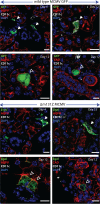
Phenotype of MCMV+ cells in the salivary gland during acute and persistent infection. Salivary glands from wild-type MCMV-GFP or Δm131Z taken during acute (day 4 p.i.) or persistent (day 12 p.i.) phases of infection were stained for CD11c and the acinar cell marker aquaporin V (AQPV) or cadherin (CAD). Infected cells are depicted by GFP expression (wild type) or β-galactosidase (Δm131Z). WIthin each virus group, the top row indicates sections stained with both CD11c and AQPV on the indicated days p.i. Colocalization of infected cells with CD11c cells is indicated by solid arrows. Colocalization of MCMV+ cells with AQPV and CAD in salivary glands taken on day 12 p.i. is indicated in the bottom row of each virus group. Closed arrowheads indicate colocalization between infected cells and the indicated acinar cell marker. Bars = 10 μm.
FIG 4
FIG 4
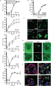
Probing in vitro correlates of Δm131/m129 in vivo attenuation. (A). Multistep growth comparisons of GFP-tagged wild-type and Δm131 viruses were performed in NIH 3T3 cells, NMuMG cells, primary acinar cells, BMDCs, and RAW cells. The MOI for nonmyeloid cells was 0.05 to 0.1; for myeloid cells, the PFU input was adjusted to achieve the same number of cells infected at time zero. (B) Single-step growth comparisons in NMuMG cells infected at an MOI of 5. For all growth comparisons, each point represents the mean from triplicate samples. Dotted lines indicate the sensitivity of the plaque assays. (C) Comparison of the spread of wild-type GFP-tagged and Δm131-GFP viruses on NMuMG cells infected at an MOI of 0.05. Images of GFP+ plaques were taken on days 3 and 5 p.i. (D) Example infection of acinar explant cultures taken at 4 days p.i. showing multiple infected cells per acinus. (E, left) Frozen sections (7 μm) of primary acinar cultures infected with either MCMV-GFP or Δm131-GFP depicted in panel D were counterstained with E-cadherin (CAD). (Right) Additional sections were also counterstained for MCMV to confirm lytic infection. Arrows indicate cytoplasmic MCMV+ vesicles. Bars = 10 μm.
FIG 5
FIG 5
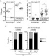
T-cell depletion does not rescue Δm131 salivary gland persistence. BALB/c mice were given anti-Thy1 antibody or not on days −1, +1, +3, +5, +9, and +11 relative to MCMV-GFP or Δm131Z MCMV lung infection (3 × 105 PFU). (A) Virus was quantified in the salivary glands at 12 days p.i. Symbols denote individual mice, and the bars are the means for each group. Experimental groups were analyzed by Student’s two-tailed t test with Welch’s correction (**, P < 0.01; ***, P < 0.001). The dotted line shows the assay sensitivity limit. (B) Number of MCMV+ cells in salivary glands from control or Thy1-treated mice (n = 5/group) taken at day 12 p.i. At least 20 random fields were counted. Symbols denote individual counts per section, and the bars are the means for each group. Experimental groups were analyzed by Student’s two-tailed t test with Welch’s correction (**, P < 0.01; ns, not significant). (C) Proportions of CD11c+ to CD11c cells in undepleted and depleted mice. The effect of anti-Thy1 treatment was analyzed by Fisher’s exact test; there was no significant effect of anti-Thy1 treatment on the proportion of CD11c+ to CD11c cells in MCMV-GPF- or Δm131Z-infected mice. Thus, the relative proportion of CD11c+ to CD11c cells between the MCMV-GFP and Δm131Z groups remained significant, regardless of anti-Thy1 treatment.
FIG 6
FIG 6
MCK-2 is dispensable for MCMV spread and monocyte-dependent lung infection following i.p. challenge. BALB/c mice were inoculated i.p. with 106 PFU wt K181 or the Δm131-stop mutant. (A and B) Viral loads were assessed in the lungs and salivary glands by plaque assays on the days indicated (A) and in the blood by qPCR on day 5 p.i. (B). Differences in virus loads between viruses were detected in the salivary glands but not the lungs or blood. Symbols denote individual mice, and bars show means (n = 6 mice/group). The dashed line indicates the limit of sensitivity. (C) BALB/c mice were infected i.p. with either MCMV-GFP or Δm131-GFP (106 PFU/mouse i.p.). Lungs and salivary glands were harvested on days 5 and 12 p.i. For both viruses, lung infection comprised >90% CD68+ CD11b+ CD11c monocytes/macrophages at day 5 p.i. Solid arrows show colocalization of infected GFP+ cells with CD11b or CD68 in the lungs at day 5 p.i. (top 2 panels, filled arrows). We detected uninfected CD11c+ cells in the lung at day 5 p.i. (bottom panel, closed arrows), but GFP+ cells were CD11c (open arrows). (D) In the salivary glands, we detected >95% CD11c+ infected cells at day 5 p.i. for both virus groups (top) (closed arrows indicate punctate CD11c). By day 12 p.i., wt MCMV-GFP-infected cells in the salivary glands were CAD+ CD11c (middle) (open arrows), but Δm131-GFP-infected counterparts were CD11c+ (filled arrows). The dichotomy of CD11c expression between wild-type MCMV- and Δm131-infected cells in the salivary gland at day 12 p.i. was confirmed using wt K181- and Δm131-stop-infected mice as described above for panel A (bottom). We detected uninfected CD11c+ cells in wt K181-infected salivary glands (filled arrows), but most MCMV+ cells were CD11c (open arrows). In contrast, MCMV+ cells from Δm131-stop infection were CD11c+ (filled arrows). Bars, 10 μm.

Similar articles

Cited by

References

    1. Ingersoll MA, Platt AM, Potteaux S, Randolph GJ. 2011. Monocyte trafficking in acute and chronic inflammation. Trends Immunol 32:470–477. 10.1016/j.it.2011.05.001. - DOI - PMC - PubMed
    1. McSharry BP, Avdic S, Slobedman B. 2012. Human cytomegalovirus encoded homologs of cytokines, chemokines and their receptors: roles in immunomodulation. Viruses 4:2448–2470. 10.3390/v4112448. - DOI - PMC - PubMed
    1. Akter P, Cunningham C, McSharry BP, Dolan A, Addison C, Dargan DJ, Hassan-Walker AF, Emery VC, Griffiths PD, Wilkinson GWG, Davison AJ. 2003. Two novel spliced genes in human cytomegalovirus. J Gen Virol 84:1117–1122. 10.1099/vir.0.18952-0. - DOI - PubMed
    1. Auerbach M, Yan D, Fouts A, Xu M, Estevez A, Austin CD, Bazan F, Feierbach B. 2013. Characterization of the guinea pig CMV gH/gL/GP129/GP131/GP133 complex in infection and spread. Virology 441:75–84. 10.1016/j.virol.2013.03.008. - DOI - PubMed
    1. MacDonald MR, Burney MW, Resnick SB, VirginHW, IV.. 1999. Spliced mRNA encoding the murine cytomegalovirus chemokine homolog predicts a beta chemokine of novel structure. J Virol 73:3682–3691. 10.1128/JVI.73.5.3682-3691.1999. - DOI - PMC - PubMed

Publication types

MeSH terms