. 2021 Jun 10;16(6):e0252674.
doi: 10.1371/journal.pone.0252674. eCollection 2021.

De novo activated transcription of inserted foreign coding sequences is inheritable in the plant genome

Affiliations

De novo activated transcription of inserted foreign coding sequences is inheritable in the plant genome

Takayuki Hata et al. PLoS One. .

Abstract

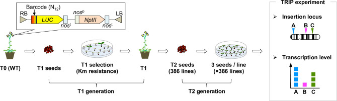
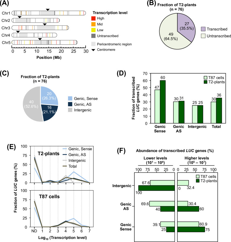
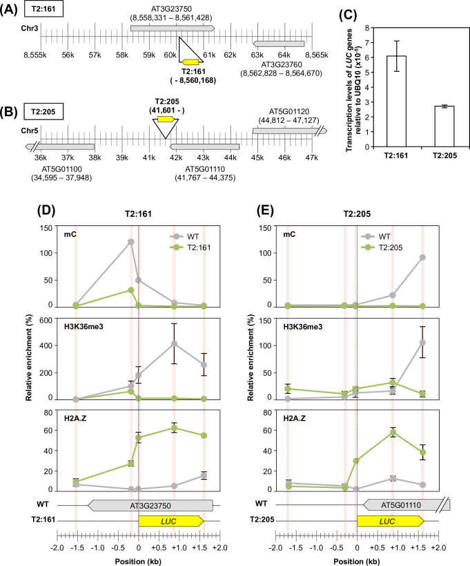
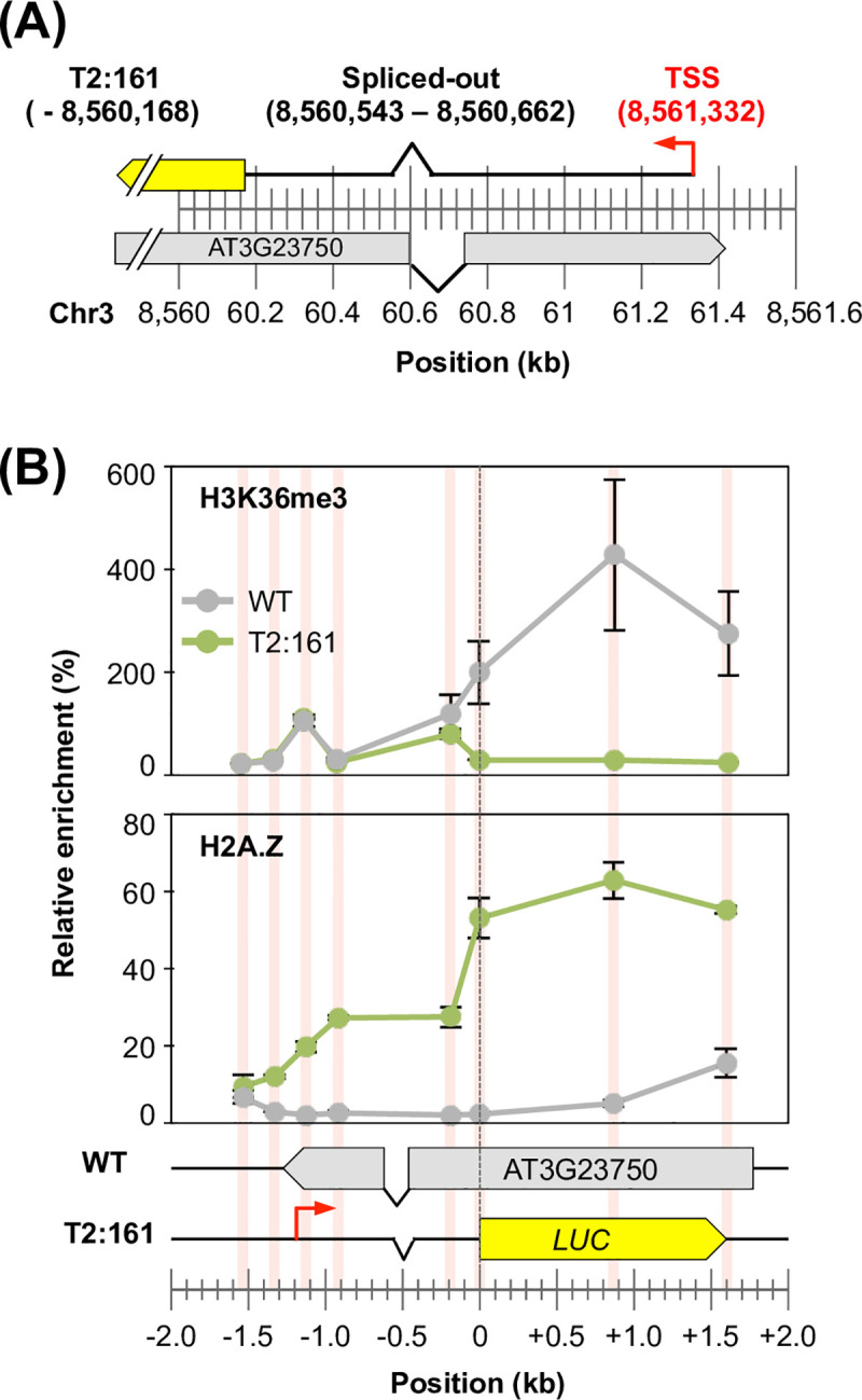
The manner in which inserted foreign coding sequences become transcriptionally activated and fixed in the plant genome is poorly understood. To examine such processes of gene evolution, we performed an artificial evolutionary experiment in Arabidopsis thaliana. As a model of gene-birth events, we introduced a promoterless coding sequence of the firefly luciferase (LUC) gene and established 386 T2-generation transgenic lines. Among them, we determined the individual LUC insertion loci in 76 lines and found that one-third of them were transcribed de novo even in the intergenic or inherently unexpressed regions. In the transcribed lines, transcription-related chromatin marks were detected across the newly activated transcribed regions. These results agreed with our previous findings in A. thaliana cultured cells under a similar experimental scheme. A comparison of the results of the T2-plant and cultured cell experiments revealed that the de novo-activated transcription concomitant with local chromatin remodelling was inheritable. During one-generation inheritance, it seems likely that the transcription activities of the LUC inserts trapped by the endogenous genes/transcripts became stronger, while those of de novo transcription in the intergenic/untranscribed regions became weaker. These findings may offer a clue for the elucidation of the mechanism by which inserted foreign coding sequences become transcriptionally activated and fixed in the plant genome.


Conflict of interest statement

The authors have declared that no competing interests exist.

Figures

Fig 1
Fig 1. Experimental design of the promoter-trap experiment in A. thaliana plants.
Schematic illustration of the TRIP experiment performed in the T2 generation of A. thaliana transgenic lines. T-DNA including a barcode, a promoterless LUC gene and an expression cassette with a Km-resistance gene was introduced into A. thaliana via Agrobacterium-mediated transformation. T2 seeds were harvested from Km-resistant T1 lines. Three seeds per T2 transgenic line were grown under the non-selective condition and subjected to subsequent locus and transcription-level analysis based on the TRIP method. NptII, neomycin phosphotransferase II; nosp, nopaline synthase promoter; nost, nopaline synthase terminator.
Fig 2
Fig 2. An artificial evolutionary experiment revealed the genetic behaviours of the activated transcription of coding sequences inserted in A. thaliana plants.
(A) The insertion loci and transcription levels of determined T2-plants (n = 76) were mapped on the A. thaliana chromosomes. The coloured bars indicate individual insertion sites and corresponding transcription levels based on their percentiles (High: 100–67, Mid: 66–34, and Low: 33–1). (B) Classification of T2-plants according to their transcription. (C) Number of T2-plants according to their insertion types: Genic sense, AS (antisense) or intergenic. The definition of each type is provided in Materials and Methods. (D) Fraction of transcribed LUC genes among T2-plants (n = 76) and T87 cultured cells (n = 4,443) (Satoh et al., 2020) against each insertion type, as in (C). (E) Fraction of LUC genes in T2-plants (upper panel) and T87 cells (lower panel) (Satoh et al., 2020) against their transcription levels, as normalized using the total number of each insertion type as 100%. ND, untranscribed LUC genes. (F) The abundance of transcribed LUC genes in each insertion type was classified according to their transcription levels; lower (101–104) and higher (105–107), as in (E). Each frequency was normalized to the number of transcribed LUC genes in each insertion type, which was set as 100%.
Fig 3
Fig 3. Transcription status of LUC insertion loci in T2-plants and WT plants.
(A) LUC insertion loci were classified by the combination of the transcription status of the LUC gene and the corresponding WT locus as follows; (i) LUC gene and the corresponding WT locus were both transcribed; (ii) LUC gene was untranscribed, but WT locus was transcribed; (iii) LUC gene was transcribed, but WT locus was untranscribed; and (iv) LUC gene and WT locus were both untranscribed. Upper panel, T2-plants; lower panel, T87 cells (Satoh et al., 2020). (B) Relative fractions of types (iii) and (iv) normalized to the sum of their abundances in (A), as 100%. (C) Relative fractions of types (i) and (ii) normalized to the sum of their abundances in (A), as 100%. (D) The transcription levels of T2-plants and corresponding inherent transcripts in WT plants were compared. R, Spearman’s correlation test.
Fig 4
Fig 4. Localization analysis of chromatin marks in selected T2-plants.
(A and B) Locus details of the (A) T2:161 and (B) T2:205 lines. The genomic loci of LUC inserts are represented as the individual position of the RB–genome junction. (C) Transcription levels of the T2:161 and T2:205 lines relative to the endogenous UBQ10 gene (AT4G05320). (D and E) Localization patterns of three chromatin marks (mC: upper panel; H3K36me3: middle panel; and H2A.Z: lower panel) around individual LUC insertion loci of the (D) T2:161 and (E) T2:205 lines. Individual localization signals were normalized to the enrichment of the control locus of each chromatin mark (see Materials and Methods) as 100%. The red bars indicate the analysed positions, which were normalized to the genomic position of the start codon of LUC inserts as zero. Error bar, ±SD of two biological replicates.
Fig 5
Fig 5. Localization of chromatin marks around the transcription start site of the T2:161 line.
(A) Transcription start site of the T2:161 line, as determined using a template-switching-based method. (B) Localization analysis of H2A.Z and H3K36me3 in T2:161 and WT plants, as in Fig 4D.

Similar articles

Cited by

References

    1. Kaessmann H. Origins, evolution, and phenotypic impact of new genes. Genome Res. 2010;20(10):1313–1326. doi: 10.1101/gr.101386.109 - DOI - PMC - PubMed
    1. Cardoso-Moreira M, Long M. The origin and evolution of new genes. Methods Mol Biol. 2012;856:161–186. doi: 10.1007/978-1-61779-585-5_7 - DOI - PubMed
    1. McLysaght A, Guerzoni D. New genes from non-coding sequence: the role of de novo protein-coding genes in eukaryotic evolutionary innovation. Philos Trans R Soc Lond B Biol Sci. 2015;370(1678):20140332. doi: 10.1098/rstb.2014.0332 - DOI - PMC - PubMed
    1. Van Oss SB, Carvunis AR. De novo gene birth. PLoS Genet. 2019;15(5):e1008160. doi: 10.1371/journal.pgen.1008160 - DOI - PMC - PubMed
    1. Haberle V, Stark A. Eukaryotic core promoters and the functional basis of transcription initiation. Nat Rev Mol Cell Biol. 2018;19(10):621–637. doi: 10.1038/s41580-018-0028-8 - DOI - PMC - PubMed

Publication types

MeSH terms

Grants and funding

This work was supported by the Japan Society for the Promotion of Science (JSPS) KAKENHI Grant Number: 17J04887 to Takayuki Hata, 26660008 to Soichirou Satoh, and 17K19358, 23125512, 23657040, 23117006, and 25650131 to Junichi Obokata, and by the Strategic Research Funds at Kyoto Prefectural University. The funders had no role in study design, data collection and analysis, decision to publish, or preparation of the manuscript.