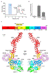
Structural analysis of the full-length human LRRK2
- PMID: 34107286
- PMCID: PMC8887629
- DOI: 10.1016/j.cell.2021.05.004
Structural analysis of the full-length human LRRK2
Abstract
Mutations in leucine-rich repeat kinase 2 (LRRK2) are commonly implicated in the pathogenesis of both familial and sporadic Parkinson's disease (PD). LRRK2 regulates critical cellular processes at membranous organelles and forms microtubule-based pathogenic filaments, yet the molecular basis underlying these biological roles of LRRK2 remains largely enigmatic. Here, we determined high-resolution structures of full-length human LRRK2, revealing its architecture and key interdomain scaffolding elements for rationalizing disease-causing mutations. The kinase domain of LRRK2 is captured in an inactive state, a conformation also adopted by the most common PD-associated mutation, LRRK2G2019S. This conformation serves as a framework for structure-guided design of conformational specific inhibitors. We further determined the structure of COR-mediated LRRK2 dimers and found that single-point mutations at the dimer interface abolished pathogenic filamentation in cells. Overall, our study provides mechanistic insights into physiological and pathological roles of LRRK2 and establishes a structural template for future therapeutic intervention in PD.
Keywords: LRRK2; LRRK2 dimer; LRRK2 mutations; Parkinson's disease; kinase.
Copyright © 2021 Elsevier Inc. All rights reserved.
Conflict of interest statement
Declaration of interests The authors declare no competing interests.
Figures


Similar articles
-
Structural model of the dimeric Parkinson's protein LRRK2 reveals a compact architecture involving distant interdomain contacts.Proc Natl Acad Sci U S A. 2016 Jul 26;113(30):E4357-66. doi: 10.1073/pnas.1523708113. Epub 2016 Jun 29. Proc Natl Acad Sci U S A. 2016. PMID: 27357661 Free PMC article.
-
The dynamic switch mechanism that leads to activation of LRRK2 is embedded in the DFGψ motif in the kinase domain.Proc Natl Acad Sci U S A. 2019 Jul 23;116(30):14979-14988. doi: 10.1073/pnas.1900289116. Epub 2019 Jul 10. Proc Natl Acad Sci U S A. 2019. PMID: 31292254 Free PMC article.
-
Kinase activity of mutant LRRK2 manifests differently in hetero-dimeric vs. homo-dimeric complexes.Biochem J. 2019 Feb 8;476(3):559-579. doi: 10.1042/BCJ20180589. Biochem J. 2019. PMID: 30670570
-
Understanding the GTPase Activity of LRRK2: Regulation, Function, and Neurotoxicity.Adv Neurobiol. 2017;14:71-88. doi: 10.1007/978-3-319-49969-7_4. Adv Neurobiol. 2017. PMID: 28353279 Free PMC article. Review.
-
Molecular Insights and Functional Implication of LRRK2 Dimerization.Adv Neurobiol. 2017;14:107-121. doi: 10.1007/978-3-319-49969-7_6. Adv Neurobiol. 2017. PMID: 28353281 Review.
Cited by
-
Association between inflammatory bowel disease and Parkinson's disease: a prospective cohort study of 468,556 UK biobank participants.Front Aging Neurosci. 2024 Jan 15;15:1294879. doi: 10.3389/fnagi.2023.1294879. eCollection 2023. Front Aging Neurosci. 2024. PMID: 38288279 Free PMC article.
-
Structure-Based Virtual Screening and De Novo Design to Identify Submicromolar Inhibitors of G2019S Mutant of Leucine-Rich Repeat Kinase 2.Int J Mol Sci. 2022 Oct 24;23(21):12825. doi: 10.3390/ijms232112825. Int J Mol Sci. 2022. PMID: 36361616 Free PMC article.
-
RAB12-LRRK2 complex suppresses primary ciliogenesis and regulates centrosome homeostasis in astrocytes.Nat Commun. 2024 Sep 29;15(1):8434. doi: 10.1038/s41467-024-52723-6. Nat Commun. 2024. PMID: 39343966 Free PMC article.
-
Phosphorylation of AQP4 by LRRK2 R1441G impairs glymphatic clearance of IFNγ and aggravates dopaminergic neurodegeneration.NPJ Parkinsons Dis. 2024 Jan 31;10(1):31. doi: 10.1038/s41531-024-00643-z. NPJ Parkinsons Dis. 2024. PMID: 38296953 Free PMC article.
-
Functional Analyses of Two Novel LRRK2 Pathogenic Variants in Familial Parkinson's Disease.Mov Disord. 2022 Aug;37(8):1761-1767. doi: 10.1002/mds.29124. Epub 2022 Jun 16. Mov Disord. 2022. PMID: 35708213 Free PMC article.
References
-
- Christensen KV, Hentzer M, Oppermann FS, Elschenbroich S, Dossang P, Thirstrup K, Egebjerg J, Williamson DS, and Smith GP (2018). LRRK2 exonic variants associated with Parkinson’s disease augment phosphorylation levels for LRRK2-Ser1292 and Rab10-Thr73. BioRxiv.
Publication types
MeSH terms
Substances
Grants and funding
LinkOut - more resources
Full Text Sources
Other Literature Sources
Research Materials

