. 2021 May 20:9:671210.
doi: 10.3389/fcell.2021.671210. eCollection 2021.

Sorting Nexin 10 as a Key Regulator of Membrane Trafficking in Bone-Resorbing Osteoclasts: Lessons Learned From Osteopetrosis

Affiliations

Sorting Nexin 10 as a Key Regulator of Membrane Trafficking in Bone-Resorbing Osteoclasts: Lessons Learned From Osteopetrosis

Ari Elson et al. Front Cell Dev Biol. .

Abstract

Bone homeostasis is a complex, multi-step process, which is based primarily on a tightly orchestrated interplay between bone formation and bone resorption that is executed by osteoblasts and osteoclasts (OCLs), respectively. The essential physiological balance between these cells is maintained and controlled at multiple levels, ranging from regulated gene expression to endocrine signals, yet the underlying cellular and molecular mechanisms are still poorly understood. One approach for deciphering the mechanisms that regulate bone homeostasis is the characterization of relevant pathological states in which this balance is disturbed. In this article we describe one such "error of nature," namely the development of acute recessive osteopetrosis (ARO) in humans that is caused by mutations in sorting nexin 10 (SNX10) that affect OCL functioning. We hypothesize here that, by virtue of its specific roles in vesicular trafficking, SNX10 serves as a key selective regulator of the composition of diverse membrane compartments in OCLs, thereby affecting critical processes in the sequence of events that link the plasma membrane with formation of the ruffled border and with extracellular acidification. As a result, SNX10 determines multiple features of these cells either directly or, as in regulation of cell-cell fusion, indirectly. This hypothesis is further supported by the similarities between the cellular defects observed in OCLs form various models of ARO, induced by mutations in SNX10 and in other genes, which suggest that mutations in the known ARO-associated genes act by disrupting the same plasma membrane-to-ruffled border axis, albeit to different degrees. In this article, we describe the population genetics and spread of the original arginine-to-glutamine mutation at position 51 (R51Q) in SNX10 in the Palestinian community. We further review recent studies, conducted in animal and cellular model systems, that highlight the essential roles of SNX10 in critical membrane functions in OCLs, and discuss possible future research directions that are needed for challenging or substantiating our hypothesis.

Keywords: ARO; SNX10; bone resorption; osteoclast; osteopetrosis; sorting nexin.


Conflict of interest statement

The authors declare that the research was conducted in the absence of any commercial or financial relationships that could be construed as a potential conflict of interest.

Figures

FIGURE 1
FIGURE 1
Osteoclasts: form and function. (A) Outline of osteoclastogenesis in culture. M-CSF and RANKL induce mononucleated monocytes to differentiate and fuse to form bi-nucleated cells, which continue to fuse and form immature, multi-nucleated cells characterized by their random organization of podosomes (small black dots). The immature cells then mature and become rounder, and display a podosomal sealing zone-like belt at their periphery. (B) Schematic drawing of a mature, bone-resorbing osteoclast. OCLs adhere to bone using podosomes, actin-rich adhesion structures that, when grown on mineralized matrix, form a closed array (sealing zone) that isolates the area of bone that is to be degraded. The cells then secrete from their ventral ruffled border membrane protons, which dissolve the mineral components of the bone matrix, and proteases [such as cathepsin K (CathK) and matrix metalloproteases (MMPs)] that degrade its protein components. This combined activity results in formation of a resorption lacuna, or pit, in the bone surface. OCLs then endocytose the bone degradation products through their ruffled border membrane, transport them through the cell, and secrete them from the apical Functional Secretory Domain (FSD). (C) Mature mouse OCLs grown on glass coverslips, stained for actin (green, highlights the closed podosomal sealing zone-like array) and DNA (blue, nuclei). Note much smaller cells that have not fused and are located between the larger OCLs. Panels (B,C) were modified with permission from Shalev and Elson (2018).
FIGURE 2
FIGURE 2
Osteopetrotic mutations in SNX10. Three mutations that affect exon splicing and a large scale deletion in the SNX10 locus are shown in green. Non-sense mutations leading to an early stop codon, which may produce a highly truncated and potentially unstable protein product are shown in red. Known missense mutations, which may affect the stability of the SNX10 protein or its ability to bind lipids and proteins, are shown in blue. A compound heterozygous nonsense mutation of p.Q62X with p.R16L has also been described. The PX and Pxe (extended PX domain, Xu et al., 2014 Proteins 82, 3483-3489, 2014) are marked in yellow and orange, respectively.
FIGURE 3
FIGURE 3
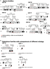
Pedigrees of Palestinian families displaying ARO. (A) Karma village families displaying SNX10 p.R51Q genotype of sampled individuals; arrows indicate probands. Highly consanguineous marital relationships produce offspring with two copies of the mutant SNX10 allele that segregate with ARO phenotype. B/III-15, G/III-9, and I/II-2 ARO patients underwent hematopoietic stem cell transplantation, and thus their allele compositions reflect those of their unaffected donors. The only exception is the affected individual H/II-3 in OST-H Family, who was wildtype for SNX10_p.R51Q variant as discussed in the text. (B) Pedigrees of four Palestinian families from other regions of the West Bank with affected individuals diagnosed with osteopetrosis displaying different genetic mutations. The affected individual of a family from Ramallah (OST-J family) was homozygous for a frameshift mutation (p.N462del) in the TCIRG1 gene, and the affected individual from the Bethlehem family (OST-L family) was homozygous for a non-synonymous mutation (p.R660C) in the same gene. The affected individual from the family from Yatta (OST-A family) was homozygous for a missense mutation in ClCN7 gene: CLCN7 p.G521R. A second family from Ramallah (OST-K family) carries a deletion mutation of exon 8 in TNFSF11 gene.
FIGURE 4
FIGURE 4
Haplotype analysis of the SNX10 locus in affected Palestinian families from the village of Karma. Six microsatellite repeat markers that flank the SNX10 gene, three on either side, were selected and amplified by PCR using fluorescent labeled primers and examined by capillary electrophoresis (CE). Haplotype analyses were analyzed using GeneScan. Selected markers were tested on SNX10 positive families on the affected individual and the parents, one heterozygous sibling (not shown here). Haplotype analysis revealed that all Karma patients who are homozygous for p.R51Q mutation were also homozygous for all studied markers. BMT denotes patients who had undergone hematopoietic stem cell transplantation prior to analysis. Different haplotypes were found in unrelated OST-J Family (Figure 3), as expected. The minimum shared haplotype block (orange) linked to the mutation was found in all the affected SNX10 patients. For each repeat the following parameters are provided: its position [according to the Human Genome Assembly GRCh37 (hg19), https://www.ncbi.nlm.nih.gov/assembly/GCF_000001405.13/], the length and composition of the nucleotide repeat, and its distance in bps from the R51Q mutation in the SNX10 gene.
FIGURE 5
FIGURE 5
Uncontrolled fusion of mature R51Q SNX10 OCLs. (A) Outline of osteoclastogenesis in culture, similar to Figure 1A. Top: WT mature OCLs do not fuse with each other. Bottom: Mature R51Q SNX10 OCLs readily fuse with each other and continue to fuse with other mature OCLs. (B) Phase light microscopy images of cultures of WT (top) and R51Q SNX10 (bottom) spleen-derived monocytes undergoing osteoclastic differentiation on glass coverslips in the presence of M-CSF and RANKL. Note development of mature, round WT OCLs that remain juxtaposed with each other, vs. continuous fusion that generates giant R51Q SNX10 OCLs (Barnea-Zohar et al., 2021). Times are noted from start of imaging; OCL boundaries are indicated by dashed yellow lines in the final image of each series. Each image is a composite of nine smaller images. Scale bars: 250 μm.
FIGURE 6
FIGURE 6
Overview of the known functions of SNX10 along the plasma membrane-to-ruffled border axis. Functions or abilities indicated include support of endocytosis (references Ye et al., 2015 and Barnea-Zohar et al., 2021 noted in the figure), binding to PI3P and PI(3,5)P2 (Barnea-Zohar et al., 2021), association with PIKfyve, EEA1, and early endosomal markers (Qin et al., 2006; Battaglino et al., 2019; Sultana et al., 2020), inhibition of chaperone mediated autophagy (You et al., 2018; Zhang et al., 2018, 2020), association with LAMP1 and ATG5 and promotion of lysosome-autophagosome fusion (Zhang et al., 2018, 2020), and ruffled border formation (Ye et al., 2015; Stein et al., 2020; Barnea-Zohar et al., 2021). See the section “Cell-Based Studies,” for additional details. PI3P, phosphatidylinositol-3-phosphate; PI(3,5)P2, phosphatidylinositol-3,5-diphosphate; Atp6i, the a3 subunit of the V0 domain of the OCL ATP-dependent proton pump; CLCN7, voltage-gated chloride/proton antiporter; OSTM1, beta subunit of the voltage-gated chloride/proton antiporter; CathK, cathepsin K; MMPs, matrix metalloproteases.

Similar articles

Cited by

References

    1. Aker M., Rouvinski A., Hashavia S., Ta-Shma A., Shaag A., Zenvirt S., et al. (2012). An SNX10 mutation causes malignant osteopetrosis of infancy. J. Med. Genet. 49 221–226. 10.1136/jmedgenet-2011-100520 - DOI - PubMed
    1. Amirfiroozy A., Hamidieh A. A., Golchehre Z., Rezamand A., Yahyaei M., Beiranvandi F., et al. (2017). A novel mutation in SNX10 gene causes malignant infantile osteopetrosis. Avicenna J. Med. Biotechnol. 9 205–208. - PMC - PubMed
    1. Antonarakis S. E. (2019). Carrier screening for recessive disorders. Nat. Rev. Genet. 20 549–561. 10.1038/s41576-019-0134-2 - DOI - PubMed
    1. Baer S., Schaefer E., Michot C., Fischbach M., Morelle G., Bendavid M., et al. (2019). Intermediate autosomal recessive osteopetrosis with a large noncoding deletion in SNX10: a case report. Pediatr. Blood Cancer 66:e27751. 10.1002/pbc.27751 - DOI - PubMed
    1. Balemans W., Van Wesenbeeck L., Van Hul W. (2005). A clinical and molecular overview of the human osteopetroses. Calcif. Tissue Int. 77 263–274. 10.1007/s00223-005-0027-6 - DOI - PubMed