Role of krüppel-like factor 8 for therapeutic drug-resistant multi-organ metastasis of breast cancer
- PMID: 34094677
- PMCID: PMC8167685
Role of krüppel-like factor 8 for therapeutic drug-resistant multi-organ metastasis of breast cancer
Abstract
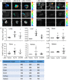
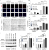
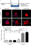
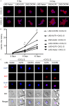
Metastasis and drug resistance are intertwined processes that are responsible for the vast majority of patient deaths from breast cancer. The underlying mechanisms remain incompletely understood. We previously demonstrated that KLF8 activates CXCR4 transcription in metastatic breast cancer. Here, we report a novel role of KLF8-CXCR4 signaling for converting single organ metastasis into multiple organ metastasis associated with chemotherapeutic resistance. We show that KLF8 expression in metastatic breast cancer cells can be over-induced by chemotherapeutic drugs. Analysis of data from large-cohorts of patients indicates that post-chemotherapy there is a close correlation between the aberrant high levels of KLF8 and CXCR4 and that this correlation is well associated with drug resistance, metastasis, and poor prognosis. To mimic their aberrant high levels, we overexpressed KLF8 or CXCR4 in a human breast cancer cell line known to metastasize only to the lungs after intravenous injection in nude mice. As expected, these cells become more resistant to chemotherapeutic drugs. Surprisingly, these KLF8 or CXCR4 overexpressing cells, even implanted orthotopically, metastasized extensively to multiple organs particularly the CXCL12-rich organs. Tube formation assay, Ki67 staining and Western blotting revealed that KLF8 or CXCR4 overexpression enhanced angiogenesis involving increased expression and secretion of VEGF protein. We also found that KLF8 or CXCR4 overexpression strongly enhanced formation of filopodium-like protrusions and proliferation via CXCR4 stimulation in a 3D culture model mimicking the colonization step of metastasis. Taken together, these results suggest that the chemo-induction of KLF8 upregulation be critical for drug resistance and systemic metastasis through enhanced tumor angiogenesis and colonization via CXCR4 over-activation and that KLF8-CXCR4 signaling axis may be a new therapeutic target for drug-resistant breast cancer metastasis.
Keywords: CXCR4; KLF8; breast cancer; drug resistance; metastasis.
AJCR Copyright © 2021.
Conflict of interest statement
None.
Figures


Similar articles
-
KLF8 promotes invasive outgrowth of breast cancer by inducing filopodium-like protrusions via CXCR4.Am J Transl Res. 2022 Feb 15;14(2):1220-1233. eCollection 2022. Am J Transl Res. 2022. PMID: 35273724 Free PMC article.
-
Krüppel-like factor 8 activates the transcription of C-X-C cytokine receptor type 4 to promote breast cancer cell invasion, transendothelial migration and metastasis.Oncotarget. 2016 Apr 26;7(17):23552-68. doi: 10.18632/oncotarget.8083. Oncotarget. 2016. PMID: 26993780 Free PMC article.
-
Identification of epidermal growth factor receptor and its inhibitory microRNA141 as novel targets of Krüppel-like factor 8 in breast cancer.Oncotarget. 2015 Aug 28;6(25):21428-42. doi: 10.18632/oncotarget.4077. Oncotarget. 2015. PMID: 26025929 Free PMC article.
-
The role of Krüppel-like factor 8 in cancer biology: Current research and its clinical relevance.Biochem Pharmacol. 2021 Jan;183:114351. doi: 10.1016/j.bcp.2020.114351. Epub 2020 Nov 27. Biochem Pharmacol. 2021. PMID: 33253644 Review.
-
Role of the CXCR4/CXCL12 signaling axis in breast cancer metastasis to the brain.Clin Exp Metastasis. 2010 Feb;27(2):97-105. doi: 10.1007/s10585-008-9210-2. Epub 2008 Sep 24. Clin Exp Metastasis. 2010. PMID: 18814042 Review.
Cited by
-
Sensitization of breast cancer to Herceptin by redox active nanoparticles.Am J Cancer Res. 2021 Oct 15;11(10):4884-4899. eCollection 2021. Am J Cancer Res. 2021. PMID: 34765298 Free PMC article.
-
KLF8 promotes invasive outgrowth of breast cancer by inducing filopodium-like protrusions via CXCR4.Am J Transl Res. 2022 Feb 15;14(2):1220-1233. eCollection 2022. Am J Transl Res. 2022. PMID: 35273724 Free PMC article.
References
-
- Siegel RL, Miller KD, Jemal A. Cancer statistics, 2020. CA Cancer J Clin. 2020;70:7–30. - PubMed
-
- Gonzalez-Angulo AM, Morales-Vasquez F, Hortobagyi GN. Overview of resistance to systemic therapy in patients with breast cancer. Adv Exp Med Biol. 2007;608:1–22. - PubMed
-
- Jones SE. Metastatic breast cancer: the treatment challenge. Clin Breast Cancer. 2008;8:224–233. - PubMed
-
- Jackson JK, Gleave ME, Gleave J, Burt HM. The inhibition of angiogenesis by antisense oligonucleotides to clusterin. Angiogenesis. 2005;8:229–238. - PubMed
Grants and funding
LinkOut - more resources
Full Text Sources
Research Materials
