. 2021 Jun 5;22(1):33.
doi: 10.1186/s12860-021-00371-9.

Effect of Voacamine upon inhibition of hypoxia induced fatty acid synthesis in a rat model of methyln-nitrosourea induced mammary gland carcinoma

Affiliations

Effect of Voacamine upon inhibition of hypoxia induced fatty acid synthesis in a rat model of methyln-nitrosourea induced mammary gland carcinoma

Lakhveer Singh et al. BMC Mol Cell Biol. .

Abstract

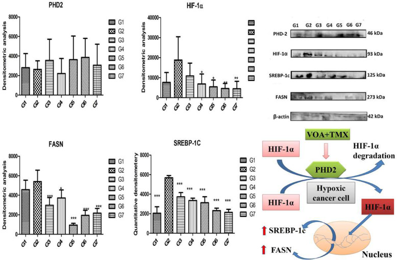
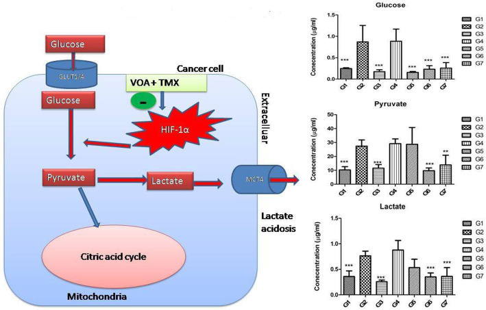
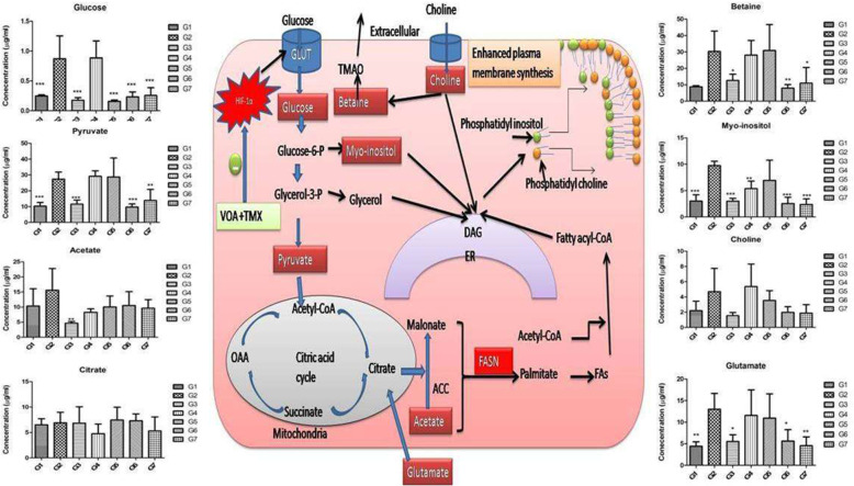
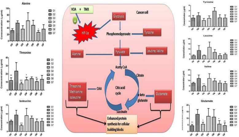
Background: In the present study, fatty acid synthesis is targeted to combat mammary gland carcinoma by activating prolyl hydroxylase-2 with Voacamine alone and in combination with Tamoxifen. It was hypothesized that the activation of prolyl hydroxylase-2 would inhibit the hypoxia-induced fatty acid synthesis and mammary gland carcinoma. Mammary gland carcinoma was induced with a single dose administration of N-methyl-N-nitrosourea (50 mg/kg,i.p.) and treatment with Voacamine and Tamoxifen 15 days after carcinogen administration.

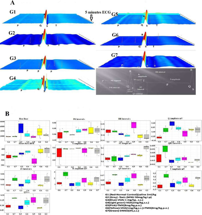
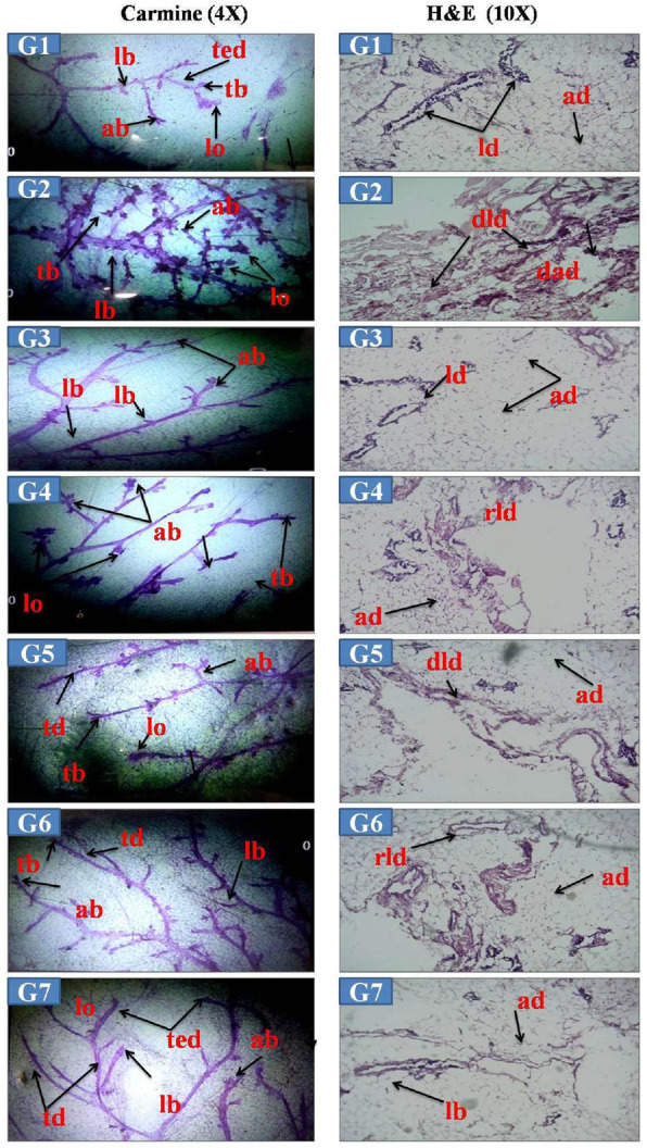
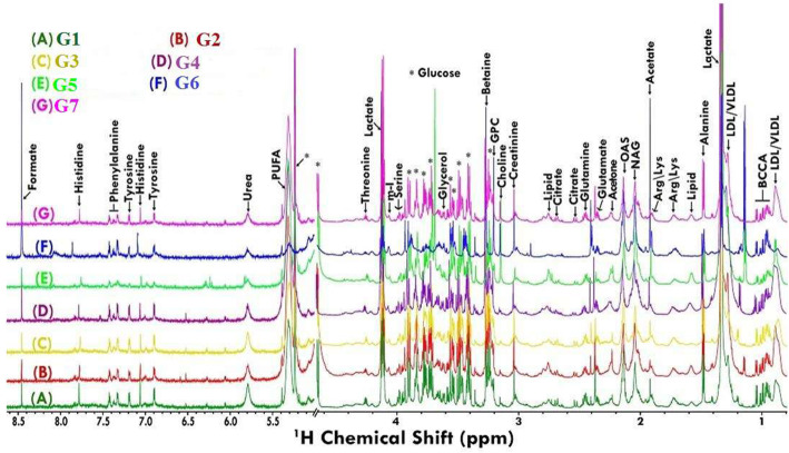
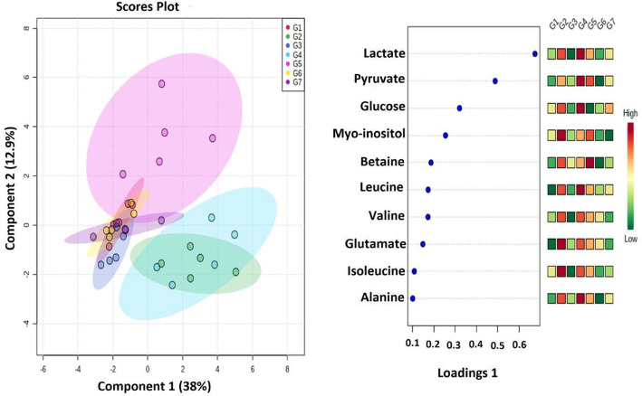
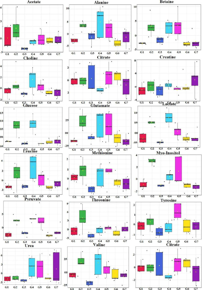
Results: At the end of the study, hemodynamic profiling of animals was recorded to assess the cardiotoxic potential of the drug. Blood serum was separated and subjected to nuclear magnetic resonance spectroscopy. Carmine staining and histopathology of mammary gland tissue were performed to evaluate the anti-angiogenic potential of the drug. The antioxidant potential of the drug was measured with antioxidant markers. Western blotting was performed to study the effect of the drug at the molecular level.

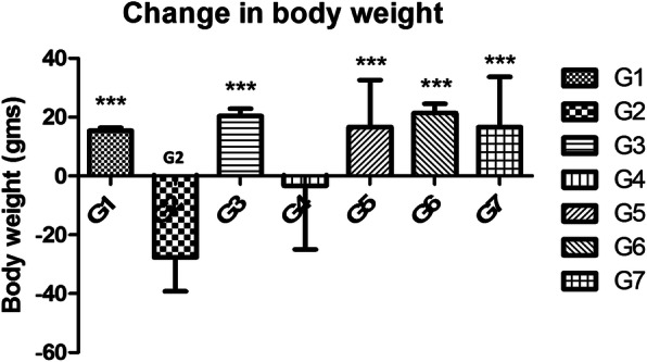
Conclusion: Results of the study have shown that Voacamine treatment stopped further decrease in body weight of experimental animals. The hemodynamic study evidenced that Voacamine at a low dose is safe in cardiac patients. Microscopic evaluation of mammary gland tissue documented the anti-angiogenic potential of Voacamine and Tamoxifen therapy. Perturbed serum metabolites were also restored to normal along with antioxidant markers. Immunoblotting of mammary gland tissue also depicted restoration of proteins of the hypoxic and fatty acid pathway. Conclusively, Voacamine and its combination with Tamoxifen activated prolyl hydroxylase-2 to combat mammary gland carcinoma.

Keywords: Fatty acid synthase; Hypoxia-inducible factor-1α; Mammary gland carcinoma; Prolyl hydroxylase-2; Tamoxifen.


Conflict of interest statement

The authors declare that they have no competing interests.

Figures

Fig. 1
Fig. 1
Docked pose of VOA with PHD-2 and in silico pharmacokinetics. VOA was docked with PHD2 protein (PDB ID: 2G19) with Autodock 4.2, and calculated binding energy was found to be −9.46 kCal/mol. The drug has hydrogen bonding interaction with LEU271, THR218, LEU193, LEU191, HIS282, ARG282, ARG281, ALA190, LEU188, PHE213, GLY213, LEU214, GLU217, ASP278, SER275, and SER214 type of amino acids. In silico, ADME was tested with pkCSM software (http://biosig.unimelb.edu.au/pkcsm/prediction_single/adme_1604411699.32), and results are presented in Table 1
Fig. 2
Fig. 2
A: Waterfall map presentation of 5 min ECG recording of experimental animals. G1(normal control receives normal saline 2 ml/kg, p.o.); G2 (toxic control receives MNU 50 mg/kg, i.p); G3(MNU 50 mg/kg, i.p. + VOA 1 mg/kg, s.c.); G4 (MNU 50 mg/kg, i.p. + VOA 2 mg/kg, s,c.); G5 (MNU 50 mg/kg, i.p. + TMX 8 mg/kg, p.o.); G6 (MNU 50 mg/kg,i.p. + VOA 1 mg/kg, s.c. + TMX 1 mg/kg s.c.) and G7 (dummy control receives 3% DMSO solution s.c.). B: Box –Cum whisker plot of ECG recording of VOA treated on experimental animals. Red-G1-Normalcontrol (saline 2 ml/kg), Grey-G2-Toxic control-MNU (50 mg/kg, i.p), Blue-G3-VOA low dose (1 mg/kg, s.c), Light green-G4-VOA high dose (2 mg/kg, s.c.), Pink-G5-TMX (8 mg/kg,p.o.) Yellow-G6- MNU (50 mg/kg, i.p.) + VOA (1 mg/kg,s.c.) + TMX (8 mg/kg,p.o.), GreenG7-DMSO 3% (3 ml/kg, s.c)
Fig. 3
Fig. 3
Effect of VOA on the morphology of mammary gland carmine staining. G1&G7-Carmine staining of normal control (G1) and DMSO (G7) administered rats showing very less branching of ducts, terminal end duct (ted), terminal bud (tb), normal lateral bud (lb), alveolar buds (ab), B-Carmine staining toxic control (G2) rats showing excessive branching of mammary gland ducts, a high number of ab and lobules (lo) along with tb and lb. C-G3 rats treated with a low dose of VOA showing very less branching of mammary gland ducts and few numbers of ab and no lo formation was noted, D-G4-Rats treated with a high dose of VOA showing tb, lb, ab, and lo, E-G5-Rats treated with TMX showing lb, tb, ab, and lo, F-G6-Rats treated with combination therapy of VOA and TMX showing along with tb, lb, ted and ab only. H&E staining: G1-G7 -Histology of G1 and G7 rats were showing the normal shape and of lactiferous duct (ld) and adipocytes, B-G2-Histology of rats showing damaged lactiferous duct (dld) and distorted shape of adipocytes (ad), C-G3-Rats were showing normal shape of ld, and ad, D-G4-VOA high dose treated rats showing regenerated lactiferous duct (rld) and ad, E-Rats treated with TMX showing rld and ad, F-G6-VOA and TMX treated rats showing the normal shape of ld and ad
Fig. 4
Fig. 4
Effect of VOA/TMX on body weight of experimental animals. Cachexia is the hallmark of cancer. Continuous loss in body weight after MNU treatment was observed in toxic control (G2). Treatment with a low dose of VOA (G3), TMX treatment (G5), and combination therapy of VOA and TMX (G6) stopped the further loss of body weight like normal control rats (G1, G7). Continuous loss in body weight was also observed in VOA high dose (2 mg/kg,s.c.) treatment which indicates VOA has toxic consequences. G1(normal control receives normal saline 2 ml/kg, p.o.); G2 (toxic control receives MNU 50 mg/kg, i.p); G3(MNU 50 mg/kg, i.p. + VOA 1 mg/kg, s.c.); G4 (MNU 50 mg/kg, i.p. + VOA 2 mg/kg, s,c.); G5 (MNU 50 mg/kg, i.p. + TMX 8 mg/kg, p.o.); G6 (MNU 50 mg/kg,i.p. + VOA 1 mg/kg, s.c. + TMX 1 mg/kg s.c.) and G7 (dummy control receives 3% DMSO solution s.c)
Fig. 5
Fig. 5
Stack plot representation of H 1 NMR peaks of serum metabolites. The peaks annotated in the figure show the assignments of serum metabolites. The abbreviations used are: LDL/VLDL: Low/very-low-density lipoproteins; PUFA: polyunsaturated fatty acids; Ile: isoleucine; Leu: leucine; Val: Valine, Pyr: pyruvate; Ch: choline; GPC: glycerophosphocholine; Glucose resonances have been indicated using symbol asterisk “*”
Fig. 6
Fig. 6
2D-PLS-DA score plot of rat serum metabolites treated with VOA and TMX. The recorded serum metabolomics data were first analyzed with combined 2D PLS-DA to see the variation in the principal components. The 2D PLS score plots of toxic control (Group 2) and that Group 4 animals treated with MNU dose depicting a clear separation from the normal, VOA low dose (G3), TMX treated (Group 5), and with a combination of both of these drugs (Groups 6). A metabolic profile of Group 4 animals was observed like that of toxic control animals after treatment with a high dose of VOA, which indicates its toxicity at this dose. This means VOA low dose and its combination with somehow resetting back the perturbed metabolites due to MNU administration excluded from the data matrix to evaluate the discriminatory significance of aromatic residues
Fig. 7
Fig. 7
Box cum whisker plot of rat serum metabolites of experimental animals after treatment with VOA/TMX. Representative box-cum whisker plots showing the quantitative variation of relative signal integrals for serum metabolites. For presented metabolite entities, the VIP score > 1 and statistical significance are at the level of p ≤ 0.05. In the box plots, the boxes denote the interquartile ranges; horizontal lines inside the boxes denote the median and bottom and top boundaries of boxes are 25th and 75th percentiles, respectively. Lower and upper whiskers are 5th and 95th percentiles, respectively
Fig. 8
Fig. 8
Effect of VOA/TMX on proteins of the hypoxic pathway. Hypoxia in cancer cells activated HIF-1α, which enhanced the cellular expression of SREBP-1c and FASN and thus fatty acid synthesis in toxic control. PHD-2 is a negative regulator of HIF-1α, and its expression is downregulated in a hypoxic environment. Treatment with VOA upregulated PHD-2 activity and downregulated HIF-1α and reverted its downstream effects. G1(normal control receives normal saline 2 ml/kg, p.o.); G2 (toxic control receives MNU 50 mg/kg, i.p); G3(MNU 50 mg/kg, i.p. + VOA 1 mg/kg, s.c.); G4 (MNU 50 mg/kg, i.p. + VOA 2 mg/kg, s,c.); G5 (MNU 50 mg/kg, i.p. + TMX 8 mg/kg, p.o.); G6 (MNU 50 mg/kg,i.p. + VOA 1 mg/kg, s.c. + TMX 1 mg/kg s.c.) and G7 (dummy control receives 3% DMSO solution s.c)
Fig. 9
Fig. 9
Effect of VOA/TMX therapy on hypoxia-induced metabolic reprogramming of serum metabolites involved in glucose metabolism. In response to hypoxia, cancer cells utilize more and more glucose which is not used in oxidative phosphorylation in mitochondria, and as a result, excess pyruvate accumulates in the cells. Accumulated pyruvate is then converted into lactate which is expelled out through MCT4 transporter into the tumor microenvironment and makes the extracellular environment exploited in various ways by the cancer cell. Treatment with VOA/TMX caused activation of PHD-2, which carries out the hydroxylation and finally proteolytic degradation of HIF-1α. Inactivation of HIF-1α ultimately reduced the glucose utilization and lactate acidosis in the tumor microenvironment. G1(normal control receives normal saline 2 ml/kg, p.o.); G2 (toxic control receives MNU 50 mg/kg, i.p); G3(MNU 50 mg/kg, i.p. + VOA 1 mg/kg, s.c.); G4 (MNU 50 mg/kg, i.p. + VOA 2 mg/kg, s,c.); G5 (MNU 50 mg/kg, i.p. + TMX 8 mg/kg, p.o.); G6 (MNU 50 mg/kg,i.p. + VOA 1 mg/kg, s.c. + TMX 1 mg/kg s.c.) and G7 (dummy control receives 3% DMSO solution s.c)
Fig. 10
Fig. 10
Effect of VOA/TMX on hypoxia-induced metabolic reprogramming of serum fatty acids. Cancer cells remain in high demand of fatty acids required for plasma membrane synthesis of cancer cells. Consequently, glycolysis is upregulated to enhance de novo fatty acid synthesis. Glutamate from extracellular regions also helps in de novo fatty acid synthesis. To form plasma phospholipids, choline, and myo-inositol processes in the endoplasmic reticulum. Phosphatidylcholine and phosphatidylinositol thus formed incorporated into the newly synthesized plasma membrane. Glycerol and myo-inositol are supplied by glycolysis, and choline is supplied from the dietary sources or degradation of plasma lipids. Treatment with low dose of VOA, and its combination with TMX, restored the perturbed metabolic profile of experimental animals. Also, metabolic profiles like that toxic control animals were observed in VOA high dose and with monotherapy of TMX
Fig. 11
Fig. 11
Effect of VOA /TMX therapy on hypoxia-induced metabolic reprogramming of serum amino acids. After MNU administration, hypoxia developed in the cancer cells, which enhanced the glycolytic pathways. As a result of this, amino acid metabolism was reprogrammed. Due to the increase in biosynthesis of amino acids like alanine, threonine, tyrosine, leucine, isoleucine, and glutamate, excess polypeptides are formed to be incorporated into the plasma membrane of rapidly dividing cells. Glutamate also acts as substrates in fatty acid synthesis. G1(normal control receives normal saline 2 ml/kg, p.o.); G2 (toxic control receives MNU 50 mg/kg, i.p); G3(MNU 50 mg/kg, i.p. + VOA 1 mg/kg, s.c.); G4 (MNU 50 mg/kg, i.p. + VOA 2 mg/kg, s,c.); G5 (MNU 50 mg/kg, i.p. + TMX 8 mg/kg, p.o.); G6 (MNU 50 mg/kg,i.p. + VOA 1 mg/kg, s.c. + TMX 1 mg/kg s.c.) and G7 (dummy control receives 3% DMSO solution s.c)

Similar articles

Cited by

References

    1. Aranda-Gutierrez A, Diaz-Perez HM. Histology, Mammary Glands. 2019. - PubMed
    1. Seyfried TN, Huysentruyt LC. On the origin of cancer metastasis. Crit Rev Oncog. 2013;18(1–2):43–73. doi: 10.1615/CritRevOncog.v18.i1-2.40. - DOI - PMC - PubMed
    1. Dey S, Roy S, Deb N, Sen KK, Besra SE. Anti-carcinogenic activity of Ruellia tuberosa L.(Acanthaceae) leaf extract on hepatoma cell line & increased superoxide dismutase activity on macrophage cell lysate. Int J Pharm Pharm Sci. 2013;5(3):854–861.
    1. Mishra A, Roy S, Maity S, Yadav RK, Keshari A, Saha S. Antiproliferative effect of flower extracts of Spilanthes paniculata on hepatic carcinoma cells. Int J Pharm Sci. 2015;7:130–134.
    1. Graham K, Unger E. Overcoming tumor hypoxia as a barrier to radiotherapy, chemotherapy and immunotherapy in cancer treatment. Int J Nanomedicine. 2018;13:6049–6058. doi: 10.2147/IJN.S140462. - DOI - PMC - PubMed

MeSH terms

LinkOut - more resources