. 2021 Jun 2;11(1):11603.
doi: 10.1038/s41598-021-91105-6.

Setd4 controlled quiescent c-Kit+ cells contribute to cardiac neovascularization of capillaries beyond activation

Affiliations

Setd4 controlled quiescent c-Kit+ cells contribute to cardiac neovascularization of capillaries beyond activation

Sheng Xing et al. Sci Rep. .

Abstract

Blood vessels in the adult mammal exist in a highly organized and stable state. In the ischemic heart, limited expansion capacity of the myocardial vascular bed cannot satisfy demands for oxygen supply and the myocardium eventually undergoes irreversible damage. The predominant contribution of endogenous c-Kit+ cells is understood to be in the development and homeostasis of cardiac endothelial cells, which suggests potential for their targeting in treatments for cardiac ischemic injury. Quiescent cells in other tissues are known to contribute to the long-term maintenance of a cell pool, preserve proliferation capacity and, upon activation, facilitate tissue homeostasis and regeneration in response to tissue injury. Here, we present evidence of a Setd4-expressing quiescent c-Kit+ cell population in the adult mouse heart originating from embryonic stages. Conditional knock-out of Setd4 in c-Kit-CreERT2;Setd4f/f;Rosa26TdTomato mice induced an increase in vascular endothelial cells of capillaries in both neonatal and adult mice. We show that Setd4 regulates quiescence of c-Kit+ cells by the PI3K-Akt-mTOR signaling pathway via H4K20me3 catalysis. In myocardial infarction injured mice, Setd4 knock-out resulted in attenuated cardiomyocyte apoptosis, decreased infarction size and improved cardiac function. Lineage tracing in Setd4-Cre;Rosa26mT/mG mice showed that Setd4+ cells contribute to each cardiac lineage. Overall, Setd4 epigenetically controls c-Kit+ cell quiescence in the adult heart by facilitating heterochromatin formation via H4K20me3. Beyond activation, endogenous quiescent c-Kit+ cells were able to improve cardiac function in myocardial infarction injured mice via the neovascularization of capillaries.


Conflict of interest statement

The authors declare no competing interests.

Figures
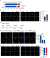
Figure 1
Figure 1
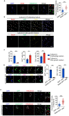
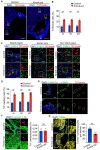
Identification of quiescent c-Kit+ cells. (A) FMC cell-cycle analysis of FACS-sorted c-Kit+ cells. (B, C) Representative immunofluorescence and quantification for c-Kit+ cells with EdU incorporation and proliferation marker, Ki67, H3pS10. n = 5 mice. (D) Experimental outline for BrdU retention assay. (EG) Representative immunofluorescence and quantification for c-Kit+ cells with retained BrdU in P1, P28 and P56 hearts (E), Setd4 retained BrdU (F) and the cell proliferation marker, Ki67 in P56 hearts (G). n = 4 mice. Nuclei were stained with DAPI. Scale bars = 50 μm. All data are represented as mean ± SEM. Unpaired t test for (B) and (F), one-way ANOVA with Bonferroi’s correction for multiple comparisons test for (E). ***p < 0.001, ns not significant.
Figure 2
Figure 2
Setd4 regulates the quiescence of c-Kit+ cells by facilitating heterochromatin formation. (A) Identification and quantification for FACS-sorted c-Kit+ cells with high levels of Setd4 and H4K20me3. Scale bar = 50 μm. n = 173 cells from 3 mice. (B, C) Representative immunofluorescence and quantification for c-Kit+ cells cultured in proliferating medium or cardiac stem cell growth medium with Setd4 incorporation of EdU and Ki67. Scale bar = 50 μm. n = 4 mice. (D, E) Representative immunofluorescence and quantification for activated c-Kit+ cells affected by Ad-GFP and Ad-Setd4 with incorporation of EdU, Ki67 (D), n = 4 mice and H4K20me3 (E), n = 105 cells from 3 mice. Scale bars = 10 μm. Nuclei were stained with DAPI. All data are represented as mean ± SEM. Comparison of independent variables was conducted by two-tailed unpaired Student’s t test. **p < 0.01, ***p < 0.001.
Figure 3
Figure 3
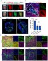
Setd4+ cells contribute to cardiac development in embryos. (A), Fluorescent images of hearts of Setd4-Cre+;Rosa26mT/mG mice and the control Setd4-Cre;Rosa26mT/mG mice. Scale bars = 500 μm. (B, C) Representative immunofluorescence for recombinant mG+ cells in E6.5 germ layers (B), E15.5 and P14 hearts (C). PS: primitive streak; ExE: extra embryonic ectoderm. Scale bars = 500 μm. (D) Quantification for recombinant mG+ cells in E6.5 PS, E15.5 and P14 hearts. (E) Representative immunofluorescence for recombinant mG+ cells with cardiomyocyte, EC, smooth muscle cell and fibroblast markers, Troponin T, CD31, α-SMA and PDGFRα, respectively, in the P14 heart. Scale bars = 50 μm. Nuclei were stained with DAPI. All data are represented as mean ± SEM. n = 4 mice. One-way ANOVA with Bonferroi’s correction for multiple comparisons test for (D). ***p < 0.001, ns: not significant.
Figure 4
Figure 4
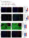
Knock-out of Setd4 induced generation of newborn ECs in adult mice. (A) Experimental outline for lineage tracing of adult mice after knock-out of Setd4. (B) Representative immunofluorescence and quantification for recombinant cells after four weeks of Setd4 knock-out. Scale bars = 200 μm. (C–E) Identification and quantification for c-Kit+ cell-derived ECs (Td+CD31+) (C), cardiomyocytes (Td+Troponin T+) (D) and fibroblasts (Td+PDGFRα+) (E) after four weeks of Setd4 knock-out. Scale bars = 50 μm. Arrows indicate recombined cells and positive staining. Nuclei were stained with DAPI. All data are represented as mean ± SEM. n = 4 mice. Comparison of independent variables was conducted by two-tailed unpaired Student’s t test. ***p < 0.001, ns: not significant.
Figure 5
Figure 5
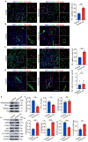
Knock-out of Setd4 contributes to neovascularization of capillaries in adult heart. (A) Representative immunofluorescence and quantification for recombinant cells with coronary endothelial cell marker FABP4 in adult mice after four weeks of Setd4 knock-out. n = 4 mice. (B) Immunostaining for Td and smooth muscle cell marker of α-SMA shows no Td+ coronary arteries generated in adult mice after four weeks of Setd4 knock-out. n = 4 mice. (C, D) Immunostaining for Td and FABP4 (C) and α-SMA (D) and quantification for recombinant cells of capillary and coronary artery potential in neonatal mice after four weeks of Setd4 knock-out. n = 4 mice. Scale bars = 50 μm. Nuclei were stained with DAPI. (E, F) Representative western blot analysis (E) and relative band densitometric analysis (F) of H4K20me3, HP1α and H3K9ac. n = 3. (G) Representative western blot analysis of PI3K, Akt, mTOR with their phosphorylated forms and PTEN. (H) The relative band densitometric analysis of P-PI3K, P-Akt, P-mTOR and PTEN. n = 3. All data are represented as mean ± SEM. n = 4 mice. Comparison of independent variables was conducted by two-tailed unpaired Student’s t test. ***p < 0.001.
Figure 6
Figure 6
Knock-out of Setd4 improves cardiac function in MI-injured mice. (A) Diagram of MI-induced injury. (B) Experimental outline for injured hearts of c-Kit-CreERT2(Cre+);Setd4f/f and c-Kit-CreERT2−(Cre-);Setd4f/f mice. (C) Masson trichrome staining and quantification for infarction size in hearts of Setd4 knock-out and control mice. Scale bars = 2 mm. n = 4 mice. (D) Detection of left ventricular function by transthoracic 2-dimensional echocardiography of Setd4 knock-out and control mice. n = 3 mice. All data are represented as mean ± SEM. Unpaired t test for (C), two-way ANOVA with Bonferroi’s correction for multiple comparisons test for (D). **p < 0.01.
Figure 7
Figure 7
Knock-out of Setd4 inhibits cardiomyocyte apoptosis in MI-injured mice. (A, B) Representative immunofluorescence (A) and quantification (B) for recombinant cells. Scale bars = 500 μm. In the three areas of the MI-injured left ventricle after four weeks of Setd4 knock-out. Scale bars = 500 μm. (C, D) Immunostaining (C) and quantification (D) for recombinant cells of capillary (Td+FABP4+) in the three areas of the MI-injured left ventricle. Scale bars = 50 μm. (E, F) Immunostaining for α-SMA (Scale bars = 200 μm) (E) and Troponin T (Scale bars = 50 μm) (F) after four weeks of Setd4 knock-out. (G) Representative immunofluorescence and quantification for apoptotic cardiomyocytes by performing TUNEL assay after two weeks of Setd4 knock-out. Scale bars = 50 μm. Nuclei were stained with DAPI. All data are represented as mean ± SEM. n = 4 mice. Unpaired t test for (F, G), two-way ANOVA with Bonferroi’s correction for multiple comparisons test for (B, D). **p < 0.01. ns not significant, IR infarction region, BZ border zone, NIR non-infarction region.

Similar articles

Cited by

References

    1. Maliken BD, Molkentin JD. Undeniable evidence that the adult mammalian heart lacks an endogenous regenerative stem cell. Circulation. 2018;138:806–808. doi: 10.1161/CIRCULATIONAHA.118.035186. - DOI - PMC - PubMed
    1. Liu QZ, Yang R, Huang XZ, Zhang H, He LJ, Zhang LB, Tian XY, Nie Y, Hu SS, Yan Y, et al. Genetic lineage tracing identifies in situ Kit-expressing cardiomyocytes. Cell Res. 2016;26:119–130. doi: 10.1038/cr.2015.143. - DOI - PMC - PubMed
    1. He LJ, Li Y, Li Y, Pu WJ, Huang XZ, Tian XY, Wang Y, Zhang H, Liu QZ, Zhang LB, et al. Enhancing the precision of genetic lineage tracing using dual recombinases. Nat. Med. 2017;23:1488–1498. doi: 10.1038/nm.4437. - DOI - PMC - PubMed
    1. Vagnozzi RJ, Maillet M, Sargent MA, Khalil H, Johansen AKZ, Schwanekamp JA, York AJ, Huang V, Nahrendorf M. An acute immune response underlies the benefit of cardiac stem cell therapy. Nature. 2020;577:405–409. doi: 10.1038/s41586-019-1802-2. - DOI - PMC - PubMed
    1. Deng JD, Ni ZC, Gu WD, Chen QS, Nowak WN, Chen T, Bhaloo SI, Zhang ZY, Hu YH, Zhou B. Single-cell gene profiling and lineage tracing analyses revealed novel mechanisms of endothelial repair by progenitors. Cell. Mol. Life Sci. 2020;77:5299–5320. doi: 10.1007/s00018-020-03480-4. - DOI - PMC - PubMed

Publication types

MeSH terms