

Aberrant B Cell Receptor Signaling in Naïve B Cells from Patients with Idiopathic Pulmonary Fibrosis
- PMID: 34073225
- PMCID: PMC8226954
- DOI: 10.3390/cells10061321
Aberrant B Cell Receptor Signaling in Naïve B Cells from Patients with Idiopathic Pulmonary Fibrosis
Abstract
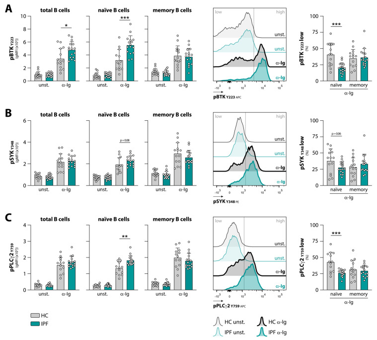
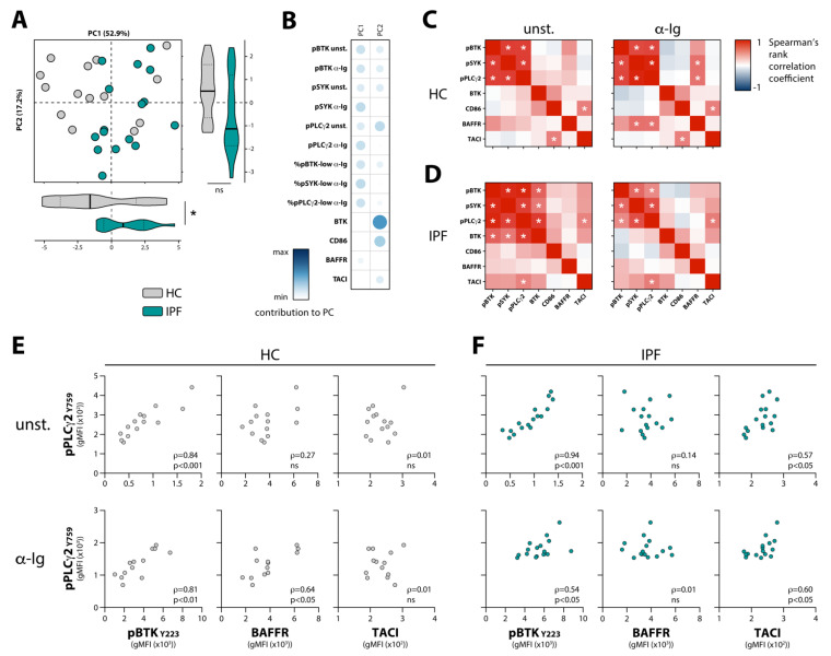
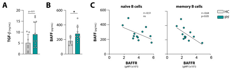
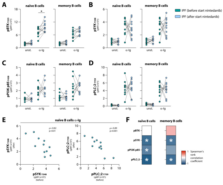
Idiopathic pulmonary fibrosis (IPF) is a chronic and ultimately fatal disease in which an impaired healing response to recurrent micro-injuries is thought to lead to fibrosis. Recent findings hint at a role for B cells and autoimmunity in IPF pathogenesis. We previously reported that circulating B cells from a fraction of patients, compared with healthy controls, express increased levels of the signaling molecule Bruton's tyrosine kinase (BTK). However, it remains unclear whether B cell receptor (BCR) signaling is altered in IPF. Here, we show that the response to BCR stimulation is enhanced in peripheral blood B cells from treatment-naïve IPF patients. We observed increased anti-immunoglobulin-induced phosphorylation of BTK and its substrate phospholipase Cγ2 (PLCγ2) in naïve but not in memory B cells of patients with IPF. In naïve B cells of IPF patients enhanced BCR signaling correlated with surface expression of transmembrane activator and calcium-modulator and cyclophilin ligand interactor (TACI) but not B cell activating factor receptor (BAFFR), both of which provide pro-survival signals. Interestingly, treatment of IPF patients with nintedanib, a tyrosine kinase inhibitor with anti-fibrotic and anti-inflammatory activity, induced substantial changes in BCR signaling. These findings support the involvement of B cells in IPF pathogenesis and suggest that targeting BCR signaling has potential value as a treatment option.
Keywords: B cell receptor (BCR) signaling; Bruton’s tyrosine kinase (BTK); autoimmunity; idiopathic pulmonary fibrosis (IPF); nintedanib.
Conflict of interest statement
S.F.H.N., P.H., J.A.C.v.H., J.R., R.W.H. and O.B.J.C. declare no conflicts of interest. M.S.W. reports grants and consultancy fees from Boehringer Ingelheim and Hoffman la Roche and consultancy fees from Galapagos, Respivant, Novartis, BMS, Horizon and Safara, all outside this study. All grants and fees were paid to her institution.
Figures
Similar articles
-
Bruton's Tyrosine Kinase mediates platelet receptor-induced generation of microparticles: a potential mechanism for amplification of inflammatory responses in rheumatoid arthritis synovial joints.Immunol Lett. 2013 Feb;150(1-2):97-104. doi: 10.1016/j.imlet.2012.12.007. Epub 2012 Dec 21. Immunol Lett. 2013. PMID: 23266841
-
Noncatalytic Bruton's tyrosine kinase activates PLCγ2 variants mediating ibrutinib resistance in human chronic lymphocytic leukemia cells.J Biol Chem. 2020 Apr 24;295(17):5717-5736. doi: 10.1074/jbc.RA119.011946. Epub 2020 Mar 17. J Biol Chem. 2020. PMID: 32184360 Free PMC article.
-
Enhanced Bruton's tyrosine kinase in B-cells and autoreactive IgA in patients with idiopathic pulmonary fibrosis.Respir Res. 2019 Oct 24;20(1):232. doi: 10.1186/s12931-019-1195-7. Respir Res. 2019. PMID: 31651327 Free PMC article.
-
Bruton's tyrosine kinase inhibitors for the treatment of rheumatoid arthritis.Drug Discov Today. 2014 Aug;19(8):1200-4. doi: 10.1016/j.drudis.2014.03.028. Epub 2014 Apr 12. Drug Discov Today. 2014. PMID: 24721226 Review.
-
Role of Bruton's tyrosine kinase in B cells and malignancies.Mol Cancer. 2018 Feb 19;17(1):57. doi: 10.1186/s12943-018-0779-z. Mol Cancer. 2018. PMID: 29455639 Free PMC article. Review.
Cited by
-
Potential of resveratrol in the treatment of interstitial lung disease.Front Pharmacol. 2023 Apr 6;14:1139460. doi: 10.3389/fphar.2023.1139460. eCollection 2023. Front Pharmacol. 2023. PMID: 37089962 Free PMC article. Review.
-
B Cell Signaling and Activation in Autoimmunity.Cells. 2023 Feb 3;12(3):499. doi: 10.3390/cells12030499. Cells. 2023. PMID: 36766841 Free PMC article.
-
Aberrant B Cell Signaling in Autoimmune Diseases.Cells. 2022 Oct 27;11(21):3391. doi: 10.3390/cells11213391. Cells. 2022. PMID: 36359789 Free PMC article. Review.
-
Identification of key immune-related genes in dilated cardiomyopathy using bioinformatics analysis.Sci Rep. 2023 Feb 1;13(1):1820. doi: 10.1038/s41598-022-26277-w. Sci Rep. 2023. PMID: 36725968 Free PMC article.
-
Single-cell sequencing analysis fibrosis provides insights into the pathobiological cell types and cytokines of radiation-induced pulmonary fibrosis.BMC Pulm Med. 2023 Apr 28;23(1):149. doi: 10.1186/s12890-023-02424-5. BMC Pulm Med. 2023. PMID: 37118713 Free PMC article.
References
-
- King T.E., Bradford W.Z., Castro-Bernardini S., Fagan E.A., Glaspole I., Glassberg M.K., Gorina E., Hopkins P.M., Kardatzke D., Lancaster L., et al. A Phase 3 Trial of Pirfenidone in Patients with Idiopathic Pulmonary Fibrosis. N. Engl. J. Med. 2014;370:2083–2092. doi: 10.1056/NEJMoa1402582. - DOI - PubMed
Publication types
MeSH terms
Substances
LinkOut - more resources
Full Text Sources
Other Literature Sources
