. 2021 May 27;11(6):1572.
doi: 10.3390/ani11061572.

H3K4me2 Promotes the Activation of lncCPSET1 by Jun in the Chicken PGC Formation

Affiliations

H3K4me2 Promotes the Activation of lncCPSET1 by Jun in the Chicken PGC Formation

Chen Zhang et al. Animals (Basel). .

Abstract

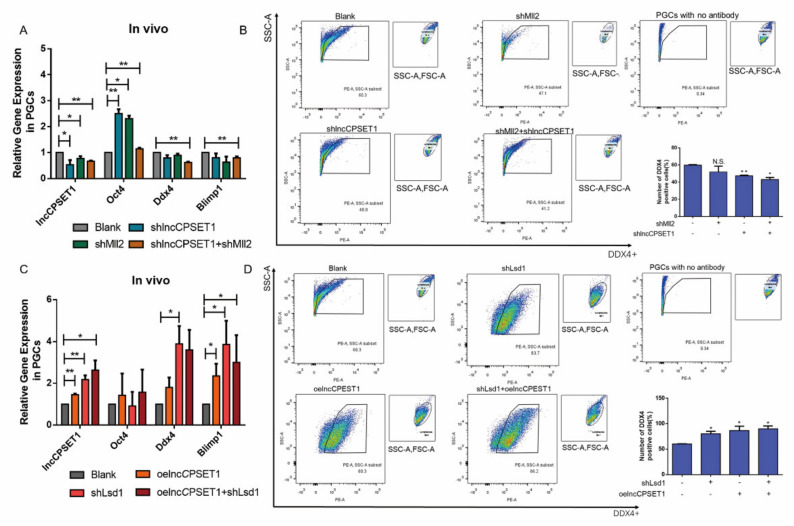
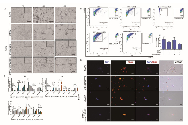
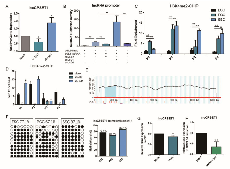
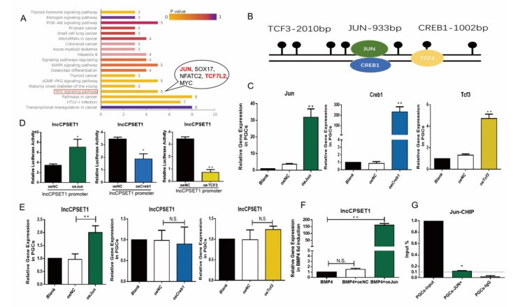
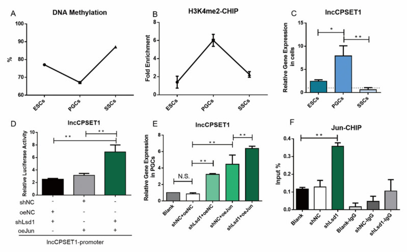
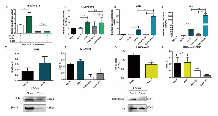
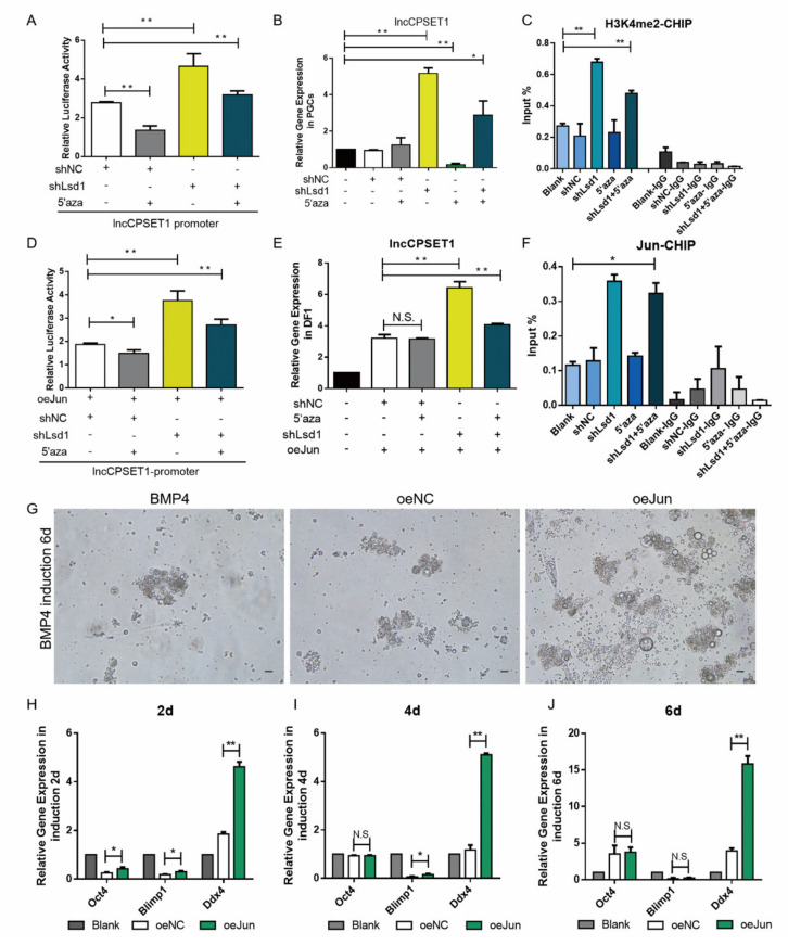
Primordial germ cells are the ancestors of female and male cells. Current research has shown that long non-coding RNA (lncRNA) and Histone methylation are the pivotal epigenetic factors in the PGC formation. However, there are few studies on the regulatory mechanism of lncRNA in the formation of PGC. Here, we define the lncRNA highly expressed in chicken PGC, lncCPSET1 (chicken-PGC-specifically-expressed transcript 1) This study found that compared with the interference of lncCPSET1/histone methylase Mll2 alone, the PGC formation was severely inhibited with the interference of lncCPSET1 and histone methylase Mll2 jointly in vivo and in vitro. Studies on the transcription level of lncCPSET1 found that H3K4me2 and transcription factor Jun have a positive effect on the activation of lncCPSET1; while DNA hypomethylation inhibits the expression of lncCPSET1. In terms of mechanism, compared with DNA methylation, H3K4me2 dominates lncCPSET1 activation. H3K4me2 can be enriched in the lncCPSET1 promoter, change its chromosome conformation, recruit the transcription factor Jun, and activate the expression of lncCPSET1. Taken together, we confirmed the model that H3K4me2 rather than DNA hypomethylation mediates Jun to regulate lncCPSET1 transcription, which broadens the study of lncCPSET1 pre-transcriptional mechanism.

Keywords: DNA methylation; H3K4me2; chicken; lncRNA; primordial germ cells.


Conflict of interest statement

The authors declare no conflict of interest.

Figures

Figure 1
Figure 1
H3K4me2 combined with lncCPSET1 promotes the formation of PGC in vivo. (A) The relative mRNA expression of genes (lncCPSET1, Oct4, Ddx4, and Blimp1) after knockdown of lncCPSET1 and Mll2. (B) Flow cytometry analysis of DDX4+ positive cells was determined after knockdown of lncCPSET1 and Mll2. (C) The relative mRNA expression of genes (lncCPSET1, Oct4, Ddx4, and Blimp1) after knockdown of Lsd1 and overexpression of lncCPSET1. (D) Flow cytometry analysis of DDX4+ positive cells was determined after knockdown of Lsd1 and overexpression of lncCPSET1. The experiments were repeated three times. Error bars indicate the means ± S.E.M. * p < 0.05, ** p < 0.01. 3.2 H3K4me2 combined with lncCPSET1 promoted the formation of PGC-like in vitro.
Figure 2
Figure 2
Knockdown of Mll2 and lncCPSET1 inhibits PGCLC formation in vitro. (A) Cell morphology was observed at different days of induction (2d, 4d, and 6d) under knockdown of Mll2 and lncCPSET1. (B) The relative mRNA expression of genes (lncCPSET1, Oct4, Ddx4, Blimp1, Gata6, and Ecomes) after knockdown of lncCPSET1 and Mll2 in different induction days (2d, 4d, and 6d) (C) Flow cytometry analysis of DDX4+ positive cells was determined after knockdown of lncCPSET1 and Mll2 in 6d of BMP4 induction. (D) Indirect immunofluorescence showed the DDX4+ positive cells in different groups in 6d of BMP4 induction. The experiments were repeated three times. Error bars indicate the means ± S.E.M. * p < 0.05, ** p < 0.01.
Figure 3
Figure 3
Dual epigenetics (H3k4me2 and DNA methylation) regulate the expression of lncCPSET1. (A) The relative mRNA expression of lncCPSET1 after knockdown of Mll2 and Lsd1 in PGC. (B) The initiation activity of lncCPSET1 was inhibited after interference with Mll2 and was enhanced after interference with Lsd1 by luciferase reporter assays. (C) CHIP-qPCR assay revealed the fold enrichment of H3K4me2 in lncCPSET1 promoter in different cells (ESC, PGC, and SSC) (D) CHIP-qPCR assay was used to detect the H3K4me2 fold enrichment after knockdown of Mll2 or Lsd1 in lncCPSET1 promoter. (E) CPG island prediction in the promoter region of lncCPSET1. (F) The dot plot shows the DNA methylation level of the lncCPSET1 promoter region in three cells (ESC, PGC, and SSC) by bisulfite sequencing. (G) The relative mRNA expression of lncCPSET1 treated with 5′aza in DF1. (H) The relative mRNA expression of lncCPSET1 treated with 5′aza in BMP4 6d induction. The experiments were repeated three times. Error bars indicate the means ± S.E.M. * p < 0.05, ** p < 0.01.
Figure 4
Figure 4
The transcription factor Jun is enriched in the promoter region to regulate the expression of lncCPSET1. (A) KEGG analysis showed the critical signaling pathway of transcription factor enrichment in the lncCPSET1 promoter. (B) The position and length of the three transcription factors on lncCPSET1 promoter. (C) qRT-PCR showed the corresponding gene expression after overexpression of transcription factor (Jun, Creb1, and Tcf3) in PGC. (D) The initiation activity of lncCPSET1 was detected by luciferase reporter assays after overexpression of transcription factor (Jun, Creb1, and Tcf3) (E) The relative mRNA expression of lncCPSET1 after overexpression of transcription factor (Jun, Creb1 and Tcf3) in PGC. (F) The relative mRNA expression of lncCPSET1 after overexpression of Jun in BMP4 6d induction. (G) CHIP-qPCR examined the enrichment of Jun in the lncCPSET1 promoter. The experiments were repeated three times. Error bars indicate the means ± S.E.M. * p < 0.05, ** p < 0.01.
Figure 5
Figure 5
H3K4me2 promotes transcription factor Jun binding promoter region to activate the expression of lncCPSET1. A, B. Expression profile of DNA methylation (A) and H3K4me2 (B) in three cells (ESC, PGC, and SSC) (C) The relative mRNA expression of lncCPSET1 in three cells (ESC, PGC, and SSC) (D) The initiation activity of lncCPSET1 increased after knockdown of Lsd1 and overexpression of Jun. (E) The relative mRNA expression of lncCPSET1 in PGCs after knockdown of Lsd1 and overexpression of Jun. (F) CHIP-qPCR was used to detect the enrichment of Jun after knockdown of Lsd1. The experiments were repeated three times. Error bars indicate the means ± S.E.M. * p < 0.05, ** p < 0.01.
Figure 6
Figure 6
DNA hypomethylation affects H3K4me2 level instead of affects Jun level to inhibit lncCPSET1 expression. (A) The initiation activity of lncCPSET1 increased after overexpression of Jun and 5′aza treatment. (B) The relative mRNA expression of lncCPSET1 in PGC after overexpression of Jun and 5′aza treatment. (C,D) The relative mRNA expression of Jun in DF1 (C) and PGC (D) after overexpression of Jun and 5′aza treatment. (E) The protein expression of JUN was detected by western blot after 5′aza treatment in PGC. (F) The enrichment of Jun in the lncCPSET1 promoter was examined by CHIP-qPCR after 5′aza treatment in PGC. (G) The H3K4me2 level was detected by western blot after 5′aza treatment in PGC. (H) CHIP-qPCR was used to evidence the enrichment change of H3K4me2 in the lncCPSET1 promoter after 5′aza treatment. The experiments were repeated three times. Error bars indicate the means ± S.E.M. * p < 0.05, ** p < 0.01. Original western blot figures in Figure S4.
Figure 7
Figure 7
H3K4me2 participates in the formation of PGC as the dominant epigenetic factor regulating Jun activation of lncCPSET1. (A) The initiation activity of lncCPSET1 increased after the knockdown of Lsd1 and 5′aza treatment. (B) The relative mRNA expression of lncCPSET1 in PGC after knockdown of Lsd1 and 5′aza treatment. (C) CHIP-qPCR was used to evidence the enrichment change of H3K4me2 in the lncCPSET1 promoter after knockdown of Lsd1 and 5′aza treatment. (D) The initiation activity of lncCPSET1 increased after knockdown of Lsd1, overexpression of Jun and 5′aza treatment. (E) The relative mRNA expression of lncCPSET1 in DF1 after knockdown of Lsd1, overexpression of Jun and 5′aza treatment. (F) The enrichment of Jun in the lncCPSET1 promoter was examined by CHIP-qPCR after knockdown of Lsd1 and 5′aza treatment in PGC. (G) Cell morphology was observed at different days of induction (2d, 4d, and 6d) after overexpression of Jun. (H) The relative mRNA expression of Oct4, Blimp1, and Ddx4 in different induction days after overexpression of Jun. The experiments were repeated three times. Error bars indicate the means ± S.E.M. * p < 0.05, ** p < 0.01.

Similar articles

Cited by

References

    1. Motono M., Yamada Y., Hattori Y., Nakagawa R., Nishijima K.I., Iijima S. Production of transgenic chickens from purified primordial germ cells infected with a lentiviral vector. J. Biosci. Bioeng. 2010;109:315–321. doi: 10.1016/j.jbiosc.2009.10.007. - DOI - PubMed
    1. Park T.S., Han J.Y. piggyBac transposition into primordial germ cells is an efficient tool for transgenesis in chickens. Proc. Natl. Acad. Sci. USA. 2012;109:9337–9341. doi: 10.1073/pnas.1203823109. - DOI - PMC - PubMed
    1. Liu X., Li N., Hu X., Zhang R., Li Q., Cao D., Liu T., Zhang Y., Liu X. Efficient production of transgenic chickens based on piggyBac. Transgenic Res. 2013;22:417–423. doi: 10.1007/s11248-012-9642-y. - DOI - PubMed
    1. Cheng S.Z., Wang M., Wang Y.L., Zhang C., Wang Y.J., Song J.Z., Zuo Q.S., Zhang Y.N., Li B.C. RXRG associated in PPAR signal regulated the differentiation of primordial germ cell. J. Cell. Biochem. 2018;119:6926–6934. doi: 10.1002/jcb.26891. - DOI - PubMed
    1. Wang M., Zhang C., Huang C., Cheng S., He N., Wang Y., Ahmed M.F., Zhao R., Jin J., Zuo Q., et al. Regulation of fibroblast growth factor 8 (FGF8) in chicken embryonic stem cells differentiation into spermatogonial stem cells. J. Cell. Biochem. 2018;119:2396–2407. doi: 10.1002/jcb.26402. - DOI - PubMed

LinkOut - more resources