. 2021 May 19;10(5):622.
doi: 10.3390/pathogens10050622.

Replication of Influenza A Virus in Secondary Lymphatic Tissue Contributes to Innate Immune Activation

Affiliations

Replication of Influenza A Virus in Secondary Lymphatic Tissue Contributes to Innate Immune Activation

Sarah-Kim Friedrich et al. Pathogens. .

Abstract

The replication of viruses in secondary lymphoid organs guarantees sufficient amounts of pattern-recognition receptor ligands and antigens to activate the innate and adaptive immune system. Viruses with broad cell tropism usually replicate in lymphoid organs; however, whether a virus with a narrow tropism relies on replication in the secondary lymphoid organs to activate the immune system remains not well studied. In this study, we used the artificial intravenous route of infection to determine whether Influenza A virus (IAV) replication can occur in secondary lymphatic organs (SLO) and whether such replication correlates with innate immune activation. Indeed, we found that IAV replicates in secondary lymphatic tissue. IAV replication was dependent on the expression of Sialic acid residues in antigen-presenting cells and on the expression of the interferon-inhibitor UBP43 (Usp18). The replication of IAV correlated with innate immune activation, resulting in IAV eradication. The genetic deletion of Usp18 curbed IAV replication and limited innate immune activation. In conclusion, we found that IAV replicates in SLO, a mechanism which allows innate immune activation.

Keywords: Influenza virus; enforced viral replication; innate immune activation.


Conflict of interest statement

The authors declare no conflict of interest.

Figures
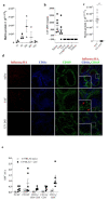
Figure 1
Figure 1
IAV replicates in the spleen during systemic infection. (a) Relative expression of IAV Matrix Protein 2 in spleens of C57BL/6 mice infected with 106 PFU of IAV i.v. normalized to naïve spleen values (n = 3). (b) Viral titers of indicated organs in C57BL/6 mice 24 h p.i. with 107 PFU IAV i.v (n = 6). (c) Quantitative analysis of Matrix Protein 2 expression via rtPCR of mice normalized to naïve spleen values (n = 6). (d) Immunofluorescense of spleen sections of naïve C57BL/6 or C57BL/6 i.v. infected with 107 PFU live or UV inactivated IAV stained for CD169 (green), CD11c (blue) and IAV HA (red). Figure is representative of 2 independent experiments (n = 6). (e) Intracellular staining of IAV+ cells of indicated myeloid cells from naïve C57BL/6 mice (black), mice infected with 107 PFU IAV i.v. (white) or mice pretreated with Oseltamivir (c, dotted white) infected with 107 PFU IAV i.v. 24 h p.i. (n = 6). # p < 0.0001 (Student’s t-test, ac). Data are representative of two (ac,e) experiments (mean ± s.e.m. (ac)). The scale bar represents 100 μm.
Figure 2
Figure 2
Replication of IAV in the spleen correlated with IFN-I induction. (a) Relative expression of Ifnα4 and Ifnβ1 of C57BL/6 mice i.v. infected with 107 PFU live or UV inactivated IAV (n = 6). (b) IAV titers of spleen samples obtained from mice pretreated with and without Oseltamivir and infected with 107 PFU IAV i.v. 24 h p.i. (n = 5–9). (c) Relative expression of Ifnα2, Ifnα4, Ifnα5 and Ifnβ1 of spleen samples obtained from mice pretreated with and without Oseltamivir and infected with 107 PFU IAV i.v. 24 h p.i. (n = 9). (d) IFNα levels in serum from mice pretreated with and without Oseltamivir and infected with 107 PFU IAV i.v. 24 h p.i. (n = 6): * p < 0.05; ** p < 0.01; # p < 0.0001 (Student’s t-test, ad). Data are representative of two (a,b) or three (c,d) independent experiments (mean ± s.e.m. (ad)).
Figure 3
Figure 3
Sialic acid residues are essential for IAV replication in the spleen. (a) Representative histograms of myeloid cell populations binding to secondary antibody control (black line) or MAL II lectin (α2,3-SA). One mouse from two independent experiments (n = 3 mice per experiment) is shown. (b) Intracellular staining of IAV+ cells of indicated myeloid cells from naïve C57BL/6 mice (black), mice infected with 107 PFU IAV i.v. (white) or mice pretreated with Sialidase i.v. and infected with 107 PFU IAV i.v. 24 h p.i. (n = 6). (c) IAV titers of spleen samples from mice with and without pretreatment with Sialidase i.p. and then infected with 107 PFU IAV i.v. 24 h p.i. (d,e) Relative expression of Ifnα2, Ifnα4, Ifnα5 and Ifnβ1 (n = 6–9). (d) or serum IFNα levels (n = 9). (e) mice with and without pre-treatment with Sialidase and then infected with 107 PFU IAV i.v. 24 h p.i. (n = 6). Dotted line represents the detection level for IFNα ELISA. * p < 0.05, (Student’s t-test, ce). Data are representative of two (ac) or three (d,e) independent experiments (mean ± s.e.m. (a,c).
Figure 4
Figure 4
Usp18 promotes replication of IAV in antigen-presenting cells. (a) Intracellular staining of IAV+ cells of indicated myeloid cells from naïve C57BL/6 mice (black) or WT or Ifnar−/− mice, infected with 107 PFU IAV i.v. analyzed 24 h p.i. (n = 6). (b) IAV titers from spleens of Cag-Cre+Usp18fl/fl and Cag-CreUsp18fl/fl mice infected with 107 PFU IAV i.v. after 24 h (n = 6–7). c+d Relative Ifnα4 and Ifnβ1 expression in the spleen (c) -Serum IFNα levels. (d) Samples derived from CAG+Usp18fl/fl and CAGUsp18fl/fl mice infected with 107 PFU IAV i.v. after 24 h (n = 6–8). * p < 0.05, and # p < 0.001 (Student’s t-test, c,d). Data are representative of two (ad) experiments (mean ± s.e.m. (ad)).
Figure 5
Figure 5
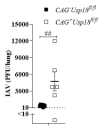
Usp18-dependent replication controls virus propagation in the lung. IAV titers from the lung of Cag-Cre+Usp18fl/fl and Cag-CreUsp18fl/fl mice infected with 107 PFU IAV i.n. after 24 h. ## p < 0.0001 (n = 6–7). Data are representative of two experiments (mean ± S.E.M.).

Similar articles

References

    1. Hopp A.K., Rupp A., Lukacs-Kornek V. Self-antigen presentation by dendritic cells in autoimmunity. Front. Immunol. 2014;5:55. doi: 10.3389/fimmu.2014.00055. - DOI - PMC - PubMed
    1. Lehuen A., Diana J., Zaccone P., Cooke A. Immune cell crosstalk in type 1 diabetes. Nat. Rev. Immunol. 2010;10:501–513. doi: 10.1038/nri2787. - DOI - PubMed
    1. Turley S.J., Fletcher A.L., Elpek K.G. The stromal and haematopoietic antigen-presenting cells that reside in secondary lymphoid organs. Nat. Rev. Immunol. 2010;10:813–825. doi: 10.1038/nri2886. - DOI - PubMed
    1. Schabbauer G., Luyendyk J., Crozat K., Jiang Z., Mackman N., Bahram S., Georgel P. TLR4/CD14-mediated PI3K activation is an essential component of interferon-dependent VSV resistance in macrophages. Mol. Immunol. 2008;45:2790–2796. doi: 10.1016/j.molimm.2008.02.001. - DOI - PMC - PubMed
    1. Yeon S.H., Song M.J., Kang H.-R., Lee J.Y. Phosphatidylinositol-3-kinase and Akt are required for RIG-I-mediated anti-viral signalling through cross-talk with IPS-1. Immunology. 2015;144:312–320. doi: 10.1111/imm.12373. - DOI - PMC - PubMed

LinkOut - more resources