. 2021 May 6;13(9):2217.
doi: 10.3390/cancers13092217.

Low RECK Expression Is Part of the Cervical Carcinogenesis Mechanisms

Affiliations

Low RECK Expression Is Part of the Cervical Carcinogenesis Mechanisms

Suellen Herbster et al. Cancers (Basel). .

Abstract

Human papillomavirus (HPV)-induced carcinogenesis comprises alterations in the expression and activity of matrix metalloproteinases (MMP) and their regulators. Reversion-inducing Cysteine-rich protein with Kazal motifs (RECK) inhibits the activation of specific metalloproteinases and its expression is frequently lost in human cancers. Here we analyzed the role of RECK in cervical carcinogenesis. Cervical cancer derived cell lines over expressing RECK were used to determine tumor kinetics as well as, cellular, immune and molecular properties in vivo. Besides, we analyzed RECK expression in cervical cancer samples. RECK over expression (RECK+) delayed tumor growth and increased overall survival in vivo. RECK+ tumors displayed an increase in lymphoid-like inflammatory infiltrating cells, reduced number and viability of tumor and endothelial cells and lower collagenase activity. RECK+ tumors exhibited an enrichment of cell adhesion processes both in the mouse model and cervical cancer clinical samples. Finally, we found that lower RECK mRNA levels were associated with cervical lesions progression and worse response to chemotherapy in cervical cancer patients. Altogether, we show that increased RECK expression reduced the tumorigenic potential of HPV-transformed cells both in vitro and in vivo, and that RECK down regulation is a consistent and clinically relevant event in the natural history of cervical cancer.

Keywords: HPV; MMP inhibitors; RECK; cervical cancer; tumorigenesis.


Conflict of interest statement

The authors declare no conflict of interest. The funders had no role in the design of the study; in the collection, analyses, or interpretation of data; in the writing of the manuscript, or in the decision to publish the results.

Figures
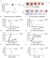
Figure 1
Figure 1
RECK over expression is associated with delayed SiHa and SW756 derived tumor establishment, reduced tumor growth and increased survival in nude mice. (A). Differential tumor growth experiment in nude mice. Here, the experiment ended once most of the animals from the control group presented tumors with 500 mm3. (BG). The effect of RECK over expression on cervical cancer derived cells tumor establishment and overall survival was assessed in vivo. SiHa (B,D,F) and SW756 (C,E,G) cells over expressing RECK were subcutaneously injected in nude mice (2 × 10⁶ cells/mouse). Tumors were identified as established once they were measurable with a manual caliper and the kinetics of tumor establishment for each of the cell lines tested are illustrated in B and C. D and E. Representative data from a tumor growth kinetics experiment. Tumor volume was measured weekly for at least 150 days. Overall survival was determined as each animal presented a tumor volume of 500 mm3 during the 150 days of experiment (F and G). * p < 0.05 by Mann–Whitney (1A), Log rank (1B, 1C, 1F and 1G) or two-way ANOVA (1D and 1E) test.
Figure 2
Figure 2
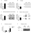
RECK over expression reduces SiHa and SW756 cell lines invasion potential and 3D growth. (A,B) Chamber migration and invasion assays. Cells present in 10 independent fields per chamber were counted. (C) Anchorage independent growth assay. After 30 days in culture, three-dimensional colonies were stained using MTT and all visible colonies were counted. (D,E) Spheroid formation assay. After 7 days in culture, spheroids were dissociated, stained with Trypan blue and counted. (F) Gelatinase activity assay. Zymography was carried out by subjecting 30 ug of protein to SDS-PAGE supplemented with 5% gelatin. After electrophoresis, the gel was incubated in an activation solution at 37 °C for 48 h, the gel was then stained with 0.5% crystal violet and images were developed with use of MultiDoc-ItTM 120 Imaging System (Analytic Jena, Upland, CA, USA). All experiments were carried out in triplicates. * p < 0.05 by Mann–Whitney (2A–E) test.
Figure 3
Figure 3
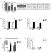
RECK over expression is associated with alterations in tumor microenvironment content. The injection protocol for tumor establishment was the same as described in Figure 1A. Here, the experiment ended once most of the animals from the control group presented tumors with 500 mm3. (A). Intra-tumoral populations were characterized by immunostaining with anti-CD31 and CD45 antibodies, followed by flow cytometry. Tumor fractions of at least three animals from each group were used for the analysis and at least 1 × 10⁵ cells/events were acquired. The EGFP+ population was identified as tumor cells. CD31+ and CD45+ populations identified endothelial cells and inflammatory infiltrate, respectively. Significant differences in population proportion are indicated in bold font in the table. (B,C,E) Tumor (B), endothelial (C) and inflammatory (E) cell populations stained with DAPI were evaluated on hypodiploid DNA content (SubG1) using flow cytometry. (D) Local inflammatory infiltrate population relative to 100,000 tumor cells. * p < 0.05 by Kruskal–Wallis (3A) or Mann–Whitney (3B–E) test.
Figure 4
Figure 4
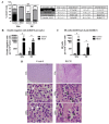
RECK over expression is associated with increased proportion of specific inflammatory infiltrate cell populations. (A). Intra-tumoral populations were characterized by immunostaining with CD45, Ly6C, Ly6G, F4/80 and CD49b antibodies, followed by flow cytometry. Tumor fractions of at least three animals from each group were used for the analysis and at least 1 × 10⁵ cells/events were acquired. Significant differences in population proportion are indicated in bold font in the table. (B,C). Proportion of Double negative (B) and NK cells (C) relative to 100,000 tumor cells. (D). Representation of hematoxylin/eosin tumor sections used for morphology evaluation of the inflammatory infiltrate in SW756 RECK+ tumors. White arrows identify cells with monocyte and macrophage morphology and white arrow heads indicate lymphoid phenotype cells. * p < 0.05 by Kruskal–Wallis (4A) or Mann–Whitney (4B and 4C) test.
Figure 5
Figure 5
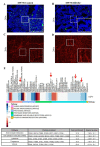
SW756 RECK+ tumors present collagenase activity, higher collagen content and increased levels of collagenase inhibitors. (A,B). In situ zymography assay carried out with SW756 tumors using DQ Collagen IV (green signal represents collagen breakdown). (C,D) Representative images showing Picro Sirius fluorescent staining of SW756 tumors (red signal shows collagen fibers). All fluorescent assays were carried out in the presence of DAPI staining and the merged images were processed using Image J software. (E). Heatmap representation of differentially expressed proteins in RECK+ tumors when compared to control. The fold change is expressed in log2 scale; where blue and red scale represents increased and decreased relative expression, respectively. Antibody arrays were depicted in different colors on the far-left side, below the heatmap. Proteins were identified as differentially expressed when fold change, expressed in log2, was over 0.58 or under −0.58. Red arrows show differentially expressed proteins detected in more than one antibody array. The table displays the enrichment analysis in KEGG pathway database with use of DAVID web tool. Statistical analysis was performed with Fisher’s exact test (bottom).
Figure 6
Figure 6
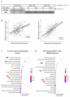
Extracellular matrix remodeling is one of the most enriched biological processes associated with RECK expression in both cervical cancer and HNSCC patients. (A). Correlation between SerpinE1 or TIMP2 mRNA levels and RECK mRNA expression in cervical cancer and HNSCC clinical samples deposited in TCGA platform. (B). Representative image of SerpinE1 and TIMP2 correlation with RECK expression in cervical cancer. Spearman’s correlation analyses between RECK and TIMP2 or RECK and SERPINE1/PAI1 mRNA levels in both cervical cancer and HNSCC patients from TCGA database. (C,D). GO enrichment analysis focused on biological processes most associated with RECK expression in cervical cancer (C) and HPV positive HNSCC clinical samples (D). Red arrows show biological processes enriched in cervical cancer and HNSCC. Orange arrow indicates the enrichment of the cell adhesion process in agreement with animal model findings.
Figure 7
Figure 7
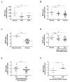
RECK expression is down regulated in cervical intraepithelial neoplasias (CIN) and invasive carcinomas being associated with worse clinical outcome of cervical cancer patients. (A). RECK gene expression is down regulated in CIN1 and CIN3 when compared with normal cervical tissues (GSE51993). (B,C). RECK gene expression is decreased in CIN and invasive carcinomas, when compared to normal cervical tissues (GSE7803 and GSE39001). (D). RECK relative expression is down regulated in women with HPV positive CIN that progressed to CIN3 or worse, when compared to women with no HPV infection (GSE75132). (E) RECK expression is down regulated in early-stage cervical carcinoma patients who presented pelvic lymph node metastasis, when compared to the same stage cervical carcinoma patients without metastasis (GSE26511). (F). RECK expression is up regulated in cervical cancer patients who presented better response to chemotherapy when compared to patients with no treatment response (GSE70035). * p < 0.05 by Kruskal–Wallis (7A, 7B and 7D) or Mann–Whitney (7C, 7E and 7F) test.
Figure 8
Figure 8
Proposed global effects of RECK expression in a cervical cancer model. Animals s.c. injected with cervical cancer derived cells transduced to over express RECK present delayed establishment and reduced tumor volume when compared to control. RECK over expressing cells are represented with light orange cell membrane and the red circles in mice lateral side represent the induced tumors. RECK+ tumors also showed reduced proportion of tumor and endothelial cells, the latter was depicted here in dark red blood vessels. Moreover, we observed specific increased proportion of Double negative inflammatory infiltrate (pie charts in the top) and higher collagen content (bright red linear structures) in RECK+ tumors. Finally, RECK over expression correlated with higher levels of SerpinE1/PAI-1, TIMP-2 and CD40/TNFRSF5 proteins, while presented an inverse correlation with IL-8 protein expression (on the far-right side).

Similar articles

Cited by

References

    1. Bray F., Ferlay J., Soerjomataram I., Siegel R.L., Torre L.A., Jemal A. Global cancer statistics 2018: GLOBOCAN estimates of incidence and mortality worldwide for 36 cancers in 185 countries. CA Cancer J. Clin. 2018;68:394–424. doi: 10.3322/caac.21492. - DOI - PubMed
    1. Ferlay J., Colombet M., Soerjomataram I., Mathers C., Parkin D.M., Piñeros M., Znaor A., Bray F. Estimating the global cancer incidence and mortality in 2018: GLOBOCAN sources and methods. Int. J. Cancer. 2019;144:1941–1953. doi: 10.1002/ijc.31937. - DOI - PubMed
    1. Schlecht N.F., Kulaga S., Robitaille J., Ferreira S., Santos M., Miyamura R.A., Duarte-Franco E., Rohan T.E., Ferenczy A., Villa L.L., et al. Persistent human papillomavirus infection as a predictor of cervical intraepithelial neoplasia. JAMA. 2001;286:3106–3114. doi: 10.1001/jama.286.24.3106. - DOI - PubMed
    1. Zur Hausen H. Papillomaviruses and cancer: From basic studies to clinical application. Nat. Rev. Cancer. 2002;2:342–350. doi: 10.1038/nrc798. - DOI - PubMed
    1. Tommasino M. The human papillomavirus family and its role in carcinogenesis. Semin. Cancer Biol. 2014;26:13–21. doi: 10.1016/j.semcancer.2013.11.002. - DOI - PubMed

LinkOut - more resources