. 2021 Sep;142(3):515-536.
doi: 10.1007/s00401-021-02333-z. Epub 2021 Jun 1.

DDX17 is involved in DNA damage repair and modifies FUS toxicity in an RGG-domain dependent manner

Affiliations

DDX17 is involved in DNA damage repair and modifies FUS toxicity in an RGG-domain dependent manner

Tyler R Fortuna et al. Acta Neuropathol. 2021 Sep.

Abstract

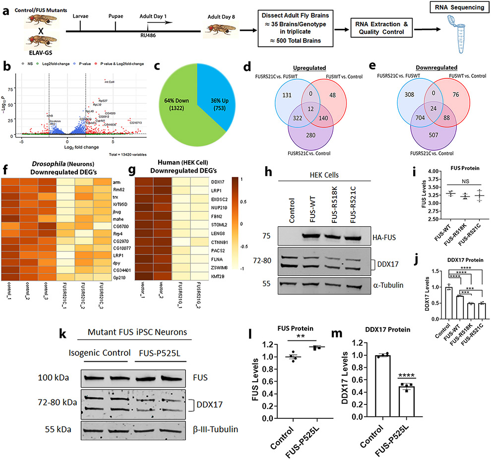
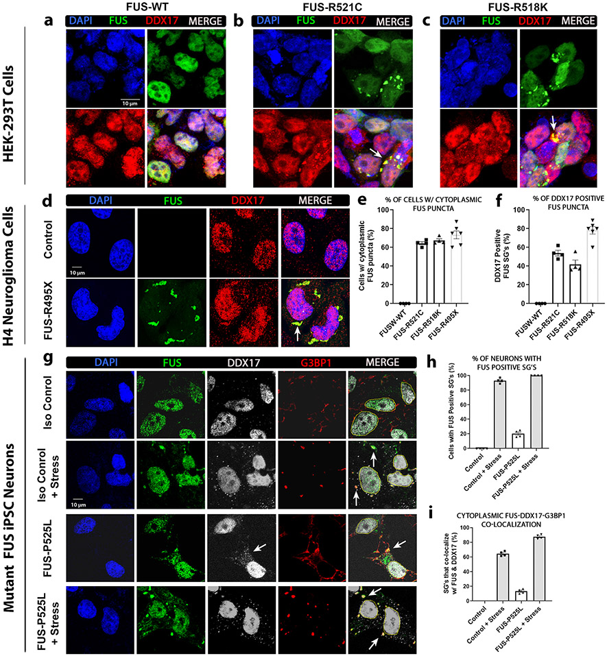
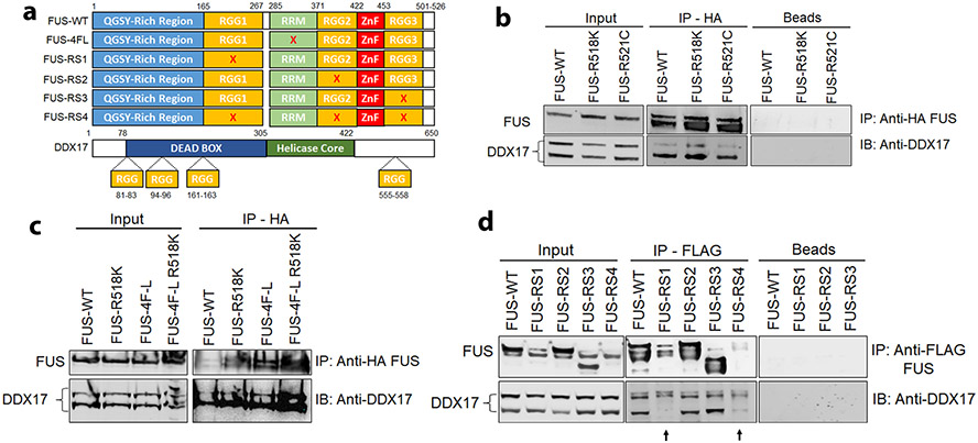
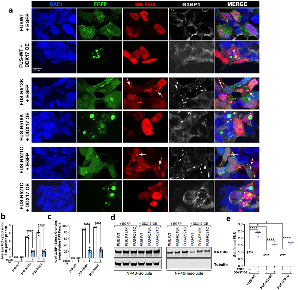
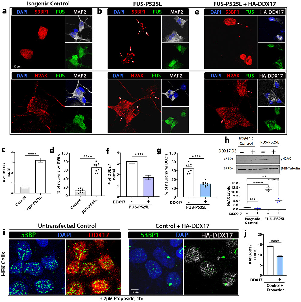
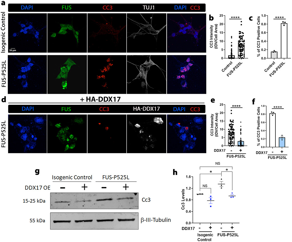
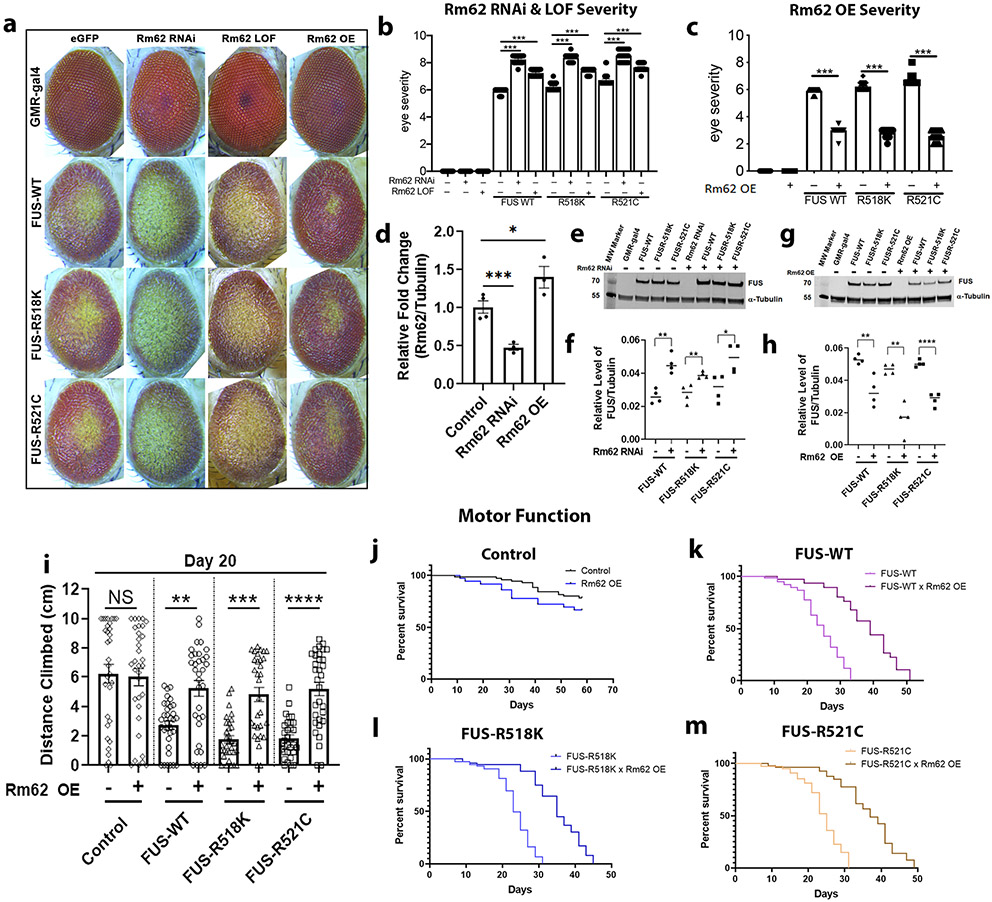
Mutations in the RNA binding protein, Fused in Sarcoma (FUS), lead to amyotrophic lateral sclerosis (ALS), the most frequent form of motor neuron disease. Cytoplasmic aggregation and defective DNA repair machinery are etiologically linked to mutant FUS-associated ALS. Although FUS is involved in numerous aspects of RNA processing, little is understood about the pathophysiological mechanisms of mutant FUS. Here, we employed RNA-sequencing technology in Drosophila brains expressing FUS to identify significantly altered genes and pathways involved in FUS-mediated neurodegeneration. We observed the expression levels of DEAD-Box Helicase 17 (DDX17) to be significantly downregulated in response to mutant FUS in Drosophila and human cell lines. Mutant FUS recruits nuclear DDX17 into cytoplasmic stress granules and physically interacts with DDX17 through the RGG1 domain of FUS. Ectopic expression of DDX17 reduces cytoplasmic mislocalization and sequestration of mutant FUS into cytoplasmic stress granules. We identified DDX17 as a novel regulator of the DNA damage response pathway whose upregulation repairs defective DNA damage repair machinery caused by mutant neuronal FUS ALS. In addition, we show DDX17 is a novel modifier of FUS-mediated neurodegeneration in vivo. Our findings indicate DDX17 is downregulated in response to mutant FUS, and restoration of DDX17 levels suppresses FUS-mediated neuropathogenesis and toxicity in vivo.

Keywords: ALS/FTD; DDX17; DNA-damage repair; Drosophila; FUS; Motor neuron disease; Neurodegeneration; RGG-domain; iPSC neurons.


Figures

Fig. 1
Fig. 1
Mutant FUS downregulates DDX17 in Drosophila brains and human cells. a Schematic showing the experimental setting used for sample preparation for RNA-sequencing. Wild-type and mutant FUS expression in Drosophila brains was achieved by a conditional neuronal driver, ELAV-GS, under the presence of the RU486 ligand. Drosophila were placed on treated RU486 drug food for 8 days, followed by brain dissection, RNA extraction, and RNA sequencing. b Volcano plot showing differentially expressed genes between FUS-R521C and control. Data for non-significant DEGs are plotted in black. Data for significant DEGs (FDR adjusted p value < 0.05) are plotted in blue. Data for significant DEGs (Bonferroni corrected p value < 0.05) and log2 fold changes less than or equal to – 1 or greater than or equal to 1 are plotted in red. c Pie chart showing the percentage and number of upregulated and downregulated genes in respect to FUS-R521C vs. control. d Venn diagram illustrating the number of upregulated DEGs (FDR adjusted p value < 0.05) between control, FUS-WT, and FUS-R521C. e Venn diagram illustrating the number of downregulated DEGs (FDR adjusted p value < 0.05) between control, FUS-WT, and FUS-R521C. f, g Heat maps of the 14-common downregulated DEGs of FUS-R521C mutation in Drosophila and FUS-R521G in HEK cells. Protein expression levels of endogenous DDX17 in (h) HEK cells transfected with FUS constructs and (k) FUS iPSC neurons harboring the FUS-P525L mutation. i, j Quantification of FUS and endogenous DDX17 protein levels from HEK cells (panel h) normalized to tubulin, indicating equivalent FUS expression in all groups and downregulated DDX17 protein levels in response to FUS constructs (n = 4 blots per condition, One-way ANOVA w/ Turkey’s multiple comparisons). l, m Quantification of endogenous FUS and DDX17 protein levels from mutant FUS iPSC neurons (panel k) normalized to the neuronal marker β-tubulin III (TUJ1). DDX17 protein levels are significantly downregulated in mutant FUS-P525L neurons compared to isogenic control (n = 4 blots, unpaired student t-test). Error bars indicate S.E.M. **p < 0.01; ***p < 0.001; ****p < 0.0001
Fig. 2
Fig. 2
DDX17 is recruited into cytoplasmic stress granules in response to mutant FUS. HEK or H4 cells were transfected with the indicated FUS constructs (wild-type, R518K, R521C, or R495X). ac For IF in HEK cells, the cells were transfected with HA tagged FUS-WT and mutants FUS-R521C and FUS-R518K. Representative confocal images showing the distributions of FUS and endogenous DDX17 in HEK cells. DAPI was used as a nuclear marker. White arrows indicate representative cytoplasmic FUS puncta in both FUS mutants that co-localize with DDX17. d For IF in H4 cells, the cells were transfected with EGFP tagged mutant FUS-R495X. Representative images showing the distributions of EGFP and endogenous DDX17 in H4 cells. DAPI was used as a nuclear marker. White arrows indicate representative cytoplasmic FUS puncta in mutant FUS-R495X that co-localize with DDX17. e Quantification of the percentage of cells with cytoplasmic FUS puncta per each FUS condition (n = 90–100 cells per FUS group). f Quantification of the percentage of DDX17 positive cytoplasmic FUS puncta. (n = 90–100 cells per FUS group). g Mutant FUS iPSC neurons expressing endogenous wild-type and mutant FUS with and without stress. Representative images showing the distributions of endogenous FUS, DDX17, and the stress granule marker G3BP1. White arrows in the 3rd column indicate cytoplasmic DDX17 in response to mutant FUS-P525L. White arrows in the last column indicate representative cytoplasmic colocalization of FUS, DDX17, and G3BP1. h Quantification of the percentage of neurons with cytoplasmic FUS puncta per each condition (n = 65–75 neurons per group). i Quantification of the percentage of G3BP1 positive SG’s that colocalize with both FUS and DDX17 (n = 65–75 neurons per group)
Fig. 3
Fig. 3
DDX17 and FUS physically interact via the RGG1 domain of FUS. a Schematic illustration of FUS constructs and DDX17 each harboring RGG domains. Mutated FUS 4F-L (F305L, F341L, F359L, F368L) disrupts the conserved RRM domain of FUS. Mutated FUS-RS1, RS2, and RS3 convert all arginine amino acids of each separate RGG domain to serine amino acids, thus disrupting individual RGG domains of FUS. In mutant FUS-RS4, arginine amino acids in all RGG domains were converted to serines, thus disrupting all RGG binding domains of FUS. b Immunoblot of co-immunoprecipitation of HA tagged FUS-WT, FUS-R518K, and FUS-R521C from HEK cells. Immunoprecipitation with HA antibody showed endogenous DDX17 (p72) in the input control samples and pulled down with HA-FUS-WT, FUS-R518K, and FUS-R521C, but absent from the negative control containing beads only. c Immunoblot of co-immunoprecipitation of HA-FUS-WT, FUS-R518K, FUS 4F-L, and FUS-R518K 4F-L from HEK cells expressing HA-FUS. Immunoprecipitation with HA antibody showed endogenous DDX17 (p72) in the input control samples and pulled down with HA-FUS-WT, FUS-R518K, FUS 4F-L, and FUS-R518K 4F-L. d Immunoblot of co-immunoprecipitation of FLAG-FUS-WT, FUS-RS1, FUS-RS2, FUS-RS3, and FUS-RS4 from HEK cells expressing FLAG-FUS. Immunoprecipitation with FLAG antibody showed endogenous DDX17 (p72) in the input control samples and pulled down with FLAG-FUS-WT, FUS-RS2, and FUS-RS3, but absent from FUS-RS1, FUS-RS4, and the negative control containing beads only. Interestingly, RGG1 disruption of FUS disrupts the interaction between FUS and DDX17
Fig. 4
Fig. 4
Ectopic DDX17 expression reduces insoluble cytoplasmic FUS formation and sequestration into stress granules. HEK cells were co-transfected with the indicated HA tagged FUS constructs (wild-type, R518K, or R521C) and either control EGFP or DDX17-EGFP over expression. a Representative confocal images showing FUS, DDX17 OE, and stress granule marker G3BP1 distributions in HEK cells. For IF, cells were co-transfected with HA tagged FUS (wild-type and mutant) and control EGFP, or EGFP tagged DDX17 OE at equal concentrations. White arrows indicate representative cytoplasmic FUS puncta in both FUS mutants that also co-localize with the stress granule marker, G3BP1. (b) Quantification of the average number of cytoplasmic FUS puncta per cell confirms that over expression of DDX17 significantly reduced the number of cytoplasmic FUS puncta in the R518K and R521C FUS mutants. (n = 55–70 cells per FUS group, One-way ANOVA w/ Turkey’s multiple comparisons). c Quantification of the percentage of G3BP1 positive cytoplasmic stress granules sequestering mutant FUS. Interestingly, overexpression of DDX17 significantly reduced mutant FUS integration into SGs (n = 55–70 cells per FUS group, One-way ANOVA w/ Turkey’s multiple comparisons). d Representative blots of soluble–insoluble fractionation of FUS protein for FUS-WT, FUS-R518K, and FUS-R521C plus EGFP control and FUS-WT, FUS-R518K, FUS-R521C plus DDX17 OE. Tubulin was used as a soluble loading control. e Quantification of soluble/insoluble (Sol/Insol) FUS formation from representative blots shown in panel (d) (n = 4 blots per condition, One-way ANOVA w/ Turkey’s multiple comparisons). Over expression of DDX17 significantly increases the sol/insol fraction of FUS expressing groups. Error bars indicate S.E.M. *p < 0.05, ****p < 0.0001
Fig. 5
Fig. 5
Upregulation of DDX17 mitigates DNA damage exhibited in mutant FUS iPSC neurons. Representative confocal images of a isogenic control and b, e mutant FUS iPSC neurons with and without overexpression of HA-DDX17 from the same set of neuronal differentiations. Neurons were probed for FUS, the DNA damage and double-strand break (DSB) markers γH2AX and p53-binding protein 1 (53BP1), HA-DDX17, and the neuronal marker microtubule-associated protein 2 (MAP2). Nuclei were stained with DAPI. c, d Quantification of the number of DNA DSBs and the percentage of neurons with DSBs between control (panel a) and mutant FUS-P525L (panel b) (n = 60–70 neurons, unpaired students t-test). Mutant FUS-P525L neurons show a significant increase in the number of DNA DSBs per neuron compared to control. f, g Quantification of the number of DNA DSBs and the percentage of neurons with DSBs between mutant FUS-P525L (panel b) and mutant FUS-P525L with overexpression of HA-DDX17 (panel e) (n = 60–70 neurons, unpaired students t-test). Mutant FUS-P525L neurons expressing HA-DDX17 show a significant decrease in the number of DNA DSBs per neuron compared to mutant FUS-P525L alone. h Representative blots of γH2AX protein in isogenic control and mutant FUS-P525L neuronal cells with and without overexpression of DDX17. β-tubulin III was used as a loading control (n = 3 blots, One-way ANOVA w/ Turkey’s multiple comparisons). FUS-P525L neurons showed a significant increase in γH2AX protein levels compared to FUS-P525L neurons expressing HA-DDX17. i Confocal images of HEK cells exposed to the DNA damage-inducing agent, etoposide (2 μM), for 1 h after being transduced with and without HA-DDX17 for 72 h. The cellular distributions of 53BP1, HA-DDX17, and endogenous DDX17 were assessed. j Quantification of the number of DNA DSBs between HEK cells exposed to etoposide with and without over expression of HA-DDX17 (n = 70–80 cells, unpaired students t-test). Cells over expressing DDX17 show a significant decrease in the number of DNA double-strand breaks per cell compared to control after etoposide exposure. Error bars indicate S.E.M. **p < 0.01, ****p < 0.0001
Fig. 6
Fig. 6
Upregulation of DDX17 protects against apoptotic activity of mutant FUS iPSC neurons. a Representative confocal images of isogenic control and mutant FUS iPSC neurons showing FUS, the apoptotic marker cleaved caspase 3 (CC3), HA-DDX17, and the neuronal marker β-tubulin III (TUJ1). Nuclei were stained with DAPI. b, c Quantification of integrated CC3 intensity per cell normalized to the area of each neuron (panel a) and the percentage of neurons that expressed CC3 signal (panel c) between FUS-P525L and control (n = 80 neurons, unpaired students t-test). FUS-P525L neurons showed a drastic increase in CC3 signal compared to control. d Representative confocal images of the same set of differentiated ALS mutant FUS-P525L iPSC neurons from (panel a) overexpressing HA-DDX17. The neurons were probed for the apoptotic marker CC3, the neuronal marker TUJ1, and DAPI. e, f Quantification of integrated CC3 intensity per cell normalized to the area of each neuron (panel e) and the percentage of neurons that expressed CC3 signal (panel f) between FUS-P525L and FUS-P525L with HA-DDX17 expression (n = 80 neurons, unpaired students t-test). g Representative blots of CC3 protein in isogenic control and mutant FUS-P525L neuronal cells with and without overexpression of DDX17. β-tubulin III was used as a loading control (n = 3 blots, One-way ANOVA w/ Turkey’s multiple comparisons). h FUS-P525L neurons showed a drastic increase in CC3 protein levels compared to FUS-P525L neurons overexpressing HA-DDX17. Error bars indicate S.E.M. *p < 0.05, ****p < 0.0001
Fig. 7
Fig. 7
DDX17 Drosophila orthologue, Rm62, is a novel modifier of FUS toxicity in vivo. a Representative images of Drosophila eyes of wild-type and ALS-linked mutant FUS protein alone (left column), in combination with RNAi-mediated knockdown of Rm62 (middle left column), LOF-mediated Rm62 (middle right column), or with overexpression of Rm62OE (right column). b Quantification of eye degeneration severity from panel (a) confirms significant enhancement of FUS toxicity following depletion of Rm62, and c significant suppression of FUS toxicity following overexpression of Rm62 (n = 15–25 Drosophila per group, One-way ANOVA w/ Turkey’s multiple comparisons). d qPCR of RNA from (n = 4 biological replicates of 6 Drosophila heads per group) confirms significant knockdown of endogenous Rm62 in the RNAi line and significant overexpression in the Rm62OE line (One-way ANOVA w/ Turkey’s multiple comparisons). e Western blot analysis showing FUS and tubulin (housekeeping control) protein levels in Drosophila eyes expressing wild-type and mutant FUS, with and without Rm62 knockdown (n = 4 biological replicates of 5 Drosophila heads per group). f Quantification of FUS from panel e normalized to tubulin, indicating equivalent FUS expression in all FUS alone-expressing groups. Interestingly, knockdown of Rm62 further enhanced FUS protein levels (n = 4 blots per condition, One-way ANOVA w/ Turkey’s multiple comparisons). g Western blot analysis showing FUS and tubulin (housekeeping control) protein levels in Drosophila eyes expressing wild-type and mutant FUS, with and without Rm62 overexpression (n = 4 biological replicates of 5 Drosophila heads per group). h Quantification of FUS from panel g normalized to tubulin, indicating equivalent FUS expression in all FUS alone-expressing groups. Interestingly, overexpression of Rm62 significantly reduced FUS protein levels (n = 4 blots per condition, One-way ANOVA w/ Turkey’s multiple comparisons). i Quantification of the total height climbed (cm) of adult flies in the first 3 s of RING climbing assay. Over expression of Rm62 significantly improved the climbing ability of neuronal expressing FUS-WT, FUS-R518K, and FUS-R521C flies. (n = 30–35 adult flies, One-way ANOVA w/ Turkey’s multiple comparisons). jm Kaplan–Meier survival curve of adult flies conditionally expressing j control, k FUS-WT, l FUS-R518K, and m FUS-R521C under the neuronal specific driver, ELAV-GS, with and without Rm62 over expression. Over expression of Rm62 drastically improved longevity of FUS-WT, FUS-R518K, and FUS-R521C expressing flies. (n = 75 adult flies). Error bars indicate S.E.M. *p < 0.05; **p < 0.01; ***p < 0.001; ****p < 0.0001

Similar articles

Cited by

References

    1. Abdelmohsen K, Tominaga K, Lee EK, Srikantan S, Kang MJ, Kim MM et al. (2011) Enhanced translation by Nucleolin via G-rich elements in coding and non-coding regions of target mRNAs. Nucleic Acids Res 39(19):8513–8530. 10.1093/nar/gkr488 - DOI - PMC - PubMed
    1. Adams MM, Wang B, Xia Z, Morales JC, Lu X, Donehower LA et al. (2005) 53BP1 oligomerization is independent of its methylation by PRMT1. Cell Cycle 4(12):1854–1861. 10.4161/cc.4.12.2282 - DOI - PubMed
    1. Adamson B, Smogorzewska A, Sigoillot FD, King RW, Elledge SJ (2012) A genome-wide homologous recombination screen identifies the RNA-binding protein RBMX as a component of the DNA-damage response. Nat Cell Biol 14(3):318–328. 10.1038/ncb2426 - DOI - PMC - PubMed
    1. Alberti S, Hyman AA (2016) Are aberrant phase transitions a driver of cellular aging? BioEssays 38(10):959–968. 10.1002/bies.201600042 - DOI - PMC - PubMed
    1. Alonso A, Logroscino G, Jick SS, Hernan MA (2009) Incidence and lifetime risk of motor neuron disease in the United Kingdom: a population-based study. Eur J Neurol 16(6):745–751. 10.1111/j.1468-1331.2009.02586.x - DOI - PMC - PubMed

Publication types