Review
. 2021 Dec 15;9(2):eESP00112020.
doi: 10.1128/ecosalplus.ESP-0011-2020. Epub 2021 Jun 1.

Dynamics of Proteins and Macromolecular Machines in Escherichia coli

Affiliations
Review

Dynamics of Proteins and Macromolecular Machines in Escherichia coli

Maxime Leroux et al. EcoSal Plus. .

Abstract

Proteins are major contributors to the composition and the functions in the cell. They often assemble into larger structures, macromolecular machines, to carry out intricate essential functions. Although huge progress in understanding how macromolecular machines function has been made by reconstituting them in vitro, the role of the intracellular environment is still emerging. The development of fluorescence microscopy techniques in the last 2 decades has allowed us to obtain an increased understanding of proteins and macromolecular machines in cells. Here, we describe how proteins move by diffusion, how they search for their targets, and how they are affected by the intracellular environment. We also describe how proteins assemble into macromolecular machines and provide examples of how frequent subunit turnover is used for them to function and to respond to changes in the intracellular conditions. This review emphasizes the constant movement of molecules in cells, the stochastic nature of reactions, and the dynamic nature of macromolecular machines.

Keywords: DNA replication; flagellar motor; fluorescence microscopy; molecular machine; protein diffusion; protein dynamics; protein target search; replisome; ribosome.


Figures
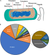
FIG 1
FIG 1
Structure and composition of Escherichia coli. (A) Schematic of the E. coli cell. Not shown are the pili and the capsule, as pili are formed only when the conjugation apparatus is expressed, and the capsule is not present in most lab-grown strains. (B) The composition of E. coli under exponential growth conditions (adapted from the Voronoi tree diagram in reference 216).
FIG 2
FIG 2
Microscope techniques to study proteins. (A) The use of fluorescent proteins like GFP allows users to view proteins within their native setting. (B) In fluorescence recovery after photobleaching (FRAP), the user photobleaches a small area of a cell. Then the recovery of fluorescence in that area is measured, reporting on the rate of movement of molecules. (C) Single-particle tracking photoactivatable localization microscopy allows the user to detect and track a single copy of a fluorescent molecule. In the example shown, activation light is used to photoconvert single proteins from the green to the red channel. This allows the user to track the movement of individual proteins. (D) In stepwise photobleaching, the user utilizes a strong laser power and fast acquisition rate settings. The stepwise drop of fluorescence intensity due to photobleaching allows the quantification of the number of molecules in an individual fluorescent spot.
FIG 3
FIG 3
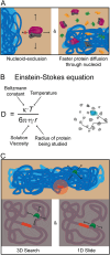
Proteins and DNA. (A) Diagram displaying both types of effects of the nucleoid on protein localization. The first is nucleoid exclusion, in which large protein assemblies (and other molecules, like plasmids) are excluded from the nucleoid region; these proteins usually diffuse to the poles. The second is the effect of the nucleoid on the diffusion of proteins. Due to greater crowding in the nucleoid free areas, proteins diffuse faster when traversing the nucleoid. (B) The Einstein-Stokes equation is generally used to describe the diffusion of proteins. (C) Proteins that search for their DNA target use two different strategies. They combine a 3D search with 1D diffusion on DNA to minimize the time to find their target.
FIG 4
FIG 4
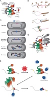
DNA replication. (A) Diagram representing the proteins involved in DNA replication. Grayscale objects represent stable component (DnaB). (B) DNA replication is initiated by DnaA, which melts DNA at a particular point of the chromosome, called origin of replication (oriC). Full occupancy of the DnaA-binding sites at oriC and the formation of DnaA oligomers (DnaA filaments) are needed for DNA unwinding. Once oriC is open, interactions between DnaA, DnaB, and the helicase loader DnaC mediate the loading of two DnaB on an ssDNA. Binding of primase and Pol III follows shortly after this. (C) The single E. coli chromosome begins its duplication during initiation at the oriC. The position of the replisome is highly variable, frequently switching from moving away to toward the cell center and often coming close to its sister replisome. However, the position of the replisome does not recapitulate the spatial organization of the chromosome in the nucleoid, staying closer to the cell center than expected if it were travel across all chromosomal DNA. (D) The components in the replisome can be split into 3 groups, stable, dynamic, and other. The helicase is the stable part of the replisome, able to bind for more than 15 min. Pol III*, which is comprised of the clamp loader (τ/γ, δ, δ′, χ, and ψ) and Pol III (α, ε, and θ), is very dynamic, lasting only about 10 s on DNA. SSB, the β-clamp and DnaG have their own dynamic behaviors. (E) Binding of Pol II and Pol IV to the replication fork is largely regulated by their concentration. These polymerases are normally present at low concentrations in the cell. The levels of the translesion polymerases are increased during the SOS response. This ultimately results in the displacement of Pol III from the β-clamp, the 3′ end in the nascent strand, or both.
FIG 5
FIG 5
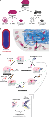
Ribosome dynamics. (A) Diagram representing the composition of the ribosome. The complete 70S ribosome is comprised of a 50S subunit and a 30S subunit. The 50S subunit is made from a 23S rRNA, 5S rRNA, and 33 ribosomal proteins. The 30S subunit is made from a 16S rRNA and 21 ribosomal proteins. (B) The complete 70S ribosome is excluded from the nucleoid. However, the individual subunits, 50S subunit and 30S subunit, can enter the nucleoid and bind to newly synthetized mRNA. Once the 70S ribosome is formed, it will quickly be pushed out of the nucleoid. (C) Schematic of the elongation cycle. The aminoacyl-tRNA, in a ternary complex with the elongation factor EF-Tu bound to GTP, interacts with the ribosome. This is the first step in the elongation cycle of the amino acid complex. Multiple instances of binding of the ternary complex to the ribosome are needed before incorporation of the correct amino acid residue in the growing chain. Each ribosome binds to at least 3 copies of the ternary complex. Binding to a new ribosome and unbinding from it occur at rates of 1 to 2 ms, with over 20 interactions before a successful incorporation. EF-P then binds to help with the peptidyl transfer. Once the new amino acid chain is formed, the EF-Tu is released from the complex. The chain is then separated from the tRNA located in the P site. The EF-P is removed and an EF-G bound to GTP interacts with the ribosome. The EF-G helps with the translocation of both tRNAs from the P and A site to the E and P site. The EF-G is released from the complex. Once complete, the cycle begins again. (D) The peptidyl transfer step is known to be a slow step. The binding of EF-P has been shown to speed up this step, especially with the incorporation of proline at sites with multiple consecutive Pro codons.
FIG 6
FIG 6
(A) The composition of the flagellar motor. The flagella have a filament and hook, which have parts outside the cell. The basal body is composed of a series of rings that form the rotor of the flagellar motor. From the outside of the cell to the inside, those rings are called the L, P, MS, and C rings. They surround a structure called the rod that is connected to the hook. The stator proteins are associated with those rings but do not copurify with them. (B) The stator complex provides the energy necessary for the rotation of the flagellum. There are about 11 copies in active flagella. However, regulation of rotation rate of the flagellum is correlated with stoichiometry of the stator. The change in the number of stators is linked to the magnitude of torque needed by the flagellum. In order to do this, E. coli needs to detect the torque requirement at a given time. This is an example of mechanosensing. This sensing is done by the stator complexes themselves. The remodeling of the stators in response to mechanical stimuli has been linked to conformational changes at higher torque that expose more binding sites. These changes strengthen the binding interaction between the stators and the rotor. (C) The C ring has also been shown to be a dynamic structure, with an exchange of the FliM subunits. FliM forms a switch complex with another protein of the C ring, FliN. Consequently, this flagellar subunit is also subject to a dynamic exchange, albeit on a different timescale.

Similar articles

References

    1. Hu P, Janga SC, Babu M, Díaz-Mejía JJ, Butland G, Yang W, Pogoutse O, Guo X, Phanse S, Wong P, Chandran S, Christopoulos C, Nazarians-Armavil A, Nasseri NK, Musso G, Ali M, Nazemof N, Eroukova V, Golshani A, Paccanaro A, Greenblatt JF, Moreno-Hagelsieb G, Emili A. 2009. Global functional atlas of Escherichia coli encompassing previously uncharacterized proteins. PLoS Biol 7:e1000096. doi:10.1371/journal.pbio.1000096. - DOI - PMC - PubMed
    1. Balzani V, Credi A, Raymo FM, Stoddart JF. 2000. Artificial molecular machines. Angew Chem Int Ed 39:3348–3391. doi:10.1002/1521-3773(20001002)39:19<3348::AID-ANIE3348>3.0.CO;2-X. - DOI - PubMed
    1. Ornes S. 2018. News Feature: What’s the best way to build a molecular machine? Proc Natl Acad Sci U S A 115:9327–9330. doi:10.1073/pnas.1811689115. - DOI - PMC - PubMed
    1. Neidhardt FCN, Ingraham JL, Schaechter M. 1990. Physiology of the bacterial cell: a molecular approach.article-title. Sinauer Associates, Sunderland, MA.
    1. Milo R. 2013. What is the total number of protein molecules per cell volume? A call to rethink some published values. Bioessays 35:1050–1055. doi:10.1002/bies.201300066. - DOI - PMC - PubMed

Publication types

Substances

LinkOut - more resources