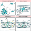
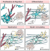
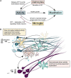
Glial Cells Promote Myelin Formation and Elimination
- PMID: 34046407
- PMCID: PMC8144722
- DOI: 10.3389/fcell.2021.661486
Glial Cells Promote Myelin Formation and Elimination
Abstract
Building a functional nervous system requires the coordinated actions of many glial cells. In the vertebrate central nervous system (CNS), oligodendrocytes myelinate neuronal axons to increase conduction velocity and provide trophic support. Myelination can be modified by local signaling at the axon-myelin interface, potentially adapting sheaths to support the metabolic needs and physiology of individual neurons. However, neurons and oligodendrocytes are not wholly responsible for crafting the myelination patterns seen in vivo. Other cell types of the CNS, including microglia and astrocytes, modify myelination. In this review, I cover the contributions of non-neuronal, non-oligodendroglial cells to the formation, maintenance, and pruning of myelin sheaths. I address ways that these cell types interact with the oligodendrocyte lineage throughout development to modify myelination. Additionally, I discuss mechanisms by which these cells may indirectly tune myelination by regulating neuronal activity. Understanding how glial-glial interactions regulate myelination is essential for understanding how the brain functions as a whole and for developing strategies to repair myelin in disease.
Keywords: astrocyte; glial-glial interactions; microglia; myelin; oligodendrocyte.
Copyright © 2021 Hughes.
Conflict of interest statement
The author declares that the research was conducted in the absence of any commercial or financial relationships that could be construed as a potential conflict of interest.
Figures


Similar articles
-
The oligodendrocyte-enriched orphan G protein-coupled receptor Gpr62 is dispensable for central nervous system myelination.Neural Dev. 2021 Nov 29;16(1):6. doi: 10.1186/s13064-021-00156-y. Neural Dev. 2021. PMID: 34844642 Free PMC article.
-
Emerging concepts in oligodendrocyte and myelin formation, inputs from the zebrafish model.Glia. 2023 May;71(5):1147-1163. doi: 10.1002/glia.24336. Epub 2023 Jan 16. Glia. 2023. PMID: 36645033 Review.
-
Fbxw7 Limits Myelination by Inhibiting mTOR Signaling.J Neurosci. 2015 Nov 4;35(44):14861-71. doi: 10.1523/JNEUROSCI.4968-14.2015. J Neurosci. 2015. PMID: 26538655 Free PMC article.
-
Axonal Regulation of Central Nervous System Myelination: Structure and Function.Neuroscientist. 2018 Feb;24(1):7-21. doi: 10.1177/1073858417703030. Epub 2017 Apr 11. Neuroscientist. 2018. PMID: 28397586 Review.
-
Myelination of Neuronal Cell Bodies when Myelin Supply Exceeds Axonal Demand.Curr Biol. 2018 Apr 23;28(8):1296-1305.e5. doi: 10.1016/j.cub.2018.02.068. Epub 2018 Apr 5. Curr Biol. 2018. PMID: 29628374 Free PMC article.
Cited by
-
Functional myelin in cognition and neurodevelopmental disorders.Cell Mol Life Sci. 2024 Apr 13;81(1):181. doi: 10.1007/s00018-024-05222-2. Cell Mol Life Sci. 2024. PMID: 38615095 Free PMC article. Review.
-
Morphogenetic theory of mental and cognitive disorders: the role of neurotrophic and guidance molecules.Front Mol Neurosci. 2024 Apr 3;17:1361764. doi: 10.3389/fnmol.2024.1361764. eCollection 2024. Front Mol Neurosci. 2024. PMID: 38646100 Free PMC article. Review.
-
Association between type 2 inflammatory diseases and neurodevelopmental disorders in low-birth-weight children and adolescents.Front Psychol. 2024 Feb 22;15:1292071. doi: 10.3389/fpsyg.2024.1292071. eCollection 2024. Front Psychol. 2024. PMID: 38455122 Free PMC article.
-
Spatial proteomics reveals human microglial states shaped by anatomy and neuropathology.Res Sq [Preprint]. 2023 Jun 2:rs.3.rs-2987263. doi: 10.21203/rs.3.rs-2987263/v1. Res Sq. 2023. PMID: 37398389 Free PMC article. Preprint.
-
Insights into myelin dysfunction in schizophrenia and bipolar disorder.World J Psychiatry. 2022 Feb 19;12(2):264-285. doi: 10.5498/wjp.v12.i2.264. eCollection 2022 Feb 19. World J Psychiatry. 2022. PMID: 35317338 Free PMC article. Review.
References
-
- Albrecht P. J., Murtie J. C., Ness J. K., Redwine J. M., Enterline J. R., Armstrong R. C., et al. (2003). Astrocytes produce CNTF during the remyelination phase of viral-induced spinal cord demyelination to stimulate FGF-2 production. Neurobiol. Dis. 13 89–101. 10.1016/S0969-9961(03)00019-6 - DOI - PubMed
Publication types
Grants and funding
LinkOut - more resources
Full Text Sources
Other Literature Sources

