Diverse Mucosal-Associated Invariant TCR Usage in HIV Infection
- PMID: 34045357
- PMCID: PMC10563122
- DOI: 10.4049/immunohorizons.2100026
Diverse Mucosal-Associated Invariant TCR Usage in HIV Infection
Abstract
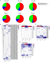
Mucosal-associated invariant T (MAIT) cells are innate-like T cells that specifically target bacterial metabolites but are also identified as innate-like sensors of viral infection. Individuals with chronic HIV-1 infection have lower numbers of circulating MAIT cells compared with healthy individuals, yet the features of the MAIT TCR repertoire are not well known. We isolated and stimulated human PBMCs from healthy non-HIV-infected donors (HD), HIV-infected progressors on antiretroviral therapy, and HIV-infected elite controllers (EC). We sorted MAIT cells using flow cytometry and used a high-throughput sequencing method with bar coding to link the expression of TCRα, TCRβ, and functional genes of interest at the single-cell level. We show differential patterns of MAIT TCR usage among the groups. We observed expansions of certain dominant MAIT clones in HIV-infected individuals upon Escherichia coli stimulation, which was not observed in clones of HD. We also found different patterns of CDR3 amino acid distributions among the three groups. Furthermore, we found blunted expression of phenotypic genes in HIV individuals; most notably, HD mounted a robust IFNG response to stimulation, whereas both HIV-infected progressors and EC did not. In conclusion, our study describes the diverse MAIT TCR repertoire of persons with chronic HIV-1 infection and suggest that MAIT clones of HIV-infected persons may be primed for expansion more than that of noninfected persons. Further studies are needed to examine the functional significance of unique MAIT cell TCR usage in EC.
Copyright © 2021 The Authors.
Conflict of interest statement
DISCLOSURES
The authors have no financial conflicts of interest.
Figures


Similar articles
-
CD161+ MAIT cells are severely reduced in peripheral blood and lymph nodes of HIV-infected individuals independently of disease progression.PLoS One. 2014 Nov 4;9(11):e111323. doi: 10.1371/journal.pone.0111323. eCollection 2014. PLoS One. 2014. PMID: 25369333 Free PMC article.
-
Human liver-derived MAIT cells differ from blood MAIT cells in their metabolism and response to TCR-independent activation.Eur J Immunol. 2021 Apr;51(4):879-892. doi: 10.1002/eji.202048830. Epub 2021 Jan 25. Eur J Immunol. 2021. PMID: 33368232
-
Functional and Activation Profiles of Mucosal-Associated Invariant T Cells in Patients With Tuberculosis and HIV in a High Endemic Setting.Front Immunol. 2021 Mar 22;12:648216. doi: 10.3389/fimmu.2021.648216. eCollection 2021. Front Immunol. 2021. PMID: 33828558 Free PMC article.
-
Mucosal-associated invariant T cells: A cryptic coordinator in HIV-infected immune reconstitution.J Med Virol. 2022 Jul;94(7):3043-3053. doi: 10.1002/jmv.27696. Epub 2022 Mar 12. J Med Virol. 2022. PMID: 35243649 Review.
-
The Immune Modulating Properties of Mucosal-Associated Invariant T Cells.Front Immunol. 2020 Aug 13;11:1556. doi: 10.3389/fimmu.2020.01556. eCollection 2020. Front Immunol. 2020. PMID: 32903532 Free PMC article. Review.
Cited by
-
Characterization of the TCRβ repertoire of peripheral MR1-restricted MAIT cells in psoriasis vulgaris patients.Sci Rep. 2023 Nov 28;13(1):20990. doi: 10.1038/s41598-023-48321-z. Sci Rep. 2023. PMID: 38017021 Free PMC article.
-
IL7RA single nucleotide polymorphisms are associated with the size and function of the MAIT cell population in treated HIV-1 infection.Front Immunol. 2022 Oct 20;13:985385. doi: 10.3389/fimmu.2022.985385. eCollection 2022. Front Immunol. 2022. PMID: 36341446 Free PMC article.
References
-
- Nicolosi A, Corrêa Leite ML, Musicco M, Arici C, Gavazzeni G, Lazzarin A, and Italian Study Group on HIV Heterosexual Transmission. 1994. The efficiency of male-to-female and female-to-male sexual transmission of the human immunodeficiency virus: a study of 730 stable couples. Epidemiology 5: 570–575. - PubMed
-
- Stieh DJ, Maric D, Kelley ZL, Anderson MR, Hattaway HZ, Beilfuss BA, Rothwangl KB, Veazey RS, and Hope TJ. 2014. Vaginal challenge with an SIV-based dual reporter system reveals that infection can occur throughout the upper and lower female reproductive tract. PLoS Pathog. 10: e1004440. - PMC - PubMed
-
- Belyakov IM, and Berzofsky JA. 2004. Immunobiology of mucosal HIV infection and the basis for development of a new generation of mucosal AIDS vaccines. Immunity 20: 247–253. - PubMed
Publication types
MeSH terms
Substances
Grants and funding
LinkOut - more resources
Full Text Sources
Other Literature Sources
Medical

