Review
. 2021 Jul 5;50(13):7820-7880.
doi: 10.1039/d0cs00729c.

The multifaceted nature of antimicrobial peptides: current synthetic chemistry approaches and future directions

Affiliations
Review

The multifaceted nature of antimicrobial peptides: current synthetic chemistry approaches and future directions

Bee Ha Gan et al. Chem Soc Rev. .

Erratum in

Abstract

Bacterial infections caused by 'superbugs' are increasing globally, and conventional antibiotics are becoming less effective against these bacteria, such that we risk entering a post-antibiotic era. In recent years, antimicrobial peptides (AMPs) have gained significant attention for their clinical potential as a new class of antibiotics to combat antimicrobial resistance. In this review, we discuss several facets of AMPs including their diversity, physicochemical properties, mechanisms of action, and effects of environmental factors on these features. This review outlines various chemical synthetic strategies that have been applied to develop novel AMPs, including chemical modifications of existing peptides, semi-synthesis, and computer-aided design. We will also highlight novel AMP structures, including hybrids, antimicrobial dendrimers and polypeptides, peptidomimetics, and AMP-drug conjugates and consider recent developments in their chemical synthesis.


Conflict of interest statement

There are no conflicts to declare.

Figures

Fig. 1
Fig. 1. Scheme of possible bacterial mechanisms of resistance. Some examples of antibiotics affected by different mechanisms of resistance are shown in brackets. ag: aminoglycosides, ma: macrolides, tet: tetracyclines, ox: oxazolidinones, lin: lincosamides, strepA: streptogramin A, strepB: streptogramin B, pleurom: pleuromutilins, quin: quinolones, nal: nalidixic acid, novo: novobiocin, sulfon: sulfonamides, trim: trimethoprim, mu: mupirocin, chloram: chloramphenicol, fosfo: fosfomycin, rifam: rifamycins, nitro: nitroimidazoles, pen: penicillins, ceph: cephalosporins, mono: monobactams, carbp: carbapenems, bet: β-lactams.
Fig. 2
Fig. 2. (A) Sources of the 3217 antimicrobial peptides in the APD3 database as of July 2020. (B and C) Structural classes of AMPs in the APD3 as of July 2020 using the α-helix/β-sheet system and using the universal classification system (UCS). Only those AMPs with the reported structural data necessary for classification have been included. (D) Classification of AMPs based on connection patterns of the polypeptide chain. An example linkage is used for class II and class III. The exact chemical nature of the linkage can vary. R is an aa side chain.
Fig. 3
Fig. 3. (A) AMPs with post-translational modifications reported in the APD3 as of July 2020. This data was collected from the APD3 by searching in the ‘chemical modification’ box with each of the 23 search terms corresponding to the modification, as outlined by Wang. (B) The number of AMPs in each of the 25 searchable peptide functions reported in the APD3 as of August 2020. Physicochemical properties of AMPs in the APD3 as of July 2020: aa length (C), net charge (D), and hydrophobicity (E).
Fig. 4
Fig. 4. Different components of cellular envelopes. The charge of the different structures is highlighted, with negatively charged groups being shown in blue and positively charged groups in red. Phospholipid building blocks are found in cell membranes of mammals and bacteria: the surface of bacterial membranes contains a higher proportion of anionic phospholipids like phosphatidylglycerol, phosphatidylserine or cardiolipin, but it also contains neutral phosphatidylethanolamine. Mammalian cells contain a higher proportion of neutral phospholipids like phosphatidylcholine or sphingolipids. In addition to the cell membrane, bacteria have a cell wall consisting of peptidoglycan, which is mostly neutral. The cell wall of Gram-positive bacteria consists of a thick layer of peptidoglycan, which also contains teichoic acid with negatively charged phosphate units. Gram-negative bacteria have a thinner layer of peptidoglycan for their cell wall, which is protected by additional outer phospholipid bilayer. The surface of this outer membrane is covered with lipopolysaccharides (LPS). LPS consist of a chain of saccharide units, which are anchored to the membrane by the lipid A subunit. Saccharides in the core and O-antigen parts of LPS, as well as saccharides and fatty acids of lipid A, vary between different strains of bacteria, hence the structures shown are only a representative sample.
Fig. 5
Fig. 5. (A) Different membrane disruption mechanisms displayed by AMPs. The membrane is shown in pale grey with negatively charged phospholipids highlighted in green. The amphipathic nature of peptides is demonstrated by the double colouring of the helices: blue represents the surface with positively charged residues and red the surface with hydrophobic ones. (i) In the barrel-stave model peptides form a pore spanning the membrane. (ii) In the toroidal model peptides cause local membrane curvature resulting in membrane disruption. (iii) Peptides may cover the surface of the membrane (carpet model). As the peptide to lipid ratio increases, this can lead to membrane dispersal (detergent model, iv). (v) Peptides may form leaky aggregates in the membrane (aggregate model). (vi) In the electroporation model, peptide accumulation on the membrane surface causes an electric potential build-up resulting in a membrane disruption. (vii) Phospholipid clustering can change the morphology of the membrane. (B) Roles of AMPs in modulating the immune system. NETs: neutrophil extracellular traps, LPS: lipopolysaccharide, PRRs: pattern recognition receptors.
Fig. 6
Fig. 6. Various modifications that can occur to bacterial membrane and cell wall components that confer resistance towards AMPs: (A) modification of lipid A with phosphoethanolamine or 4-amino-4-deoxy-l-arabinose. (B) Modification of lipids with cationic residues (e.g. Lys). (C) d-Alanine esterification of teichoic acid residues. Negatively charged groups highlighted in blue, positively charged groups highlighted in red.
Fig. 7
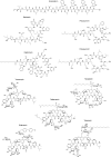
Fig. 7. Structural formulae of some marketed peptide-based antibiotics. For gramicidin D, the polymyxins, and teicoplanin, only the most abundant components of the clinically used mixtures are shown.
Fig. 8
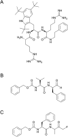
Fig. 8. The structure of LTX-109 (A) and the peptidomimetics reported by Hiromatsu and co-workers (B) and (C).
Fig. 9
Fig. 9. An antimicrobial lipopeptide, S-8, reported by Mukhopadhyay and co-workers.
Fig. 10
Fig. 10. Linear AMP IDR-1018 and its cyclic analogues. Additional residues are in red, amidation at the C-terminus in green, and C3 disulfide bridge in blue. Figure reproduced from Hancock and co-workers with permission from The American Chemical Society, Copyright (2020).
Fig. 11
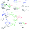
Fig. 11. (A) Different strategies for hybrid preparation. (i) Sections of parent peptides (blue and green) can be joined directly at their termini, (ii) the middle section of one of the parent peptides can be replaced by a sequence from the other parent peptide, or (iii) sections of the parent peptides can be joined using a short peptide linker (grey). (B) Hybrids from Ptaszynska and co-workers joined via peptide side chains. Spheres represent individual aa where blue spheres are of human neutrophil protein 1 (HNP1) and green spheres of lactoferrampin. The amide-linked hybrids used analogues of HNP1 with X being either l-α-aminobutyric acid (Abu) or acetaminoethyl cysteine (C-Acm) while the disulfide-linked hybrid had a cysteine residue at the same position. In all hybrids, the HNP1 component also contained 2-aminobenzoic acid (2-Abz, labelled Z). (C) Side chain–side chain joined hybrid (chimera) using unnatural aa, labelled B, for CuAAC click chemistry.
Fig. 12
Fig. 12. Frequently reported semi-synthetic modifications of glycopeptides vancomycin and eremomycin.
Fig. 13
Fig. 13. (A) Representation of G1, G2, and G3-generation dendrimers. Red: dendrimer core, orange: terminal groups, green: G1, G2, and G3 arms. (B) Divergent and convergent synthetic paths for building dendrimers.
Fig. 14
Fig. 14. Structure of the peptide dendrimers (RW)4D (A), M33 and M33-Peg (B), and 19 and 11 (C).
Fig. 15
Fig. 15. Structure of the PDs with a monolysine-core, such as lipodimeric peptide dendrimer reported by Rinaldi and co-workers (A), 4 (B), 14 (C) and 42 (D). The lysine at the core is in red.
Fig. 16
Fig. 16. Structures of glycopeptide dendrimers FD2, GalAG2/GalBG2, and Het1G2. Figure reproduced from Reymond and co-workers with permission from The Royal Society of Chemistry, Copyright (2015).
Fig. 17
Fig. 17. Structures of the peptide dendrimers G3KL (A) and TNS18 (B). (A) Structure reported by Reymond and co-workers. (B) was adapted from Reymond and co-workers with permission from The American Chemical Society, Copyright (2017).
Fig. 18
Fig. 18. Synthetic scheme of synthetic polypeptides using NCA-ROP.
Fig. 19
Fig. 19. Schematic representation of facial (A) and radial (B) amphipathicity, and the chemical structure of PHLG-Blm family (C). Figure reproduced with permission from Xiong et al., Proc. Natl. Acad. Sci. U. S. A., 2015, 112, 13155–13160.
Fig. 20
Fig. 20. Schematic illustration of the pH-responsive PL2 polypeptide that transits to a helical conformation under acidic condition. Figure reproduced with permission from Xiong et al., Proc. Natl. Acad. Sci. U. S. A., 2017, 114, 12675–12680.
Fig. 21
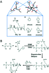
Fig. 21. Schematic representation of SNAPPs with 16- and 32-arms. The synthesis was initiated from terminal amines of PAMAM dendrimers using ROP of lysine and valine NCAs. Figure reproduced from Qiao and co-workers with permission from The American Chemical Society, Copyright (2016).
Fig. 22
Fig. 22. (A) Schematic representation and chemical structure of star-shaped polypeptides bearing different pendent groups. (B) Synthesis scheme of star-shaped co-polypeptide with 6 arms using dipentaerythritol as the initiator. Figure reproduced from Jan and co-workers with permission from The Royal Society of Chemistry, Copyright (2019).
Fig. 23
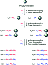
Fig. 23. Schematic representation of the synthesis of random peptide cocktails via SPPS. Three coupling steps are shown in the figure. A mixture of aa is added at each coupling step to yield peptides composed of several different sequences with the same length. Figure reproduced from Hayouka and co-workers with permission from The Royal Society of Chemistry, Copyright (2019).
Fig. 24
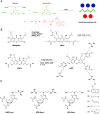
Fig. 24. Hit bicyclic peptides 27b and 62b (A), bp50 and bp56 (B), and peptide dendrimer T7 (C) identified from virtual libraries generated from the chemical space. (A) adapted from Reymond and co-workers with permission from The Royal Society of Chemistry, Copyright (2017). (B) adapted from Reymond and co-workers with permission from The Royal Society of Chemistry, Copyright (2018). (C) Structure reported by Reymond and co-workers.
Fig. 25
Fig. 25. The two approaches to peptide stapling: one-component (A) and two-component (B).
Fig. 26
Fig. 26. (A) Fmoc-triazine aa used to make AMPMs by Bang and co-workers. (B) γ-Mangostin AMPM derivative LS02. Figure adapted from Lin et al., Biochim. Biophys. Acta - Biomembr., 2020, 1862, 1–10, with permission from Elsevier, Copyright (2020). (C) The structure of azidothymidine (AZT)-based small molecule antimicrobials adapted from Chirumarry et al., Eur. J. Med. Chem., 2020, 193, 1–13, with permission from Elsevier, Copyright (2020).
Fig. 27
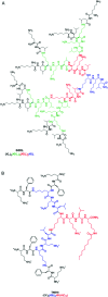
Fig. 27. Synthesis of peptidomimetic hybrids. Murepavadin derivative 2 was prepared by substitution of several aa residues (in red). The derivative 2 was further optimised and a hybrid with a polymyxin B1 analogue prepared (substituted residues in blue). The resulting hybrid 3 was further optimised to hybrid 8 (green residues modified), which contains 5 variable residues (T, U, X, Y, Z) that were not specified by the authors.
Fig. 28
Fig. 28. The structure of vancomycin-ceftazidime PDC TD-1792.
Fig. 29
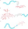
Fig. 29. The structures of PDCs. (A) A tobramycin-penetratin PDC, (B) A polyarginine CPP-vancomycin PDC. (C) A cleavable AMP-ciprofloxacin PDC. The drugs are highlighted in red, the linkers in black, and the aa are shown as a blue sphere.
Fig. 30
Fig. 30. The structures of two PDCs targeting intracellular bacteria. (A) A multi-component PDC comprising of methotrexate, a cationic AMPM and an anionic peptide linked via a cleavable cephalosporin moiety. (B) A kanamycin–AMPM conjugate containing a disulfide linkage.
Fig. 31
Fig. 31. The structure of VPP-G, a PDC targeting intracellular S. aureus reported by Jiang and co-workers. Vancomycin is shown in red, and a membrane disruptive AMPM is shown in blue.
Fig. 32
Fig. 32. A cartoon depicting the mechanisms of action of cationic AVPs. Figure reproduced from Mulder et al., Front. Microbiol., 2013, 4, 1–23, with permission from Frontiers, Copyright (2013).
Fig. 33
Fig. 33. The structure of an antiviral peptide dendrimer.
Fig. 34
Fig. 34. The structure of an oligo-acyl-lysine peptidomimetic based on dermaseptin S3.
None
Bee Ha Gan
None
Josephine Gaynord
None
Sam M. Rowe
None
Tomas Deingruber
None
David R. Spring

Similar articles

Cited by

References

    1. Walsh C., Antibiotics: actions, origins, resistance, 1st edn, ASM Press, 2003
    1. Patrick G. L., An Introduction to Medicinal Chemistry, 4th edn, Oxford University Press Inc., New York, 2009
    1. Gould K. J. Antimicrob. Chemother. 2016;71:572–575. doi: 10.1093/jac/dkv484. - DOI - PubMed
    1. Alharbi S. A. Wainwright M. Alahmadi T. A. Bin Salleeh H. Faden A. A. Chinnathambi A. Saudi J. Biol. Sci. 2014;21:289–293. doi: 10.1016/j.sjbs.2013.12.007. - DOI - PMC - PubMed
    1. 21st Model List of Essential Medicines, World Health Organisation, https://www.who.int/groups/expert-committee-on-selection-and-use-of-esse... (accessed 12 November 2020)

MeSH terms

Substances