Review
. 2021 May 13:27:323-353.
eCollection 2021.

Cutaneous and ocular rosacea: Common and specific physiopathogenic mechanisms and study models

Affiliations
Review

Cutaneous and ocular rosacea: Common and specific physiopathogenic mechanisms and study models

Daniela Rodrigues-Braz et al. Mol Vis. .

Abstract

Rosacea is a chronic inflammatory disease that affects the face skin. It is clinically classified into the following four subgroups depending on its location and severity: erythematotelangiectatic, papulopustular, phymatous, and ocular. Rosacea is a multifactorial disease triggered by favoring factors, the pathogenesis of which remains imperfectly understood. Recognized mechanisms include the innate immune system, with the implication of Toll-like receptors (TLRs) and cathelicidins; neurovascular deregulation involving vascular endothelial growth factor (VEGF), transient receptor potential (TRP) ion channels, and neuropeptides; and dysfunction of skin sebaceous glands and ocular meibomian glands. Microorganisms, genetic predisposition, corticosteroid treatment, and ultraviolet B (UVB) radiation are favoring factors. In this paper, we review the common and specific molecular mechanisms involved in the pathogenesis of cutaneous and ocular rosacea and discuss laboratory and clinical studies, as well as experimental models.


Figures

Figure 1
Figure 1
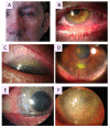
Clinical manifestations of oculocutaneous rosacea. A: Patient with phymatous rosacea–associated rhinophyma, blepharophyma, nasal and facial erythema with telangiectasia. B: Blepharophyma with thickened lid edges, lid margin telangiectasia, meibomian gland dysfunction (MGD). C: Corneal neovascularization (CNV) of ocular rosacea growing from the superior limbus with a crescent pattern forming a vascular pannus. D: Catarrhal corneal infiltrate caused by rosacea. E: Typical peripheral ulcerative keratitis (PUK) of rosacea, corresponding to sterile corneal melting of a crescentic area with newly formed stromal vessels. F: Advanced stage of ocular rosacea with white corneal infiltrates and whole corneal neovascularization, including the visual axis.
Figure 2
Figure 2
Diagram showing the factors involved in rosacea pathophysiology.
Figure 3
Figure 3
Possible mechanisms of involvement of both innate and adaptive immunity in rosacea pathophysiology. Activation of Toll-like receptor 2 and 4 (TLR-2, 4), which are expressed in different cell types in the skin and the eye, after stimulation by various external stimuli induces the production of the antimicrobial peptide human cathelicidin peptide (LL)-37 by activation of the serine protease kallikrein 5 (KLK-5), which cleaves the precursor peptide 18-kDa cationic antimicrobial protein (CAP-18) into the active form LL-37. KLK-5 is regulated by several other activators, including matrix metalloproteinase-9 (MMP-9), and repressors, such as the Kazal-type lymphoepithelial inhibitor (LEKTI). LL-37 activation by either KLK-5 or protease-activated receptor 2 (PAR-2) results in the activation and degranulation of mast cells, which release proinflammatory mediators, including MMPs, tumor necrosis factor- α (TNF-α), and interleukins (ILs), leading to the initiation or aggravation of inflammation at the cutaneous and ocular levels. Histamine release leads to fibrosis and the appearance of phyma. The activation of the nuclear factor kappa B (NF-κB) signaling pathway by TLRs or active MAPK protein 38 (p38) and extracellular signal-regulated kinase (Erk) can also activate the NF-κB pathway, leading to the release of several proinflammatory factors, including IL-33, IL-8, IL-18, and IL-1β. Their release can also be increased by the activation of the nucleotide-biding domain leucin-rich repeat and pyrin-containing receptor 3 (NLRP3) inflammasome. Endoplasmic reticulum (ER) stress participates in inflammatory mechanisms through the release of activating transcription factor 4 (ATF4), which promotes the activation of TLR-2. Sphingosine-1-phosphate (S1P) leads to the activation of enhancer-binding protein α (EBP-α), which also activates LL-37. Neutrophils release reactive oxygen species (ROS) and proteases. Macrophages, where M1 polarization appears to be favored by Adamdec1, also participate in inflammation by secreting other inflammatory factors, such as proteases or MMPs, TNF-α, ILs, and serine proteases. Plasmacytoid dendritic cells (pDCs) promote the activation of B and T cells. TH1 and TH17, polarized CD4(+) T cells of the adaptive immune system, secrete interferon-gamma (IFN-γ) and IL-17, respectively, leading to chemotaxis of other leukocytes by enhancing chemokine (C motif) ligand (CCL) and chemokine (C-X-C motif) ligand (CXCL) expression and increased activation of LL-37. TH17 cells are thought to stimulate the activity of koebnerisine (S100A15), an antimicrobial peptide that promotes the recruitment of inflammatory cells.
Figure 4
Figure 4
Possible mechanisms of neuronal and neurovascular deregulation in the rosacea pathophysiology mediated by the transient receptor potential (TRP) channel family. Vanilloid TRP (TRPV) and ankyrin TRP (TRPA), among others, are expressed in neuronal and nonneuronal cells and activated by external stimuli, such as heat, cold, or UV B (UVB) irradiation, and they are significantly regulated upwards in rosacea. An influx of Ca2+ by the activation of these receptors induces the release of neuropeptides such as substance P (SP), the peptide related to the calcitonin gene (CGRP), the polypeptide activator of the pituitary adenylates (PACAP), the intestinal vasoactive peptide (VIP), and ATP. These neuropeptides can stimulate immune cells, such as mast cells, neutrophils, macrophages, and CD4+ T cells, that release proinflammatory mediators, such as matrix metalloproteinases (MMPs), tumor necrosis factor-α (TNF-α), interferon-γ (IFN-γ), interleukins (ILs), proteases, and reactive oxygen species (ROS), leading to the initiation or aggravation of inflammation. Toll-like receptor 2 (TLR-2) activates kallikrein 5 (KLK-5), which activates the antimicrobial peptide of cathelicidin (LL-37). LL-37 is known to be an activating factor of mast cells. Furthermore, mast cells can also release VEGF, contributing to vasculogenesis. The action of neuropeptides on blood vessels leads to vasodilation, which is responsible for erythema and flushing, and to vascular permeability, which is responsible for plasma extravasation, leading to edema. The release of histamine from mast cells promotes vasodilation and the onset of fibrosis. The release of VEGF by mast cells promotes vasculogenesis. The sebaceous and meibomian glands exhibit sensory and autonomic nerve regulation with the expression of neurotransmitters. This regulation could contribute to the development of the disease.
Figure 5
Figure 5
Major mechanisms of cutaneous and ocular rosacea. Excessive activation of innate immunity by various external and internal stimuli, including hormonal dysfunctions, microbes, reactive oxygen species (ROS) or ultraviolet (UV) radiation, leads to overexpression of Toll-like receptors (TLRs), including TLR-2 and 4. The release of proinflammatory cytokines, including interleukins (ILs) and tumor necrosis factor-α (TNF-α), and chemokines, including chemokine (C-X-C motif) ligand (CXCL) 1, CXCL2, CXCL5, and CXCL6, lead to inflammation and the development of papules, pustules, erythema, and edema. If the inflammation becomes persistent, patients develop a phyma, leading to fibrosis. At the same time, TRL-2 and 4 upregulate kallikrein 5 (KLK-5), which activates the cleavage of 18-kDa cationic antimicrobial protein (hCAP-18) into a biologically active peptide, LL-37. LL-37 facilitates the degranulation of mast cells, which leads to a greater release of matrix metalloproteinases (MMPs), IL-6, TNF-α, and the vascular endothelial growth factor (VEGF) participating in the cutaneous and ocular angiogenesis processes, which can lead to corneal neovascularization and appearance of telangiectasias. Microorganisms, including Demodex, can also directly disrupt the epidermal barrier, which can lead to worsening of inflammation processes. Heat and cold and environmental stimuli overactivate receptive potential channels transient receptor potential (TRP), including s), leading to a significant release of neuropeptides, including substance P (SP), the pituitary adenylate cyclase-activating polypeptide (PACAP), the peptide linked to the calcitonin gene (CGRP), and the vasoactive intestinal peptide (VIP), which cause neurovascular dysfunction, leading to the processes of inflammation, vasodilation, and angiogenesis with the appearance of redness and worsening of erythema and edema. Patients with ocular rosacea also have a dysfunction of the meibomian glands due to an alteration of the meibum, compared with the sebum in patients with cutaneous rosacea. Overexpression of the genes coding for the small proline-rich protein (SPRR) and calcium binding proteins (S100) A8 and A9 can lead to inflammation of the eyes and of the eyelid (blepharitis).

Similar articles

Cited by

References

    1. Steinhoff M, Schauber J, Leyden JJ. New insights into rosacea pathophysiology: A review of recent findings. J Am Acad Dermatol. 2013;69(Supplement 1):S15–26. - PubMed
    1. Steinhoff M, Schmelz M, Schauber J. Facial Erythema of Rosacea - Aetiology, Different Pathophysiologies and Treatment Options. Acta Derm Venereol. 2016;96:579–86. - PubMed
    1. Gallo RL, Granstein RD, Kang S, Mannis M, Steinhoff M, Tan J, Thiboutot D. Standard classification and pathophysiology of rosacea: The 2017 update by the National Rosacea Society Expert Committee. J Am Acad Dermatol. 2018;78:148–55. - PubMed
    1. Wilkin J, Dahl M, Detmar M, Drake L, Feinstein A, Odom R, Powell F. Standard classification of rosacea: Report of the National Rosacea Society Expert Committee on the Classification and Staging of Rosacea. J Am Acad Dermatol. 2002;46:584–7. - PubMed
    1. Gold LM, Draelos ZD. New and Emerging Treatments for Rosacea. Am J Clin Dermatol. 2015;16:457–61. - PubMed