Review
. 2021 May 24;31(10):R651-R666.
doi: 10.1016/j.cub.2021.03.064.

Cellular functions of actin- and microtubule-associated septins

Affiliations
Review

Cellular functions of actin- and microtubule-associated septins

Elias T Spiliotis et al. Curr Biol. .

Abstract

Septins are an integral component of the cytoskeleton, assembling into higher-order oligomers and filamentous polymers that associate with actin filaments, microtubules and membranes. Here, we review septin interactions with actin and microtubules, and septin-mediated regulation of the organization and dynamics of these cytoskeletal networks, which is critical for cellular morphogenesis. We discuss how actomyosin-associated septins function in cytokinesis, cell migration and host defense against pathogens. We highlight newly emerged roles of septins at the interface of microtubules and membranes with molecular motors, which point to a 'septin code' for the regulation of membrane traffic. Additionally, we revisit the functions of microtubule-associated septins in mitosis and meiosis. In sum, septins comprise a unique module of cytoskeletal regulators that are spatially and functionally specialized and have properties of bona fide actin-binding and microtubule-associated proteins. With many questions still outstanding, the study of septins will continue to provide new insights into fundamental problems of cytoskeletal organization and function.


Conflict of interest statement

Declaration of interests The authors declare no competing interests.

Figures

Figure 1.
Figure 1.. Septin domains and assembly.
(A) The mammalian septin family consists of thirteen paralogs, which are classified into four groups: SEPT2 (SEPT1, SEPT2, SEPT4, SEPT5), SEPT3 (SEPT3, SEPT9, SEPT12), SEPT6 (SEPT6, SEPT8, SEPT10, SEPT11, SEPT14), and SEPT7. Septin paralogs have a highly conserved GTP-binding domain (G-domain) followed by a unique sequence termed the septin unique element (SUE). Septins of the SEPT6 group do not hydrolyze GTP, while some paralogs (e.g., SEPT9) have faster rates of GTP hydrolysis. The amino- and carboxy-terminal extensions (NTEs and CTEs) of the GTP-binding domains are highly variable. CTEs contain α-helical coiled-coil domains, and SEPT9 contains a unique and alternatively spliced NTE, which interacts directly with actin filaments and microtubules. This NTE sequence is divided into basic and acidic proline-rich domains and consists of a repeat motif (K/R-R/x-x-D/E) that interacts with the acidic carboxy-terminal tails of tubulin. Septins also contain polybasic domains and amphipathic helices, with which they bind to membrane bilayers of distinct phospholipid content and curvature. (B) Septins homodimerize and heterodimerize in tandem through two alternating interfaces of their GTP-binding domains, assembling into non-polar palindromic oligomers. The prevailing oligomeric unit is a dimer of tetramers consisting of a paralog from each of the four septin groups (SEPT2, SEPT3, SEPT6, SEPT7). Septins of the same group can replace one another, generating octamers of variable combinations. Depending on septin expression levels and rates of GTP hydrolysis, which influence the homodimeric and heterodimeric interactions of the G-domains, smaller unit oligomers can form (e.g., SEPT2–SEPT6–SEPT7 heterohexamers). Additionally, oligomeric complexes of atypical combinations that consist of paralogs of the same septin group have been reported. (C) Septin oligomers associate with actin, microtubules and cell membranes. Septins form higher-order filamentous networks on cell membranes and may also polymerize into filaments on the surface of actin and microtubules.
Figure 2.
Figure 2.. Septin interactions with regulators of actin growth, organization, contractility and disassembly.
Summary of septin interactions with animal (top) and fungal (bottom) proteins that function in actin polymerization (formins FHOD1 and Bnr1), Arp2/3-mediated nucleation (cortactin, Las17/WASP), actin organization and crosslinking (anillin, α-actinin-4, Tea1, Rvs167, coronin, Hof1), actin contractility (myosin II, myosin essential light chain (ELC) and regulatory light chain (RLC)), and actin disassembly (cofilin, MICAL-1). Septin interactions with the effector proteins of Cdc42 (Cdc42EP/BORG) and the kinases CIT-K and ROCK2, which regulate myosin and formin activities, are also shown. Mammalian and fungal (S. cerevisiae, M. oryzae) septins are shown in purple ovals. Solid lines denote direct interactions, and dotted lines represent functional interactions that might be due to indirect or direct binding. Arrows point to phosphorylation and dephosphorylation events.
Figure 3.
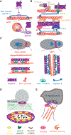
Figure 3.. Septins interact with actomyosin and spatially regulate the organization of actomyosin networks.
(A) SEPT2 interacts directly with the coiled-coil domain of the heavy chain of non-muscle myosin II. SEPT9 binds actin filaments via its NTE basic domain, which can occupy three different regions of the actin surface domains (SD) 1, 2 and 4 (purple ovals and arrows). (B) Septin association with actin involves effectors of Rho (Rhotekin, anillin and septin-associated Rho guanine nucleotide exchange factor, SA-RhoGEF) and Cdc42 (Cdc42EP3). Anillin, which interacts with the myosin regulatory light chain, can recruit septins to actin filaments, while Rhotekin and Cdc42EP3 synergize with septins in binding actin. Actin-associated septins are posited to scaffold the kinases ROCK2 and CIT-K, which activate myosin II contractility by phosphorylating the myosin regulatory light chain. (C) In the budding yeast S. cerevisiae, septins localize at the mother–bud neck cortex. In the early stages of bud growth, septins form a network of filaments oriented along the axis of cell polarity. Septins scaffold and pattern the localization of the formin Bnr1 and the F-BAR protein Hof1 into evenly spaced pillars, which enable the formation of actin cables that are spaced apart and oriented toward the growing bud. In mitosis, septins initially provide a scaffold for the recruitment of myosin II and actomyosin ring (AMR) assembly. At the onset of cytokinesis, septins associate with the AMR indirectly through Hof1, which interacts with both septins and myosin II. Subsequently, septins split into two rings, flanking the AMR. (D) Infection of rice plants by the fungal pathogen M. oryzae requires the formation of the appressorium, a pressurized hyphal structure that penetrates into the cuticle. At the base of the appressorium, a septin ring organizes the formation of a toroidal actin network by corralling the localization of the I-BAR protein Rvs167 and the WASP homolog Las17, and scaffolding the ERM protein Tea1, which promotes actin–membrane association. (E) Septins are recruited to plasma membrane sites of S. Typhimurium attachment and provide a scaffold for the phosphorylation of the formin FHOD1 by the ROCK2 kinase.
Figure 4.
Figure 4.. Septin localization and functions in the actin networks of migrating cells.
Septins associate with the actin networks of the plasma membrane, stress fibers and lamellipodia. (Top left) In migrating melanoma cells, SEPT9 and Cdc42EP5 are required for the organization of cortical actin. (Top right) In renal epithelia undergoing epithelial–to-mesenchymal transition (EMT), septins are enriched on contractile transverse arc stress fibers and at the interface of these fibers with the distal ends of the radial (dorsal) stress fibers that emanate from focal adhesions. Septins crosslink the actin filaments of these networks and are required for the maintenance of the transverse arc network and focal adhesion maturation. (Bottom right) In squamous cancer cells, SEPT1, SEPT4 and SEPT5 are distinctly enriched in lamellipodia, and in vitro SEPT6 associates with the branch points of Arp2/3-nucleated branched networks. (Bottom left) In various cells, septins associate with dorsal stress fibers that stretch in between the ventral nuclear and plasma membranes. Septin association with the stress fibers involves CDC42EP3 and the myosin II heavy chain. Septins may regulate mechanotransduction by scaffolding the phosphorylation of the myosin regulatory light chain by ROCK2 and controlling the nucleocytoplasmic shuttling of actin regulators such as Nck1, which might occur in a mechanosensitive manner. Interestingly, septins recruit Nck1 to stress fibers via SOCS7 and thereby prevent Nck1 from shuttling to the nucleus. SEPT9 isoform 2 (SEPT9_i2), which inhibits cell migration, suppresses the formation of a subset of stress fibers that localizes underneath the nucleus through an unknown mechanism.
Figure 5.
Figure 5.. Functions of microtubule-associated septins.
(A) Crystal structures of SEPT9 (PDB: 4YQF) and αβ-tubulin (PDB: 1TUB) depict their intrinsically disordered amino-terminal extension and carboxy-terminal tails (hand-drawn lines), respectively, which mediate septin–microtubule binding. Purple boxes on the amino-terminal extension of SEPT9 represent the microtubule-binding repeat motifs K/R-R/x-x-D/E. The carboxy-terminal tyrosine and polyglutamylated side chains of αβ-tubulin are denoted with the letters Y and E, respectively. (B) SEPT7 and SEPT1 promote the nucleation of microtubules from the centrosome and Golgi membranes, respectively. SEPT7 is required for the centrosomal localization of p150Glued, and SEPT1 forms a complex with GM130, CEP170 and the γ-tubulin ring complex (γ-TURC). (C) Septin oligomers (SEPT9, SEPT2–SEPT6–SEPT7) promote persistent microtubule growth and crosslink microtubules into bundles; SEPT9 recruits αβ-tubulin dimers to the microtubule lattice. At higher concentrations, microtubule-associated septins pause and stunt plus-end growth. Septins also promote the capture and zippering of microtubule plus ends with the lattices of septin-coated microtubule bundles. (D) Septins function in microtubule acetylation and polyglutamylation. SEPT7 interacts with histone deacetylase (HDAC6) and is posited to scaffold the deacetylation of α-tubulin in the cytosol. Microtubule-associated septins have been proposed to scaffold tubulin tyrosine ligase-like (TTLL) and cytosolic carboxypeptidase 1 (CCP1) enzymes that respectively elongate and trim the polyglutamylated side chains of the carboxy-terminal tails of polymerized tubulin. (E) Microtubule-associated septins differentially regulate the motility of kinesin motors and their cargo. Microtubule-associated SEPT9 inhibits kinesin-1/KIF5 and dynein and enhances the motility of kinesin-3/KIF1A.

Similar articles

Cited by

References

    1. Mostowy S, and Cossart P (2012). Septins: the fourth component of the cytoskeleton. Nat. Rev. Mol. Cell Biol 13, 183–194. - PubMed
    1. Spiliotis ET, and McMurray MA (2020). Masters of asymmetry - lessons and perspectives from 50 years of septins. Mol. Biol. Cell 31, 2289–2297. - PMC - PubMed
    1. Woods BL, and Gladfelter AS (2021). The state of the septin cytoskeleton from assembly to function. Curr. Opin. Cell Biol 68, 105–112. - PMC - PubMed
    1. Dolat L, Hu Q, and Spiliotis ET (2014). Septin functions in organ system physiology and pathology. Biol. Chem 395, 123–141. - PMC - PubMed
    1. Gladfelter AS, Pringle JR, and Lew DJ (2001). The septin cortex at the yeast mother-bud neck. Curr. Opin. Microbiol 4, 681–689. - PubMed

Publication types

LinkOut - more resources