. 2021 May 6:12:641059.
doi: 10.3389/fmicb.2021.641059. eCollection 2021.

Electrostatic Interaction Between NS1 and Negatively Charged Lipids Contributes to Flavivirus Replication Organelles Formation

Affiliations

Electrostatic Interaction Between NS1 and Negatively Charged Lipids Contributes to Flavivirus Replication Organelles Formation

Yali Ci et al. Front Microbiol. .

Abstract

Flavivirus replication occurs in membranous replication compartments, also known as replication organelles (ROs) derived from the host ER membrane. Our previous study showed that the non-structural (NS) protein 1 (NS1) is the essential factor for RO creation by hydrophobic insertion into the ER membrane. Here, we found that the association of NS1 with the membrane can be facilitated by the electrostatic interaction between NS1 and negatively charged lipids. NS1 binds to a series of negatively charged lipids, including PI4P, and a positively charged residue, R31, located on the membrane-binding face of NS1, plays important roles in this interaction. The NS1 R31E mutation significantly impairs NS1 association with negatively charged membrane and its ER remodeling ability in the cells. To interfere with the electrostatic interaction between NS1 and negatively charged lipids, intracellular phosphatidylinositol phosphates (PIPs) level was downregulated by the overexpression of Sac1 or treatment with PI3K and PI4K inhibitors to attenuate flavivirus replication. Our findings emphasize the importance of electrostatic interaction between NS1 and negatively charged lipids in flavivirus RO formation.

Keywords: ER remodeling; PIP; flavivirus; non-structural protein 1; replication organelles.


Conflict of interest statement

The authors declare that the research was conducted in the absence of any commercial or financial relationships that could be construed as a potential conflict of interest.

Figures

FIGURE 1
FIGURE 1
NS1 specifically binds to negatively charged lipids. (A) Lipid binding selectivity of ZIKV NS1. ZIKV Myc-NS1 proteins were incubated with a lipid membrane strip spotted with a series of different lipids. Antibody against the Myc tag was used to detect NS1 bound to each lipid spot. (B) NS1 preferentially binds to PI4P containing liposomes. The co-floating assay was performed to examine NS1 association with the liposomes. NS1 co-floated with the liposomes were collected from the top layer and analyzed by SDS-PAGE and silver staining. (C) Statistic analysis of NS1 co-floating efficiency in panel (B). The amount of co-floating NS1 was analyzed. The amount of NS1 bound to PI-liposomes was set as 100%. (D) NS1 remodels the liposomes in vitro. After incubation with NS1, the average diameter of liposomes was detected by DLS. (E) PI4P containing liposomes are more sensitive to NS1-induced liposome remodeling. A less amount of NS1 was incubated with liposomes containing PI or PI4P. In the presence of PI4P, the average diameter of liposomes was decreased after incubation with NS1. The data are presented as the mean ± SEM. The p-values were obtained from a two-tailed t-test. **P < 0.01, ***P < 0.001.
FIGURE 2
FIGURE 2
R31 of NS1 is important for the electrostatic interaction between NS1 and negatively charged lipids. (A) NS1 R31E mutant can hardly bind to liposomes containing PI4P. Positively charged arginine (R31) of NS1 was mutated to negatively charged glutamic acid (E). A co-floating assay was performed to examine the association of ZIKV NS1 with the liposomes. (B) NS1 R31E mutant lost the ability to remodel the PI4P liposomes. Liposomes containing PI4P were incubated with wild-type NS1 or R31E mutant. The average diameter of liposomes was detected by DLS. R31E mutant can hardly change the diameter of liposomes. (C) R31 located in the groove between β-roll and greasy finger on membrane association face. The data are presented as the mean ± SEM. The p-values were obtained from a two-tailed t-test. **P < 0.01, ***P < 0.001.
FIGURE 3
FIGURE 3
R31E mutation significantly impairs NS1 induced ER remodeling and attenuates viral replication. (A) ER morphology with the expression of WT and R31E mutant NS1. Wild type NS1 or R31E mutant with C-terminal Myc-tag was expressed in HeLa cells and then labeled with anti-Myc and anti-Calreticulin antibodies. WT NS1 expression induced apparent ER remodeling, whereas R31E mutant could rarely remodel the ER. Scale bar, 20 μm. (B) The percentage of ER-remodeled cells induced by expression of WT NS1 or R31E mutant. The ratio of ER-remodeled cells to total NS1 positive cells is defined as the percentage of ER-remodeled cells. (C) NS1 R31E mutation impairs viral replication. NS1 R31E mutation was introduced into the ZIKV replicon. Luciferase activity was measured at 10 h and 36 h post-transfection to validate the viral replication ability. (D) NS1 R31E mutation significantly impairs the production of infectious viruses. NS1 R31E mutation was introduced into ZIKV infectious clone. At about 72 h post-transfection, the culture medium was collected, and viral RNA was detected by RT-qPCR. (E) Plaque assay was performed using the recombinant ZIKV to infect Vero cells. The data are presented as the mean ± SEM. The p-values were obtained from a two-tailed t-test. ***P < 0.001.
FIGURE 4
FIGURE 4
PI4P is enriched at ZIKV replication organelles. ZIKV infected EGFP-Sec61 stable cells were stained with antibodies against PI4P and dsRNA. Scale bar, 20 μm.
FIGURE 5
FIGURE 5
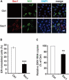
Sac1 overexpression interfered with NS1 induced ER remodeling and viral replication. (A,B) Sac1 overexpression interferes with NS1 induced ER remodeling. HeLa cells were co-transfected with Myc-NS1 and Flag-Sac1. Then cells were stained with anti-Myc and anti-Flag antibodies (A). Scale bar, 20 μm. The ratio of ER-remodeled cells to Myc and Flag double-positive cells is calculated as the percentage of ER-remodeled cells (B). (C) Sac1 overexpression impairs viral replication. BHK-21 cells were transfected with Sac1 expressing plasmid or control plasmid and then infected with ZIKV. The amount of viral RNA in the cell lysate at 48 hpi was quantified by RT-qPCR. The data are presented as the mean ± SEM. The p-values were obtained from a two-tailed t-test. ** P < 0.01, ***P < 0.001.
FIGURE 6
FIGURE 6
PIK inhibitors interfere with NS1 induced ER remodeling and viral replication. (A) PIK inhibitors decrease NS1 induced ER remodeling. NS1 expressed HeLa cells were treated with PIK inhibitors, wortmannin, or IN-9 at the indicated concentration. (B) Wortmannin or IN-9 inhibits ZIKV replication. BHK-21 cells were transfected by ZIKV replicon RNA. At 9 h post-transfection, cells were treated with indicated inhibitors. At 36 h post-transfection, luciferase activity was measured to evaluate viral replication. (C) The combination of Wortmannin and IN-9 shows improved inhibitory effects. ZIKV replicon RNA transfected BHK-21 cells were treated with Wortmannin and IN-9 at 9 h post-transfection. Luciferase activity was monitored at 36 h post-transfection. (D,E) Antiviral effects of PIK inhibitors. ZIKV infected BHK-21 cells were treated with Wortmannin (D) or IN-9 (E). Viral RNA in cell lysate was quantified by RT-qPCR at 48 hpi. The data are presented as the mean ± SEM. The p-values were obtained from a two-tailed t-test. *P < 0.05, **P < 0.01, ***P < 0.001.
FIGURE 7
FIGURE 7
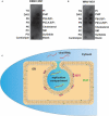
Flavivirus shares a common RC generation mechanism. (A,B) DENV and WNV NS1 possessed a similar binding pattern to negatively charged lipids as ZIKV NS1. Lipid membrane strips were used to examine the lipid-binding selectivity of DENV (A) and WNV NS1 (B). (C) Flaviviral NS1 proteins located in the ER lumen bind to the inner leaflet of the ER membrane by hydrophobic insertion, and the electrostatic interaction with negatively charged lipids (e.g., PI4P) facilitates the association between NS1 and membrane. This insertion of NS1 changes the ER membrane’s curvature, producing invaginated vesicles as the place of viral RNA synthesis, the replication compartment.

Similar articles

Cited by

References

    1. Akey D. L., Brown W. C., Dutta S., Konwerski J., Jose J., Jurkiw T. J. (2014). Flavivirus NS1 structures reveal surfaces for associations with membranes and the immune system. Science 343 881–885. 10.1126/science.1247749 - DOI - PMC - PubMed
    1. Aktepe T. E., Liebscher S., Prier J. E., Simmons C. P., Mackenzie J. M. (2017). The host protein reticulon 3.1A is utilized by flaviviruses to facilitate membrane remodelling. Cell Rep. 21 1639–1654. 10.1016/j.celrep.2017.10.055 - DOI - PubMed
    1. Apte-Sengupta S., Sirohi D., Kuhn R. J. (2014). Coupling of replication and assembly in flaviviruses. Curr. Opin. Virol. 9 134–142. 10.1016/j.coviro.2014.09.020 - DOI - PMC - PubMed
    1. Brown W. C., Akey D. L., Konwerski J. R., Tarrasch J. T., Skiniotis G., Kuhn R. J., et al. (2016). Extended surface for membrane association in Zika virus NS1 structure. Nat. Struct. Mol. Biol. 23 865–867. 10.1038/nsmb.3268 - DOI - PMC - PubMed
    1. Cerikan B., Goellner S., Neufeldt C. J., Haselmann U., Mulder K., Chatel-Chaix L. (2020). A non-replicative role of the 3’ terminal sequence of the dengue virus genome in membranous replication organelle formation. Cell Rep. 32:107859. 10.1016/j.celrep.2020.107859 - DOI - PMC - PubMed