

Positron emission tomography in the COVID-19 pandemic era
- PMID: 34013405
- PMCID: PMC8134823
- DOI: 10.1007/s00259-021-05347-7
Positron emission tomography in the COVID-19 pandemic era
Abstract
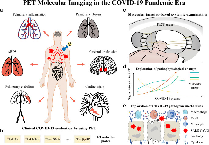
Coronavirus disease 2019 (COVID-19) has become a major public health problem worldwide since its outbreak in 2019. Currently, the spread of COVID-19 is far from over, and various complications have roused increasing awareness of the public, calling for novel techniques to aid at diagnosis and treatment. Based on the principle of molecular imaging, positron emission tomography (PET) is expected to offer pathophysiological alternations of COVID-19 in the molecular/cellular perspectives and facilitate the clinical management of patients. A number of PET-related cases and research have been reported on COVID-19 over the past one year. This article reviews the current studies of PET in the diagnosis and treatment of COVID-19, and discusses potential applications of PET in the development of management strategy for COVID-19 patients in the pandemic era.
Keywords: COVID-19; Molecular imaging; Positron emission tomography (PET); SARS-CoV-2.
© 2021. The Author(s).
Conflict of interest statement
The authors declare no competing interests.
Figures


Similar articles
-
Prior SARS-CoV-2 Infection Is Associated With Coronary Vasomotor Dysfunction as Assessed by Coronary Flow Reserve From Cardiac Positron Emission Tomography.J Am Heart Assoc. 2022 Oct 18;11(20):e025844. doi: 10.1161/JAHA.122.025844. Epub 2022 Oct 17. J Am Heart Assoc. 2022. PMID: 36250654 Free PMC article.
-
Cardiac PET and SPECT During the COVID-19 Pandemic.Semin Nucl Med. 2022 Jan;52(1):56-60. doi: 10.1053/j.semnuclmed.2021.06.020. Epub 2021 Jun 22. Semin Nucl Med. 2022. PMID: 34253333 Free PMC article. Review.
-
Unknown SARS-CoV-2 pneumonia detected by PET/CT in patients with cancer.Tumori. 2020 Aug;106(4):325-332. doi: 10.1177/0300891620935983. Epub 2020 Jun 22. Tumori. 2020. PMID: 32567505 Free PMC article.
-
Nuclear medicine in SARS-CoV-2 pandemia: 18F-FDG-PET/CT to visualize COVID-19.Nuklearmedizin. 2020 Jun;59(3):276-280. doi: 10.1055/a-1152-2341. Epub 2020 Apr 7. Nuklearmedizin. 2020. PMID: 32259853 English.
-
Imaging of Hematological Patients in the Era of COVID-19.Acta Haematol. 2022;145(3):267-274. doi: 10.1159/000522323. Epub 2022 Jan 31. Acta Haematol. 2022. PMID: 35100592 Free PMC article. Review.
Cited by
-
[18F]FDG PET/CT in Short-Term Complications of COVID-19: Metabolic Markers of Persistent Inflammation and Impaired Respiratory Function.Diagnostics (Basel). 2022 Mar 29;12(4):835. doi: 10.3390/diagnostics12040835. Diagnostics (Basel). 2022. PMID: 35453883 Free PMC article.
-
[18F]FDG-PET/CT in mechanically ventilated critically ill patients with COVID-19 ARDS and persistent inflammation.Clin Transl Imaging. 2023;11(3):297-306. doi: 10.1007/s40336-023-00550-y. Epub 2023 Mar 12. Clin Transl Imaging. 2023. PMID: 37275950 Free PMC article.
-
COVID-19 and Aspiration Pneumonia: Similar Pulmonary Findings with Different Diagnoses-a Pitfall in [18F]FDG PET/CT.SN Compr Clin Med. 2021;3(11):2322-2325. doi: 10.1007/s42399-021-01030-y. Epub 2021 Jul 29. SN Compr Clin Med. 2021. PMID: 34345767 Free PMC article.
-
2022 follow-up: impact of the COVID-19 pandemic on nuclear medicine departments in Europe.Eur J Nucl Med Mol Imaging. 2022 Aug;49(10):3309-3315. doi: 10.1007/s00259-022-05881-y. Eur J Nucl Med Mol Imaging. 2022. PMID: 35737024 Free PMC article. No abstract available.
-
T Cell Metabolism in Infection.Front Immunol. 2022 Mar 14;13:840610. doi: 10.3389/fimmu.2022.840610. eCollection 2022. Front Immunol. 2022. PMID: 35359994 Free PMC article. Review.
References
-
- Yan Y, Pan HS, Shao N, Xuan Y, Wang SF, Li WJ, et al. COVID-19 in Singapore: another story of success. International Journal of Mathematics for Industry. 2020;12(1):2050001. doi: 10.1142/S266133522050001x. - DOI
-
- Cirulli E, Barrett KMS, Riffle S, Bolze A, Neveux I, Dabe S, et al. Long-term COVID-19 symptoms in a large unselected population. medrxiv. 2020.
Publication types
MeSH terms
Grants and funding
LinkOut - more resources
Full Text Sources
Other Literature Sources
Medical
Miscellaneous
