. 2021 Jul 12;95(15):e0056321.
doi: 10.1128/JVI.00563-21. Epub 2021 Jul 12.

Cyclophilin A Inhibits Human Respiratory Syncytial Virus (RSV) Replication by Binding to RSV-N through Its PPIase Activity

Affiliations

Cyclophilin A Inhibits Human Respiratory Syncytial Virus (RSV) Replication by Binding to RSV-N through Its PPIase Activity

Wenzhang Liang et al. J Virol. .

Abstract

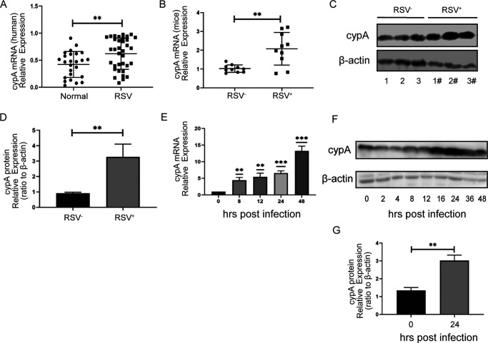
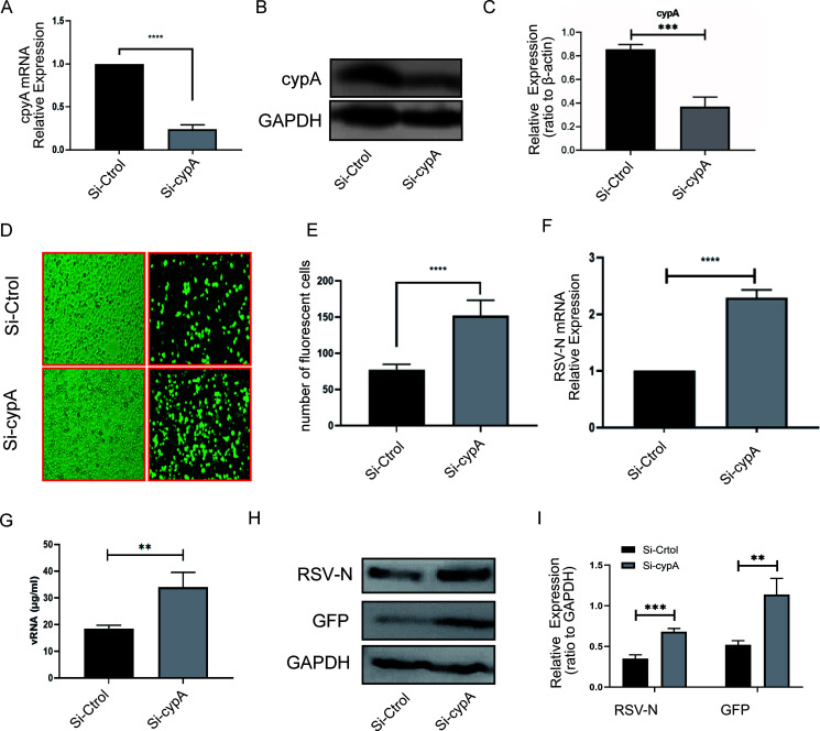
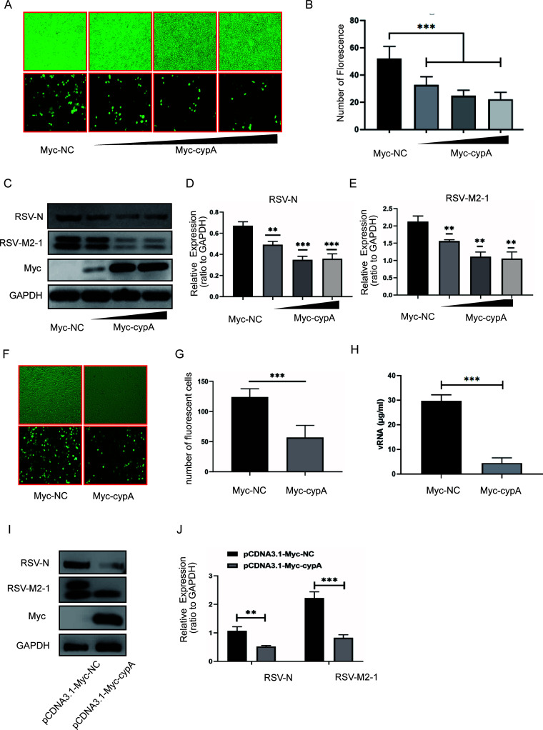
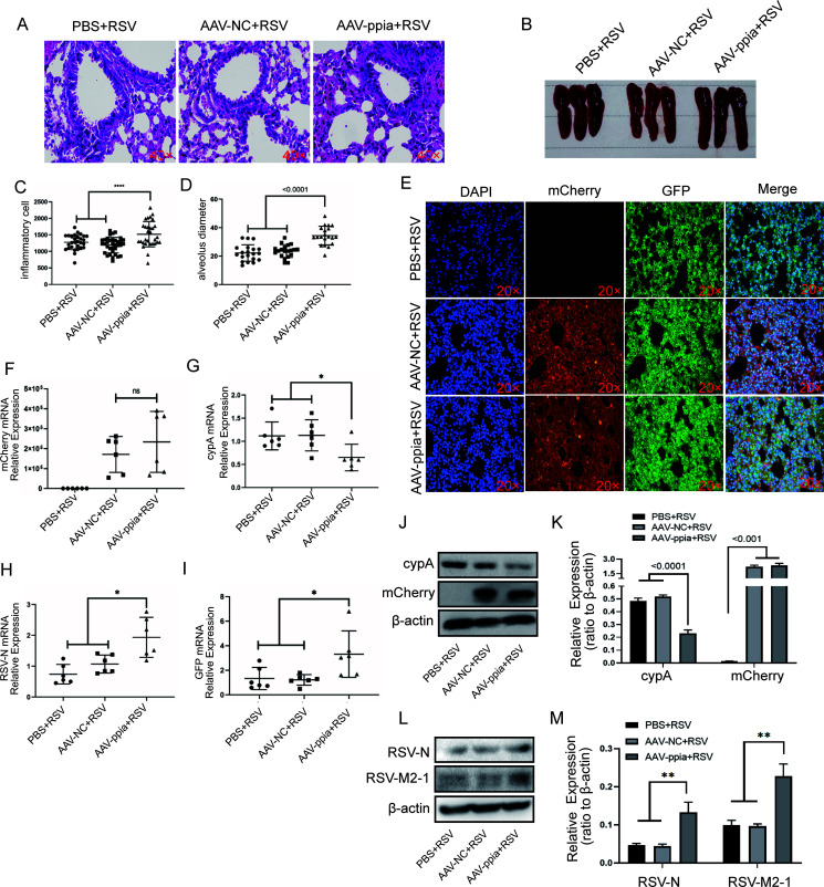
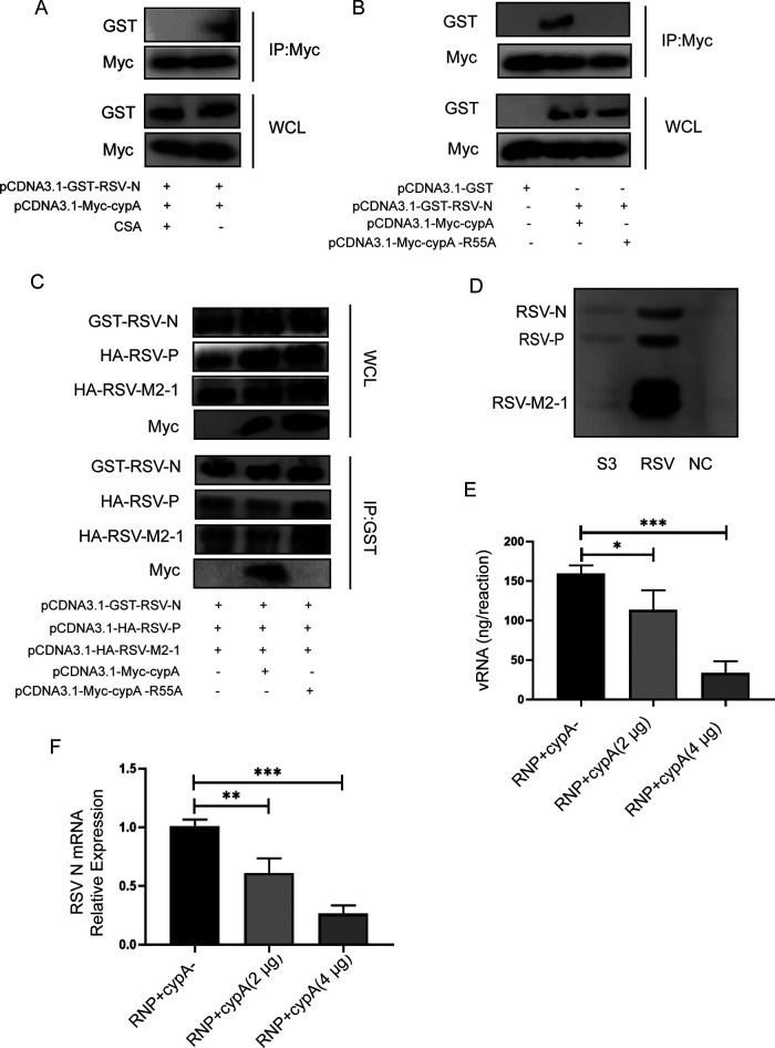
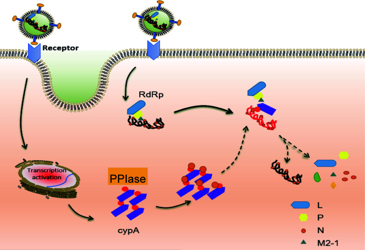
Human respiratory syncytial virus (hRSV) is the most common pathogen which causes acute lower respiratory infection (ALRI) in infants. Recently, virus-host interaction has become a hot spot of virus-related research, and it needs to be further elaborated for RSV infection. In this study, we found that RSV infection significantly increased the expression of cyclophilin A (cypA) in clinical patients, mice, and epithelial cells. Therefore, we evaluated the function of cypA in RSV replication and demonstrated that virus proliferation was accelerated in cypA knockdown host cells but restrained in cypA-overexpressing host cells. Furthermore, we proved that cypA limited RSV replication depending on its PPIase activity. Moreover, we performed liquid chromatography-mass spectrometry, and the results showed that cypA could interact with several viral proteins, such as RSV-N, RSV-P, and RSV-M2-1. Finally, the interaction between cypA and RSV-N was certified by coimmunoprecipitation and immunofluorescence. Those results provided strong evidence that cypA may play an inhibitory role in RSV replication through interaction with RSV-N via its PPIase activity. IMPORTANCE RSV-N, packed in the viral genome to form the ribonucleoprotein (RNP) complex, which is recognized by the RSV RNA-dependent RNA polymerase (RdRp) complex to initiate viral replication and transcription, plays an indispensable role in the viral biosynthesis process. cypA, binding to RSV-N, may impair this function by weakening the interaction between RSV-N and RSV-P, thus leading to decreased viral production. Our research provides novel insight into cypA antiviral function, including binding to viral capsid protein to inhibit viral replication, which may be helpful for new antiviral drug exploration.

Keywords: CSA; PPIase; RSV-N; cypA; viral replication.


Figures

FIG 1
FIG 1
cypA expression was significantly elevated when RSV infected humans, mice, or cells. (A) The results of qRT-PCR analysis of the cypA mRNA expression in clinical samples. (B) qRT-PCR analysis of the cypA mRNA expression in lung from mice infected by RSV for 3 days. (C and D) WB analysis of the cypA protein expression in lung from mice infected by RSV for 3 days (C). cypA protein levels were quantitated by densitometry and normalized to β-actin (D). (E) qRT-PCR analysis of the cypA mRNA expression in Hep2 cells infected by RSV for the indicated time. (F and G) WB analysis of the cypA protein expression in Hep2 cells infected by RSV for the indicated time (F). cypA protein levels at 24 h postinfection were quantitated by densitometry and normalized to β-actin (G). Data are means ± SD for three independent experiments. **, P < 0.01; ***, P < 0.001.
FIG 2
FIG 2
RSV replication was elevated in knockdown cypA cells. (A to C) qRT-PCR (A) and WB (B) methods were adopted to analyze the cypA mRNA or protein expression level to identify the interference efficiency of three Si-cypAs compared to Si-Ctrol. cypA protein level was quantitated by densitometry and normalized to GAPDH (C). (D and E) Fluorescence images of RSV-GFP replication in Hep2 cells transfected by Si-Ctrol or Si-cypA (D). The statistical results for the number of green fluorescent cells among Hep2 cells from 10 random microscope fields are shown (E). (F and G) qRT-PCR analysis of the RSV-N mRNA level in Hep2 cells (F) and concentration of viral RNA extracted from cell culture supernatant of Hep2 cells (G). (H and I) WB analysis of the RSV-N or GFP protein level in Hep2 cells (H). RSV-N and GFP protein levels were quantitated by densitometry and normalized to GAPDH (I). Data are means ± SD for three independent experiments. **, P < 0.01; ***, P < 0.001.
FIG 3
FIG 3
cypA could inhibit RSV replication. (A and B) Fluorescence images of RSV-GFP replication in Hep2 cells transfected with different doses of pCDNA3.1-Myc-cypA plasmid (A) and the statistical results for the number of green fluorescent cells from 10 random microscope fields for Hep2 cells transfected with plasmid as described above (B). (C to E) WB analysis of the RSV-N and RSV-M2-1 protein levels in Hep2 cells transfected with plasmid as described above (C). RSV-N (D) and RSV-M2-1 (E) protein levels were quantitated by densitometry and normalized to GAPDH. (F to H) Fluorescence images of RSV-GFP replication in A549 cells transfected with 500 ng of pCDNA3.1-Myc-cypA or 500 ng of pCDNA3.1-Myc-NC plasmid (F) and the statistical results for the number of the green fluorescent cells from 10 random microscope fields for A549 cells transfected with plasmid as described above (G). (H) Concentration of viral RNA extracted from cell culture supernatant of A549 cells. (I and J) WB analysis of the RSV-N and RSV-M2-1 protein levels in A549 cells transfected with plasmid (I). RSV-N and RSV-M2-1 protein levels were quantitated by densitometry and normalized to GAPDH (J). Data are means ± SD for three independent experiments. **, P < 0.01; ***, P < 0.001.
FIG 4
FIG 4
RSV replication was promoted in cypA knockdown BALB/c mice mediated by AAV-mCherry-ppia. (A) H&E results for the lung from three different groups of BALB/c mice infected by RSV for 3 days, after treatment with PBS, AAV-mCherry-NC, and AAV-mCherry-ppia, respectively, for 14 days (n = 6). (B) The spleens from three different groups of BALB/c mice as described above. (C and D) The statistical results for the number of inflammatory cells (C) and the diameter of alveoli (D) in H&E stain smears of three different groups of BALB/c mice as described above. (E) IF results of the lung of the BALB/c mice as described above. (F to I) The mCherry (F), cypA (G), RSV-N (H), and GFP (I) mRNA relative expressions in BALB/c mice as described above. (J and K) WB analysis of the cypA and the mCherry protein expression in three different groups of BALB/c mice as described above (J). cypA and mCherry protein levels were quantitated by densitometry and normalized to β-actin (K). (L and M) WB analysis of RSV-N and RSV-M2-1 protein in BALB/c mice as described above (L). RSV-N and RSV-M2-1protein levels were quantitated by densitometry and normalized to β-actin. Data are means ± SD. *, P < 0.05; **, P < 0.01.
FIG 5
FIG 5
cypA inhibits RSV replication via its PPIase activity. (A) Toxicity analysis of CSA on Hep2 cells measured by CCK8. (B) The results of qRT-PCR analysis of the RSV-N and RSV-F mRNA level in Hep2 cells treated with different concentration of CSA. (C and D) WB analysis of RSV-N, GFP, and cypA protein levels in Hep2 cells treated with different concentrations of CSA (24 h) (C). RSV-N and GFP protein levels were quantitated by densitometry and normalized to GAPDH (D). (E to G) WB analysis of RSV-N and RSV-M2-1 protein levels in A549 cells treated with 40 μM CSA (48 h) (E). RSV-N and RSV-M2-1 protein levels were quantitated by densitometry and normalized to GAPDH (F). (G) Virus titer in cell culture supernatant of above A549 cells. (H and I) WB analysis of GFP protein level in Hep2 cells, which were transfected with 500 ng of pCDNA3.1-Myc-NC or 500 ng of pCDNA3.1-Myc-cypA plasmid and treated with CSA (40 μM) or DMSO (H). GFP protein level was quantitated by densitometry and normalized to GAPDH (I). (J and K) WB analysis of GFP protein level in Hep2 cells, which were transfected with 500 ng of pCDNA3.1-Myc-NC, 500 ng of pCDNA3.1-Myc-cypA, and 500 ng of pCDNA3.1-Myc-cypA-R55A plasmid (J). GFP protein level was quantitated by densitometry and normalized to GAPDH (K). (L and M) WB analysis of GFP protein level in cypA knockdown Hep2 cells, which were transfected with 500 ng of pCDNA3.1-Myc-NC, 500 ng of pCDNA3.1-Myc-cypA, and 500 ng of pCDNA3.1-Myc-cypA-R55A plasmid (L). GFP protein level were quantitated by densitometry and normalized to GAPDH (M). Data are means ± SD for three independent experiments. **, P < 0.01; ***, P < 0.001.
FIG 6
FIG 6
cypA could interact with RSV-N protein. (A to D) The results of LC-MS/MS showed that cypA could interact with RSV RdRp-related proteins (A). The sequence coverage of matched peptides and protein peptide mass tolerance of nucleoprotein (B), phosphoprotein (C), and transcription elongation factor M2-1 (D) in LC-MS/MS is shown. (E) Co-IP was performed after the indicated two plasmids were cotransfected into HEK293T cells for 48 h and then WB was done to identify the interaction between cypA and RSV-N, RSV-P, or RSV-M2-1. (F) Identification of the interaction between cypA and RSV-N by confocal microscopy in HEK293T cells infected by RSV.
FIG 7
FIG 7
cypA interacts with RSV-N via its PPIase activity. (A) Co-IP analysis of the interaction between Myc-cypA and GST-RSV-N in cotransfected HEK293T cells treated or not with CSA. (B) Co-IP analysis of the interaction between cypA, cypA-R55A, and RSV-N in cotransfected HEK293T cells. (C) cypA could weaken the interaction between RSV RdRp-related proteins, such as RSV-N, RSV-P, and RSV-M2-1. (D) Analysis of RSV RNP in S3 fraction from RSV-infected Hep2 cells, taking total protein from either RSV-infected and mock-infected Hep2 cells as positive and negative controls. (E) Total RNA in the transcription reaction performed in vitro. (F) RSV N mRNA relative expression amplified by RNP with or without cypA protein. Data are means ± SD for three independent experiments. *, P < 0.05; ***, P < 0.001.
FIG 8
FIG 8
A schematic model of cypA inhibiting RSV replication through binding to RSV-N.

Similar articles

Cited by

References

    1. Shi T, et al. 2017. Global, regional, and national disease burden estimates of acute lower respiratory infections due to respiratory syncytial virus in young children in 2015: a systematic review and modelling study. Lancet 390:946–958. 10.1016/S0140-6736(17)30938-8. - DOI - PMC - PubMed
    1. Pneumonia Etiology Research for Child Health (PERCH) Study Group. 2019. Causes of severe pneumonia requiring hospital admission in children without HIV infection from Africa and Asia: the PERCH multi-country case-control study. Lancet 394:757–779. 10.1016/S0140-6736(19)30721-4. [Erratum, 394: 736, 2019, 10.1016/S0140-6736(19)32010-0.] - DOI - DOI - PMC - PubMed
    1. Chaw PS, Hua L, Cunningham S, Campbell H, Mikolajczyk R, Nair H, RESCEU Investigators . 2020. Respiratory syncytial virus-associated acute lower respiratory infections in children with bronchopulmonary dysplasia: systematic review and meta-analysis. J Infect Dis 222(Suppl 7):S620–S627. 10.1093/infdis/jiz492. - DOI - PubMed
    1. Chaw PS, Wong SWL, Cunningham S, Campbell H, Mikolajczyk R, Nair H, RESCEU Investigators . 2020. Acute lower respiratory infections associated with respiratory syncytial virus in children with underlying congenital heart disease: systematic review and meta-analysis. J Infect Dis 222(Suppl 7):S613–S619. 10.1093/infdis/jiz150. - DOI - PubMed
    1. Graham BS. 2017. Vaccine development for respiratory syncytial virus. Curr Opin Virol 23:107–112. 10.1016/j.coviro.2017.03.012. - DOI - PMC - PubMed

Publication types

MeSH terms