. 2021 May 18;118(20):e2025208118.
doi: 10.1073/pnas.2025208118.

AP-3-dependent targeting of flippase ATP8A1 to lamellar bodies suppresses activation of YAP in alveolar epithelial type 2 cells

Affiliations

AP-3-dependent targeting of flippase ATP8A1 to lamellar bodies suppresses activation of YAP in alveolar epithelial type 2 cells

Seunghyi Kook et al. Proc Natl Acad Sci U S A. .

Abstract

Lamellar bodies (LBs) are lysosome-related organelles (LROs) of surfactant-producing alveolar type 2 (AT2) cells of the distal lung epithelium. Trafficking pathways to LBs have been understudied but are likely critical to AT2 cell homeostasis given associations between genetic defects of endosome to LRO trafficking and pulmonary fibrosis in Hermansky Pudlak syndrome (HPS). Our prior studies uncovered a role for AP-3, defective in HPS type 2, in trafficking Peroxiredoxin-6 to LBs. We now show that the P4-type ATPase ATP8A1 is sorted by AP-3 from early endosomes to LBs through recognition of a C-terminal dileucine-based signal. Disruption of the AP-3/ATP8A1 interaction causes ATP8A1 accumulation in early sorting and/or recycling endosomes, enhancing phosphatidylserine exposure on the cytosolic leaflet. This in turn promotes activation of Yes-activating protein, a transcriptional coactivator, augmenting cell migration and AT2 cell numbers. Together, these studies illuminate a mechanism whereby loss of AP-3-mediated trafficking contributes to a toxic gain-of-function that results in enhanced and sustained activation of a repair pathway associated with pulmonary fibrosis.

Keywords: endosome; lung epithelium; lysosome; pulmonary fibrosis.


Conflict of interest statement

The authors declare no competing interest.

Figures

Fig. 1.
Fig. 1.
Loss of AP-3 disrupts LB localization of ATP8A1. (A) Endogenous ATP8A1 in primary hAT2 cells. Composite representative immunoblot (n = 6) of lysates from hAT2 cells and enriched LB isolates from human lung (hLB). (B) Colocalization of exogenous GFP-ATP8A1 with DC-LAMP/CD208 in primary hAT2 cells. Isolated AT2 cells transfected to express GFP-tagged ATP8A1 and myc-tagged CDC50A were immunostained for endogenous DC-LAMP/CD208 and either endogenous SFTPB or Myc. (Scale bar, 10 μm.) (C) Endogenous ATP8A1 in primary mouse AT2 cells. AT2 cells isolated from C57BL/6 (mAT2-WT) or Ap3b1pe/pe (mAT2-pearl) adult mice were immunostained for endogenous ABCA3 and ATP8A1. (Scale bar, 10 μm.) (D) ATP8A1 expression in primary mouse AT2 cells. Representative immunoblot (n = 4) using lysates from AT2 cells and enriched LB isolates from WT and pearl mice demonstrating ATP8A1 and SFTPB expression.
Fig. 2.
Fig. 2.
GFP-ATP8A1 localizes to a RAB11A-positive compartment in AP-3–defective MLE15/ΔAP3 cells. (A) Representative confocal images of MLE15/WT and MLE15/ΔAP3 cells expressing exogenous GFP-ATP8A1 and mCherry-ABCA3 and immunostained for endogenous RAB11A. (Scale bar, 10 μm.) (B) Pearson’s correlation coefficient acquired from images described in A and SI Appendix, Fig. S2, comparing GFP-ATP8A1 to either exogenous mCherry-ABCA3 or endogenous RAB5, RAB11A, STX13, GOLGIN97, and GM130 (n = 2; 10 to 20 cells per group; mean ± SE). (C) Representative confocal images comparing GFP-ATP8A1 and mCherry-ABCA3 localization in MLE15/WT cells and MLE15/ΔAP3 cells after transfection with HA-tagged mouse Ap3β1 or empty vector. (Scale bar, 10 μm.) (D) Representative composite immunoblots (n = 3) using cells described in C comparing the expression of HA, AP3B1, AP3M1, and GAPDH.
Fig. 3.
Fig. 3.
A C-terminal di-leucine motif, 1105ERAQLL, confers LB targeting of ATP8A1. (A) Schematic representation of ATP8A1 and CDC50A protein topology [adapted from ATP8A2 in Coleman et al. (41) and refined by Hiraizumi et al. (42)] and strategy used for mutagenesis (LL = di-leucine; AA = di-alanine). (B) Representative confocal images of MLE15/WT cells expressing mCherry-ABCA3, and WT GFP-ATP8A1-WT or mutagenized GFP-ATP8A1 (AA1 539ERYEAA; AA2 1105ERAQAA; AA12 539ERYEAA + 1105ERAQAA) as described in A. (Scale bar, 10 μm.) (C) Pearson’s correlation coefficients acquired from images of MLE15/WT cells described in B and D, and in SI Appendix, Fig. S3), comparing colocalization of GFP-ATP8A1-WT or GFP-ATP8A1-AA2 with organelle markers (n = 2 experiments; 15 to 20 cells; mean ± SE). (D) Representative confocal images of MLE15/WT cells expressing mCherry-ABCA3 and GFP-ATP8A1-AA2, and immunostained for RAB11A. (Scale bar, 10 μm.)
Fig. 4.
Fig. 4.
AP-3 is the dominant AP complex used to target ATP8A1 to LBs. (A) Representative composite immunoblot (n = 3) of lysates from MLE15/WT, MLE15/ΔAP3, MLE15/ΔAP1, and MLE15/ΔAP1-ΔAP3 cells. (B) Representative confocal images of MLE15/WT, MLE15/ΔAP3, MLE15/ΔAP1, and MLE15/ΔAP1-ΔAP3 cells expressing mCherry-ABCA3 and GFP-ATP8A1-WT. (Scale bar, 10 μm.) (C) Pearson’s correlation coefficients acquired from images described in B (n = 2; 10 to 20 cells per group, mean ± SD).
Fig. 5.
Fig. 5.
Preference for Glu at position -4 relative to the di-leucine of 1105ERAQLL contributes to LB targeting by AP-3. (A) Representative confocal images of MLE15/WT cells expressing mCherry-ABCA3, and WT GFP-ATP8A1, GFP-ATP8A1-AA2, GFP-ATP8A1-E1105D, or GFP-ATP8A1-Q1108A, and immunostained for endogenous RAB11A. (Scale bar, 10 μm.) (B) Pearson’s correlation coefficient acquired from images described in A (n = 2; 15 to 20 cells per group, mean ± SD).
Fig. 6.
Fig. 6.
GFP-ATP8A1 and AP-3 are closely associated with LB-like organelles in MLE15/WT cells. (A) Representative confocal images of proximity ligation assays using MLE15/WT or MLE15/ΔAP3 cells expressing exogenous GFP-ATP8A1-WT or GFP-ATP8A1-AA2. (Scale bar, 10 μm.) Arrowheads identify cytosolic PLA reaction products, and arrows indicate PLA products adjacent to organelles expressing GFP-ATP8A1-WT or GFP-ATP8A1-AA2. Cells were also transfected to express untagged ABCA3 thereby enhancing LB volume for identification. (B) Quantification of AlexaFluor594-positive PLA reaction products in cells described in Fig. 5A (n = 2; 15 to 20 cells; mean ± SD).
Fig. 7.
Fig. 7.
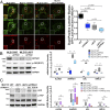
Mistargeting of ATP8A1 in the absence of AP-3 activates YAP. (A) Representative images captured from live-cell imaging of MLE15/WT, MLE15/ΔAP3, MLE15/ΔATP8A1, and MLE15/ΔAP3-ΔATP8A1 cells expressing mCherry-ABCA3 and the biosensor GFP-LactC2 (arrow: GFP-LactC2+/mCherry-ABCA3+ dual-positive LBs; arrowhead: mCherry-ABCA3+/GFP-LactC2- LBs). Live-cell imaging was obtained using identical microscope settings, and still images were derived from the first frame to avoid photobleaching. GFP-LactC2/mCherry-ABCA3 dual-positive organelles as a fraction of total mCherry-ABCA3-positive organelles (n = 2; 10 cells; box and whiskers plot showing minimum, 25th percentile, median, 75th percentile, and maximum). (Scale bar, 10 μm.) (B) ATP8A1-dependency of YAP signaling in MLE15 cells described in A. (Left) Composite representative IB (n = 3; 15 μg cell lysate per lane) for endogenous ATP8A1, phospho-YAP-Ser127, total YAP, and GAPDH. (Right) RT-qPCR for Ajuba, Ankrd1, Axl, Birc5 RNA (n = 3; box and whiskers plot as described in A; *P < 0.05, **P < 0.01, ***P < 0.001, ****P < 0.0001). (C) Flippase-dependency of YAP signaling in MLE15/ΔAP3 cells. (Left) Composite representative IB of MLE15/WT and MLE15/ΔAP3cells, and MLE15/ΔAP3-ΔATP8A1 cells expressing GFP, GFP-ATP8A1, or GFP-ATP8A1(E191Q) (n = 3; 15 μg cell lysate per lane) for GFP, p-YAP-Ser127, total YAP, and GAPDH. (Right) RT-qPCR for Ajuba, Ankrd1, Axl, Birc5 RNA (n = 3; box and whiskers plot and P values as described in B).
Fig. 8.
Fig. 8.
YAP activation in AT2 cells of pearl mice. (A) RT-qPCR using mRNA from AT2 cells of WT and pearl mice for Ajuba, Ankrd1, Axl, Birc5 (n = 3; box and whiskers plot showing minimum, 25th percentile, median, 75th percentile, and maximum). (B) Analysis of IFM shown in C of lungs from WT and pearl mice at 8 and 16 wk of age. HALO image analysis software was used to count all nuclei (DAPI-positive) and all AT2 cells (SFTPC-positive). Graphic representation of SFTPC+ AT2 cells (Left) and SFTPC+/nuclear AJUBA+ AT2 cells (Right), both as a percentage of total DAPI-positive nuclei (n = 10 nonoverlapping 40× fields of lung sections from 3 mice per group). (C) Representative images of lungs from WT and pearl mice at 8 wk of age immunostained for SFTPC (green) and AJUBA (red) and costained with DAPI (blue). Arrows point to cells that exhibit cytosolic SFTPC surrounding DAPI-positive nuclei that also exhibit nuclear AJUBA. (Scale bar, 50 μm.)

Similar articles

Cited by

References

    1. Whitsett J. A., Wert S. E., Weaver T. E., Diseases of pulmonary surfactant homeostasis. Annu. Rev. Pathol. 10, 371–393 (2015). - PMC - PubMed
    1. Vicary G. W., Vergne Y., Santiago-Cornier A., Young L. R., Roman J., Pulmonary fibrosis in hermansky-Pudlak syndrome. Ann. Am. Thorac. Soc. 13, 1839–1846 (2016). - PMC - PubMed
    1. Bowman S. L., Bi-Karchin J., Le L., Marks M. S., The road to lysosome-related organelles: Insights from Hermansky-Pudlak syndrome and other rare diseases. Traffic 20, 404–435 (2019). - PMC - PubMed
    1. Kook S., et al. ., Impaired lysosomal integral membrane protein 2-dependent peroxiredoxin 6 delivery to lamellar bodies accounts for altered alveolar phospholipid content in adaptor protein-3-deficient pearl mice. J. Biol. Chem. 291, 8414–8427 (2016). - PMC - PubMed
    1. Mahavadi P., et al. ., Epithelial stress and apoptosis underlie Hermansky-Pudlak syndrome-associated interstitial pneumonia. Am. J. Respir. Crit. Care Med. 182, 207–219 (2010). - PMC - PubMed

Publication types

MeSH terms

Supplementary concepts