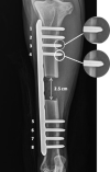
Temporal Changes in Reverse Torque of Locking-Head Screws Used in the Locking Plate in Segmental Tibial Defect in Goat Model
- PMID: 33987199
- PMCID: PMC8111000
- DOI: 10.3389/fsurg.2021.637268
Temporal Changes in Reverse Torque of Locking-Head Screws Used in the Locking Plate in Segmental Tibial Defect in Goat Model
Abstract
The objective of this study was to evaluate changes in peak reverse torque (PRT) of the locking head screws that occur over time. A locking plate construct, consisting of an 8-hole locking plate and 8 locking screws, was used to stabilize a tibia segmental bone defect in a goat model. PRT was measured after periods of 3, 6, 9, and 12 months of ambulation. PRT for each screw was determined during plate removal. Statistical analysis revealed that after 6 months of loading, locking screws placed in position no. 4 had significantly less PRT as compared with screws placed in position no. 5 (p < 0.05). There were no statistically significant differences in PRT between groups as a factor of time (p > 0.05). Intracortical fractures occurred during the placement of 151 out of 664 screws (22.7%) and were significantly more common in the screw positions closest to the osteotomy (positions 4 and 5, p < 0.05). Periosteal and endosteal bone reactions and locking screw backout occurred significantly more often in the proximal bone segments (p < 0.05). Screw backout significantly, negatively influenced the PRT of the screws placed in positions no. 3, 4, and 5 (p < 0.05). The locking plate-screw constructs provided stable fixation of 2.5-cm segmental tibia defects in a goat animal model for up to 12 months.
Keywords: animal models; biomechanics; bone healing; fracture fixation; locking screws; orthopedics; segmental bone defect.
Copyright © 2021 Grzeskowiak, Rifkin, Croy, Steiner, Seddighi, Mulon, Adair and Anderson.
Conflict of interest statement
The authors declare that the research was conducted in the absence of any commercial or financial relationships that could be construed as a potential conflict of interest.
Figures

Similar articles
-
Biomechanical evaluation of peak reverse torque (PRT) in a dynamic compression plate-screw construct used in a goat tibia segmental defect model.BMC Vet Res. 2019 Sep 5;15(1):321. doi: 10.1186/s12917-019-2058-7. BMC Vet Res. 2019. PMID: 31488151 Free PMC article.
-
Effect of cyclic loading on the stability of screws placed in the locking plates used to bridge segmental bone defects.J Orthop Res. 2021 Mar;39(3):516-524. doi: 10.1002/jor.24838. Epub 2020 Sep 9. J Orthop Res. 2021. PMID: 32844515 Free PMC article.
-
Dynamization at the near cortex in locking plate osteosynthesis by means of dynamic locking screws: an experimental study of transverse tibial osteotomies in sheep.J Bone Joint Surg Am. 2015 Feb 4;97(3):208-15. doi: 10.2106/JBJS.M.00529. J Bone Joint Surg Am. 2015. PMID: 25653321
-
The protective effect of locking screw placement on nonlocking screw extraction torque in an osteoporotic supracondylar femur fracture model.J Orthop Trauma. 2012 Sep;26(9):523-7. doi: 10.1097/BOT.0b013e318238c086. J Orthop Trauma. 2012. PMID: 22430520
-
A review of locking compression plate biomechanics and their advantages as internal fixators in fracture healing.Clin Biomech (Bristol). 2007 Dec;22(10):1049-62. doi: 10.1016/j.clinbiomech.2007.08.004. Epub 2007 Sep 27. Clin Biomech (Bristol). 2007. PMID: 17904257 Review.
Cited by
-
In vitro analysis and in vivo assessment of fracture complications associated with use of locking plate constructs for stabilization of caprine tibial segmental defects.J Exp Orthop. 2023 Apr 3;10(1):38. doi: 10.1186/s40634-023-00598-9. J Exp Orthop. 2023. PMID: 37010659 Free PMC article.
-
Changes in tibial cortical dimensions and density associated with long-term locking plate fixation in goats.J Exp Orthop. 2023 Nov 7;10(1):111. doi: 10.1186/s40634-023-00669-x. J Exp Orthop. 2023. PMID: 37934300 Free PMC article.
References
LinkOut - more resources
Full Text Sources

