. 2021 May 12;11(1):10109.
doi: 10.1038/s41598-021-89586-6.

Neurotropic influenza A virus infection causes prion protein misfolding into infectious prions in neuroblastoma cells

Affiliations

Neurotropic influenza A virus infection causes prion protein misfolding into infectious prions in neuroblastoma cells

Hideyuki Hara et al. Sci Rep. .

Abstract

Misfolding of the cellular prion protein, PrPC, into the amyloidogenic isoform, PrPSc, which forms infectious protein aggregates, the so-called prions, is a key pathogenic event in prion diseases. No pathogens other than prions have been identified to induce misfolding of PrPC into PrPSc and propagate infectious prions in infected cells. Here, we found that infection with a neurotropic influenza A virus strain (IAV/WSN) caused misfolding of PrPC into PrPSc and generated infectious prions in mouse neuroblastoma cells through a hit-and-run mechanism. The structural and biochemical characteristics of IAV/WSN-induced PrPSc were different from those of RML and 22L laboratory prions-evoked PrPSc, and the pathogenicity of IAV/WSN-induced prions were also different from that of RML and 22L prions, suggesting IAV/WSN-specific formation of PrPSc and infectious prions. Our current results may open a new avenue for the role of viral infection in misfolding of PrPC into PrPSc and formation of infectious prions.


Conflict of interest statement

The authors declare no competing interests.

Figures
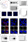
Figure 1
Figure 1
IAV/WSN infection induces conversion of PrPC to PrPSc in N2aC24 cells. (a) Cell viability of N2aC24 cells after infection with different MOIs of IAV/WSN. Cell viability was determined by counting of live cells. (b) Western blotting of IAV/WSN-uninfected (Un) N2aC24 cells and -infected N2aC24 cells at various time points with 6D11 anti-PrP antibody, anti-HA antibody, anti-NP antibody, and anti-M2 antibody after treatment with ( +) or without (-) PK (20 μg/mg of proteins). PK-untreated 15 μg proteins from each cell lysate were used for detection of total PrP, HA NP, and M2 and PK-treated 300 μg proteins for PrPSc. (c) Left panels: Immunofluorescent staining of PrPSc (green) using 132 anti-PrP antibody in IAV/WSN-uninfected and -infected N2aC24 cells at 8 dpi and in 22L prion-infected N2aC24L1-3 cells. Right panel: The percentage (%) of 132 anti-PrP antibody-positive cells to DAPI-positive cells in at least 3 fields (145 × 109 μm/field) of IAV/WSN-uninfected (Un) and -infected N2aC24 cells at various time points and in 22L prion-infected N2aC24L1-3 (L1-3) cells. Blue, DAPI; bar, 10 μm. (d) Double immunofluorescent staining for PrPSc (green) and M2 (red) using 132 anti-PrP antibody and anti-HA antibody in IAV/WSN-uninfected and -infected N2aC24 cells at 8 dpi and in 22L prion-infected N2aC24L1-3 cells. Blue, DAPI; bar, 10 μm. (e) Left panel: Western blotting with 6D11 antibody of IAV/WSN-uninfected N2aC24 (C24) cells after 20 passages in IAV/WSN-free medium and N2aC24(R1) (C24R1) cells serially passaged after IAV/WSN infection using 15 μg proteins for total PrP (Left panel) and 30 μg proteins for PrPSc (Right panel). β-actin was used as an internal control. P, passages. (f) Western blotting of 15 μg proteins from N2aC24 and N2aC24(R1) cells with anti-HA antibody, anti-NP antibody, and anti-M2 antibody. IAV/WSN-infected N2aC24 cells were used as a positive control for the viral proteins. β-actin was used as an internal control. P, passages. Full length blots of Western blot images are shown in Supplementary Figs. 5c-e, 6, and 7.
Figure 2
Figure 2
Reproducible de novo formation of PrPSc in N2aC24 and N2a cells after infection with IAV/WSN. (a) Western blotting of N2aC24 cells at 5 passages after infection with or without IAV/WSN using 6D11 antibody after treatment with different concentrations of PK. Results are shown from three other independent experiments. (b) Western blotting of N2a cells at 5 passages after infection with or without IAV/WSN using 6D11 antibody after treatment with different concentrations of PK. Full length blots of Western blot images are shown in Supplementary Fig. 8.
Figure 3
Figure 3
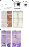
IAV/WSN infection induces de novo formation of infectious prions in N2aC24 cells. (a) The percentage of symptom-free mice after intracerebral inoculation with cell lysates from control N2aC24 and N2aC24(R1) cells. (b) Western blotting of brains from mice sacrificed at 208 dpi with the N2aC24 cell lysate and from mice symptomatic after inoculation with N2aC24(R1) cell lysate with 6D11 antibody after treatment with or without PK (20 μg/mg of proteins). PK-untreated 20 μg proteins from each brain homogenate were used for detection of total PrP and PK-treated 40 μg proteins for PrPSc. β-actin was used as an internal control. (c) Immunohistochemistry of brains from mice sacrificed at 208 dpi with the N2aC24 cell lysate and from mice terminally ill after inoculation with N2aC24(R1) cell lysate with or without 6D11 antibody. Bars, 100 μm. (d) Hematoxylin–eosin staining of brains from mice sacrificed at 208 dpi with the N2aC24 cell lysate and from mice terminally ill after inoculation with N2aC24(R1) cell lysate. Cx, cerebral cortex; Hp, hippocampus; Th, thalamus; Cb, cerebellum. Bars, 100 μm. Full length blots of Western blot images are shown in Supplementary Fig. 9a, b.
Figure 4
Figure 4
Blockage of IAV/WSN infection prevents formation of PrPSc in N2aC24 cells. (a) Western blotting with anti-HA antibody of 15 μg proteins from IAV/WSN-uninfected (-) and -infected ( +) N2aC24 cells at 4 dpi after treatment with anti-IAV/WSN serum or control serum. (b) Western blotting with 6D11 antibody after treatment with or without PK (20 μg/mg of proteins) of IAV/WSN-uninfected (-) and -infected ( +) N2aC24 cells at 5 passages after treatment with anti-IAV/WSN serum or control serum. PK-untreated 15 μg proteins from each cell lysate were used for detection of total PrP and PK-treated 30 μg proteins for PrPSc. β-actin was used as an internal control. (c) Western blotting with anti-HA antibody of 15 μg proteins from IAV/WSN-infected ( +) N2aC24 cells at 4 dpi after treatment with different concentrations of oseltamivir (Osel). (d) Western blotting with 6D11 antibody after treatment with or without PK (20 μg/mg of proteins) of IAV/WSN-uninfected and -infected N2aC24 cells at 5 (Left panels) and 10 (Right panels) passages after treatment with different concentrations of Osel. PK-untreated 15 μg proteins from each cell lysate were used for detection of total PrP and PK-treated 30 μg proteins for PrPSc. β-actin was used as an internal control. Full length blots of Western blot images are shown in Supplementary Fig. 9c, d, and 10.
Figure 5
Figure 5
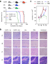
Different pathological properties of prions in N2aC24(R1) cells from RML and 22L prions. (a) The percentage of symptom-free mice after the 1st intracerebral inoculation with brain homogenates from N2aC24(R1) cell lysate-inoculated, terminally ill mice, the 2nd intracerebral inoculation with brain homogenates from terminally ill mice firstly inoculated with N2aC24(R1)-infected mouse brain homogenates, and intracerebral inoculation with RML- or 22L-infected mouse brain homogenates. (b) HE staining of the brains of terminally ill mice in (a). Bars, 100 μm. (c) Vacuole counts at different brain areas of terminally ill mice [5 fields in each brain area from each mouse group (n = 3)] in (b). Cx, cerebral cortex; Hp, hippocampus; Th, thalamus; Cb, cerebellum. Error bars, standard deviation.
Figure 6
Figure 6
Different biochemical properties of PrPSc in N2aC24(R1) cells from PrPSc in RML- and 22L-infected N2aC24 cells. (a) Western blotting of N2aC24(R1), RML-infected N2aC24Chm (Chm), and 22L-infected N2aC24L1-3 cells (L1-3) with 6D11 antibody after treatment with or without PK (20 μg/mg of proteins). PK-untreated 15 μg proteins from each cell lysate were used for detection of total PrP (Left panel) and PK-treated 30 μg proteins for PrPSc (Middle panel) Aliquots of the PK-treated samples were also treated with PNGase F before Western blotting (Right panel). β-actin was used as an internal control. (b) Ratio (%) of di-, mono-, and un-glycosylated PrPSc in the Middle panel of (a). (c) Western blotting (Left panels) of PrPSc and its signal intensities (Right panel, n = 3) in RML-infected N2aC24Chm (Chm, Upper panel), N2aC24(R1) (Middle panel), and 22L-infected N2aC24L1-3 cells (L1-3, Lower panel) with 6D11 antibody after treatment with different concentrations of GdnHCl. Full length blots of Western blot images are shown in Supplementary Fig. 11.

Similar articles

Cited by

References

    1. d'Errico P, Meyer-Luehmann M. Mechanisms of pathogenic tau and Aβ protein spreading in Alzheimer's disease. Front. Aging Neurosci. 2020;12:265. doi: 10.3389/fnagi.2020.00265. - DOI - PMC - PubMed
    1. Billings LM, Oddo S, Green KN, McGaugh JL, LaFerla FM. Intraneuronal Aβ causes the onset of early Alzheimer's disease-related cognitive deficits in transgenic mice. Neuron. 2005;45:675–688. doi: 10.1016/j.neuron.2005.01.040. - DOI - PubMed
    1. Jellinger KA. Neuropathological aspects of Alzheimer disease, Parkinson disease and frontotemporal dementia. Neurodegener. Dis. 2008;5:118–121. doi: 10.1159/000113679. - DOI - PubMed
    1. Duda JE, Lee VM, Trojanowski JQ. Neuropathology of synuclein aggregates. J. Neurosci. Res. 2000;61:121–127. doi: 10.1002/1097-4547(20000715)61:2<121::AID-JNR1>3.0.CO;2-4. - DOI - PubMed
    1. De Chiara G, et al. Infectious agents and neurodegeneration. Mol. Neurobiol. 2012;46:614–638. doi: 10.1007/s12035-012-8320-7. - DOI - PMC - PubMed

Publication types