. 2021 May 12;12(1):2750.
doi: 10.1038/s41467-021-22854-1.

S-acylation of P2K1 mediates extracellular ATP-induced immune signaling in Arabidopsis

Affiliations

S-acylation of P2K1 mediates extracellular ATP-induced immune signaling in Arabidopsis

Dongqin Chen et al. Nat Commun. .

Abstract

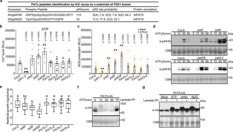
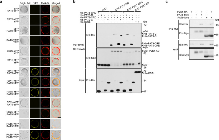
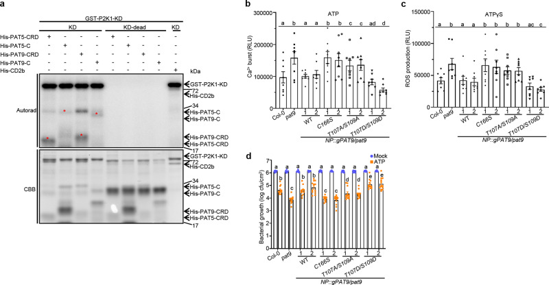
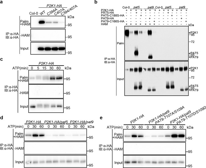
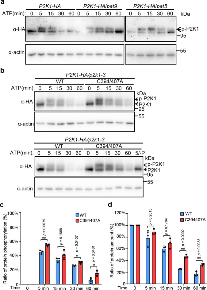
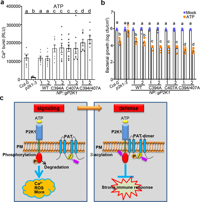
S-acylation is a reversible protein post-translational modification mediated by protein S-acyltransferases (PATs). How S-acylation regulates plant innate immunity is our main concern. Here, we show that the plant immune receptor P2K1 (DORN1, LecRK-I.9; extracellular ATP receptor) directly interacts with and phosphorylates Arabidopsis PAT5 and PAT9 to stimulate their S-acyltransferase activity. This leads, in a time-dependent manner, to greater S-acylation of P2K1, which dampens the immune response. pat5 and pat9 mutants have an elevated extracellular ATP-induced immune response, limited bacterial invasion, increased phosphorylation and decreased degradation of P2K1 during immune signaling. Mutation of S-acylated cysteine residues in P2K1 results in a similar phenotype. Our study reveals that S-acylation effects the temporal dynamics of P2K1 receptor activity, through autophosphorylation and protein degradation, suggesting an important role for this modification in regulating the ability of plants in respond to external stimuli.


Conflict of interest statement

The authors declare no competing interests.

Figures

Fig. 1
Fig. 1. PAT5 and PAT9 control PTI response triggered by eATP.
a Identification of PAT5 and PAT9 tryptic peptides as a substrate of P2K1 kinase by KiC assay. b Ligand-induced calcium influx. 5-day-old seedlings were treated with 100 μM ATP for 15 min. RLU, relative luminescence units. Error bars indicate ±SEM; n = 8 seedlings; *p < 0.05, **p < 0.01, P-values indicate significance relative to Col-0 and were determined by one-sided ANOVA with unpaired, two-tailed Student’s t test. c ROS production was measured in leaf disks after treatment with 200 μM ATPγS for 30 min. Leaf disks were taken from WT (Col-0), pat5, pat9, and pat5/9 double mutants, or their complemented lines PAT5 (NP::ATPAT5-HA/Atpat5) and PAT9 (NP::ATPAT9-HA/Atpat9). Error bars indicate ±SEM; n = 12 leaf disks; *p < 0.05, **p < 0.01, P-values indicate significance relative to Col-0 with ATPγS treatment and were determined by one-sided ANOVA with unpaired, two-tailed Student’s t test. d MAPKs activation of Arabidopsis leaf disks that treated with 200 μM ATPγS for the times indicated. Coomassie Brilliant Blue (CBB) staining of protein was used as loading control. e PAT5 and PAT9 negatively mediated bacterial invasion. Arabidopsis seedlings with the indicated genotype (x axis) were flood-inoculated with Pst. DC3000 bacteria and bacterial growth determined by plate counting 3 days post inoculation. Error bars indicate ±SEM; n = 12 (biological replicates); means with different letters are significantly different (p < 0.05); P-values indicate significance relative to Col-0 and were determined by one-sided ANOVA with multiple comparisons and adjusted using Benjamini–Hochberg post-test. Box extends from the 25th to the 75th percentile, whiskers denote minima and maxima (Boxplots, Col-0: max = 7.76; min = 5.88; center = 6.82; Q2 (25%) = 6.45; Q3 (75%) = 7.43, pat5: max = 7.76; min = 5.45; center = 6.62; Q2 (25%) = 6.13; Q3 (75%) = 7.05, pat9: max = 6.97; min = 5.3; center = 6.47; Q2 (25%) = 6.1; Q3 (75%) = 6.71, pat5/9: max = 6.55; min = 5; center = 5.9; Q2 (25%) = 5.49; Q3 (75%) = 6.21, PAT5-1: max = 7.7; min = 5.59; center = 7.05; Q2 (25%) = 6.76; Q3 (75%) = 7.25, PAT5-2: max = 7.64; min = 5.85; center = 7; Q2 (25%) = 6.51; Q3 (75%) = 7.15, PAT9-1: max = 7.72; min = 5.6; center = 7.09; Q2 (25%) = 6.58; Q3 (75%) = 7.47, PAT9-2: max = 7.7; min = 5.78; center = 7; Q2 (25%) = 6.27; and Q3 (75%) = 7.47). Experiments were repeated three times with similar results. f, g Ligand triggers PAT5 and PAT9 phosphorylation after treated with 200 μM ATP, 1 μM flg22, or 50 μg/ml chitin in their complemented lines PAT5 and PAT9. Lambda protein phosphatase (Lambda PP, − and +) was added to release phosphate groups. CBB was used as loading control. All experiments were repeated and analyzed three times with similar results.
Fig. 2
Fig. 2. PAT5 and PAT9 directly interact with P2K1 in vitro and in vivo.
a Interactions of PATs and P2K1 receptor at Arabidopsis protoplast plasma membrane. FM4-64 was used to stain the plasma membrane. Bar = 20 μm. b Directly interaction of PATs and P2K1 in vitro. Purified, recombinant proteins were incubated with Glutathione Sepharose 4B beads followed by GST-His pull-down assay. His-CD2b (At5g09390, CD2-binding protein-related, a substrate of P2K1 from KiC assay data) was used as a negative control. c PAT5 and PAT9 interact with P2K1 in vivo. The indicated constructs were transiently expressed in Arabidopsis wild-type protoplasts followed by Co-IP assays. All experiments were performed and analyzed three times with similar results.
Fig. 3
Fig. 3. P2K1 phosphorylates PAT5 and PAT9 to regulate immune responses.
a P2K1 directly phosphorylates PAT5 and PAT9. Purified P2K1 kinase domain recombinant proteins were incubated with PAT5/PAT9-CRD and -C domains in an in vitro kinase assay. Autophosphorylation and transphosphorylation were measured by incorporation of γ-[32P]-ATP. MBP and GST-CD2b were used as positive and negative controls, respectively. The protein loading was measured by CBB staining. Red stars represent the trans-phosphorylated proteins. Experiments were repeated two times with similar results. b, c Phosphorylation of PAT9 regulates ATP-induced calcium influx and ROS production. The indicated genomic PAT9 variants were transformed into the pat9 mutant, and then examined for ATP-induced calcium influx and ROS production for 30 min. RLU relative luminescence units. Error bars indicate ±SEM; n = 8 (biological replicates); means with different letters are significantly different (p < 0.05); P-values indicate significance relative to Col-0 and were determined by one-sided ANOVA with multiple comparisons and adjusted using Benjamini–Hochberg post-test. d PAT9 Phosphorylation mediates ATP-induced restriction of bacterial growth. The indicated plant leaves were syringe-infiltrated with 106 cfu/mL−1 of Pst. DC3000 after 24 h water (mock) or 400 μM ATP treatment, which is different from surface inoculation (Fig. 1e). Bacterial numbers were determined 3 days post inoculation. Error bars indicate ±SEM; n = 12 (biological replicates); means with different letters are significantly different (p < 0.05); P-values indicate significance relative to Col-0 with mock treatment and were determined by one-sided ANOVA with multiple comparisons and adjusted using Benjamini–Hochberg post-test.
Fig. 4
Fig. 4. PAT5 and PAT9 S-acylate P2K1.
a Detection of P2K1 S-acylation and their associated residues in the stable P2K1-HA transgenic plants. The S-acylation levels were detected by an acyl-resin capture (acyl-RAC) assay. Palm- palmitoylated protein, Depalm- depalmitoylated protein. HAM, hydroxylamine, for cleavage of the Cys-palmitoyl thioester linkages. Experiments were repeated three times. b PAT5 and PAT9 S-acylate P2K1 in planta. Protoplasts of Arabidopsis wild-type Col-0, Atpat5 and Atpat9 mutants were transfected with plasmids as indicated, followed by acyl-RAC assays. Experiments were repeated three times. ce Dynamic S-acylation levels of P2K1 in stable transgenic plants upon eATP addition. The P2K1-HA transgenic plant was crossed with pat5, pat9, NP::PAT9-T107/AS109A/pat9, and NP::PAT9-T107D/S109D/pat9 plants and then infiltrated with 200 μM ATP. All experiments were repeated and analyzed three times with similar results.
Fig. 5
Fig. 5. PAT9 regulates ATP-induced P2K1 phosphorylation and turnover.
a PAT5/9 controls P2K1 phosphorylation and turnover. P2K1-HA was analyzed by immunoblot in wild type and Atpat5 and Atpat9 mutants background upon addition of 400 μM ATP. p-P2K1 phosphorylation of P2K1. Actin protein was used as loading control. Experiments were repeated three times with similar results. bd Analysis of turnover and phosphorylation of P2K1 proteins modified (C → A) at the site of S-acylation. The indicated constructs were expressed into Arabidopsis p2k1-3 mutant plant and treated with 400 μM ATP. Lambda PP (-P) was added to release phosphate groups. In panel c and d, the ratio of P2K1 protein phosphorylation and total protein amount were measured from panel b and another replicate with Image J. Error bars indicate ±SE; n = 3 (biological replicates). *p < 0.05, **p < 0.01, P-values indicate significance relative to WT and were determined by one-sided ANOVA with unpaired, two-tailed Student’s t test.
Fig. 6
Fig. 6. S-acylation of P2K1 negatively regulates PTI.
a, b S-acylation of P2K1 mediates ATP-induced calcium influx and bacterial defense. The indicated 5-day-old seedlings were treated with 100 μM ATP for calcium influx in 15 min. For bacterial growth, the indicated plant leaves were pretreated with 400 μM ATP for 24 h, and then inoculated with Pst. DC3000 and bacterial growth measured by plate counting after 3 days post inoculation. Values represent the mean ± SEM, n = 8 in a, n = 12 in b (biological replicates); Means with different letters are significantly different (p < 0.05); P-values indicate significance relative to Col-0 with mock treatment and were determined by one-sided ANOVA with multiple comparisons and adjusted using Benjamini–Hochberg post-test. c Model for the role of P2K1 receptor S-acylation in eATP signaling. Upon addition of the activating ligand eATP, the P2K1 receptor is rapidly autophosphorylation and phosphorylates downstream targets, leading to PTI immune response and P2K1 protein turnover. P2K1 directly interacts with and phosphorylates PATs to activate PATs S-acylation upon ATP treatment. Activation of PATs S-acylates P2K1 and then inactivates P2K1 phosphorylation and turnover, following dampens the immune response to protect plant growth.

Similar articles

Cited by

References

    1. Dodds PN, Rathjen JP. Plant immunity: towards an integrated view of plant-pathogen interactions. Nat. Rev. Genet. 2010;11:539–548. doi: 10.1038/nrg2812. - DOI - PubMed
    1. Zipfel C. Plant pattern-recognition receptors. Trends Immunol. 2014;35:345–351. doi: 10.1016/j.it.2014.05.004. - DOI - PubMed
    1. Zhang, W. et al. Different pathogen defense strategies in Arabidopsis: more than pathogen recognition. Cells7, 252 (2018). - PMC - PubMed
    1. Gomez-Gomez L, Boller T. FLS2: an LRR receptor-like kinase involved in the perception of the bacterial elicitor flagellin in Arabidopsis. Mol. Cell. 2000;5:1003–1011. doi: 10.1016/S1097-2765(00)80265-8. - DOI - PubMed
    1. Zipfel C, et al. Bacterial disease resistance in Arabidopsis through flagellin perception. Nature. 2004;428:764–767. doi: 10.1038/nature02485. - DOI - PubMed

Publication types

MeSH terms

LinkOut - more resources