. 2021 Jul 1;81(13):2793-2807.e8.
doi: 10.1016/j.molcel.2021.04.021. Epub 2021 May 11.

Methylation of histone H3 at lysine 37 by Set1 and Set2 prevents spurious DNA replication

Affiliations

Methylation of histone H3 at lysine 37 by Set1 and Set2 prevents spurious DNA replication

Helena Santos-Rosa et al. Mol Cell. .

Abstract

DNA replication initiates at genomic locations known as origins of replication, which, in S. cerevisiae, share a common DNA consensus motif. Despite being virtually nucleosome-free, origins of replication are greatly influenced by the surrounding chromatin state. Here, we show that histone H3 lysine 37 mono-methylation (H3K37me1) is catalyzed by Set1p and Set2p and that it regulates replication origin licensing. H3K37me1 is uniformly distributed throughout most of the genome, but it is scarce at replication origins, where it increases according to the timing of their firing. We find that H3K37me1 hinders Mcm2 interaction with chromatin, maintaining low levels of MCM outside of conventional replication origins. Lack of H3K37me1 results in defective DNA replication from canonical origins while promoting replication events at inefficient and non-canonical sites. Collectively, our results indicate that H3K37me1 ensures correct execution of the DNA replication program by protecting the genome from inappropriate origin licensing and spurious DNA replication.

Keywords: H3K37methylation; Histone modifications; MCM; Origin licensing; Replication origins; Set1; Set2.


Conflict of interest statement

Declaration of interests T.K. is a co-founder and shareholder of Abcam Plc and a co-founder of Storm Therapeutics Ltd. (Cambridge, UK). T.L. is a consultant for Storm Therapeutics Ltd.

Figures
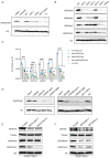
Figure 1
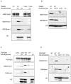
Figure 1. Set2 and Set1 (COMPASS) are necessary for H3K37me1 in vivo.
(A and B) Immunoblot analysis of total protein extracts from different yeast isogenic mutants as specified. Equal amounts of proteins were separated by SDS-PAGE in 16% acrylamide gels. Blots were probed with anti-H3K37me1 or anti-H3K4me1/me2/me3 antibodies and then re-probed with anti-H3 antibody. (C) ChIP qPCR experiments showing H3K37me1 levels at different genomic locations. Chromatin from isogenic strains was immunoprecipitated using anti-H3K37me1, anti-H3 and anti-GFP (negative control) antibodies. Statistical analysis was performed using Two-way ANOVA multiple comparisons and Tukey’s multiple comparison test (Alpha: 0.05); * - P ≤ 0.05, ** - P ≤ 0.01, *** - P ≤ 0.001, **** - P ≤ 0.0001. Error bars represent the mean ± SD of 2 independent experiments. (D) Immunoblot analysis of total protein extracts from different yeast isogenic strains as specified. Equal amounts of proteins were separated by SDS-PAGE in 16% acrylamide gels. Blots were probed with anti-H3K37me1 antibody and then re-probed with anti-H3 antibody as indicated. (E and F) Immunoblot analysis of total protein extracts from (E) SETD1A or (F) SETD2 CRISPR knockout cell pools. Human RPE1 cells stably expressing Cas9 were transfected with two guide RNAs targeting different exons of each gene or a non-targeting control. Total protein extracts were prepared 7 days after transfection, and proteins were separated by SDS-PAGE in 12% acrylamide gels or 3-8% Tris-Acetate gels.
Figure 2
Figure 2. Set2 and Set1 (COMPASS) methylate H3K37 in vitro.
(A) In vitro methyltransferase assay. Recombinant wild-type Set2p and Set2Y149Ap mutant proteins were incubated with wild-type (WT) H3.1 nucleosomes in the presence of SAM. Reactions were separated by SDS-PAGE in 16% acrylamide gels. Blots were probed with anti-H3K37me1 antibody and then re-probed sequentially with anti-H3K36me1 and anti-H3 antibodies. Finally, the membrane was blotted with anti-MBP antibody to monitor the amount of enzyme in each reaction. (B) In vitro methyltransferase assay. Equal amount of recombinant wild-type Set2p was incubated with H3.1WT or H3.1K37R nucleosomes in the presence of SAM. Reactions were separated by SDS-PAGE in 16% acrylamide gels. Blots were probed with anti-H3K37me1 antibody and then re-probed sequentially with anti-H3K36me1 and anti-H3 antibodies. (C) In vitro methyltransferase assay. Wild-type PtA-Set1p and PtA-set1ΔC92p yeast purified complexes were incubated with wild-type H3.1 nucleosomes in the presence of SAM. Reactions were separated by SDS-PAGE in 16% acrylamide. Blots were probed with anti-H3K37me1 antibody and sequentially with anti-H3K4me1 and anti-H3 antibodies in this order. * indicates Δ tail–H3. PtA-Set1p and PtA-set1ΔC92p in each reaction were detected using anti-PAP antibody. (D) In vitro methyltransferase assay. Equal amount of wild-type PtA-Set1p complex was incubated with H3.1WT or H3.1K37R nucleosomes in the presence of SAM. Reactions were separated by SDS-PAGE in 16% acrylamide gels. Blots were probed with anti-H3K37me1 antibody and sequentially with anti-H3K4me1 and anti-H3 antibodies in this order. * indicates Δ tail–H3.
Figure 3
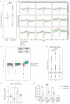
Figure 3. Histone H3K37me1 correlates with Replication Origin firing.
(A) Box-plot representing H3K37me1 enrichment over H3 (log2) of genomic windows overlapping replication origins (ARS +/- 2kb) versus non-ARS (mean of the region from Transcription Start Sites+600 to TSS+1000). (B) Coverage plot showing the mean normalised ChIP-signal (+/- s.e.m.) for H3, H3K37me1 and Input at ARS +/-5kb (total number of ARSs: 408). ARSs were grouped into 3 categories according to firing efficiency/time. (C and D) Box-plot showing the mean H3K37me1 enrichment over H3 (log2) at ARS +/-2kb (left panel) and at non-ARS control regions (TSS +600 to TSS+1000, right panel). p-values were calculated with the Mann-Whitney-Wilcoxon test. (E) ChIP qPCR experiments showing H3K37me1 levels at ARS607. Chromatin from the time points shown in Figure S4A was immunoprecipitated using anti-H3K37me1 and anti-H3 antibodies. H3K37me1/H3 levels were normalized to a non-ARS region (SPR3). Statistical analysis was performed using Two-way ANOVA multiple comparisons and Tukey’s multiple comparison test (Alpha: 0.05); * - P ≤ 0.05, ** - P ≤ 0.01, *** - P ≤ 0.001. Error bars represent the mean ± SD of 3 independent experiments. (F) ChIP qPCR experiments showing H3K37me1 enrichment at different origins of replication in G1/S and S-phase synchronized human RPE1 cells as shown in Figure S4D. Statistical analysis was performed using Two-way ANOVA corrected for the comparisons using the Holm-Sidak method (Alpha: 0.05); * - P ≤ 0.05, ** - P ≤ 0.01, *** - P ≤ 0.001. Error bars represent the mean ± SD of 3 independent experiments.
Figure 4
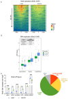
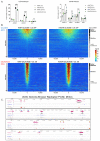
Figure 4. H3K37me1 regulates DNA replication.
(A) Heatmap showing the distribution of BrdU incorporation at replication origins in wild-type H3 and H3K37R mutant cells. Origins were aligned from highest to lowest BrdU signal in the wild-type strain and centred at ARS ACS (Ars Consensus Sequence). For visualisation purposes the heatmaps’ colour scale was saturated at the 99th percentile of the distribution of signal intensities. The plot represents the average of 4 independent experiments. (B) Box-plot showing the distribution of mean BrdU signal incorporated at the replication origins (ARS +/- 1kb) shown in (A). The p-values were calculated with the Mann-Whitney-Wilcoxon test. ARSs were classified into early/efficient, medium and late/inefficient following the BrdU distribution shown in Figure S5C. (C) ChIP qPCR experiments showing incorporation of BrdU in H3WT and H3K37R mutant cells. IPs were analysed by qPCRs at the early/efficient ARS607, late/inefficient ARS603 and “H3K37R-unique” firing locations, using specific primers. Statistical analysis was performed using Two-way ANOVA corrected for the comparisons using the Holm-Sidak method (Alpha: 0.05); * - P ≤ 0.05, ** - P ≤ 0.01, *** - P ≤ 0.001, **** - P ≤ 0.0001. Error bars represent the mean ± SD of 2 independent experiments. (D) Piechart showing proportion of H3K37R unique replication events occurring at different genomic locations.
Figure 5
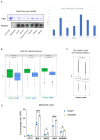
Figure 5. H3K37me1 regulates origin establishment.
(A) Left: Mcm2-His6 in vitro binding to biotinylated H3 peptides, modified as indicated. Input and peptide-bound Mcm2 were resolved by SDS-PAGE in 8% acrylamide and detected by Coomasie-Brilliant Blue (CBB) staining. 1/5 of each bead slurry was spotted onto PVDF membrane and biotin-peptides detected with anti-biotin antibody. Right: Average ImageJ quantification of assays shown in left and in Fig.S7A. Binding is represented as % of the signal corresponding to ¼ of the input. (B) Box plot of Mcm2-HA occupancy at early/efficient, medium and late/inefficient ARS +/- 1Kb. Blue: H3WT, green: H3K37R. (C) Box plot of Mcm2 occupancy at non-ARS (mean of the region from TSS+600 to TSS+1000) in H3WT and H3K37R mutant cells. The plots in (B) and (C) represent the average of the 3 independent cultures. (D) ChIP qPCR experiments showing Mcm2-HA levels at “H3K37R unique” replication sites. H3WT and H3K37R yeast cells expressing Mcm2-HA were arrested in G1, crosslinked and chromatin was immunoprecipitated (IP) with anti-HA antibody. The IP material was analyzed by qPCR with primers specific for each location. Data were normalized to the non-ARS region (SPR3). Statistical analysis was performed using Two-way ANOVA corrected for the comparisons using the Holm-Sidak method (Alpha: 0.05); * - P ≤ 0.05, ** - P ≤ 0.01, *** - P ≤ 0.001, **** - P ≤ 0.0001. Error bars represent the mean ± SD of 2 independent experiments.
Figure 6
Figure 6. Over-expression of MCM activators suppresses H3K37R replication defects.
(A) ChIP qPCR experiments showing Cdc45-HA levels at efficient ARS (left panel) and “H3K37R unique” replication sites (right panel). H3WT and H3K37R yeast cells expressing Cdc45-HA were arrested in G1 and released into HU containing medium for 30’. Samples were crosslinked and chromatin was immunoprecipitated (IP) with anti-HA antibody. The IP material was analyzed by qPCR with primers specific for each location. Data were normalized to a non-ARS region (SPR3). Statistical analysis was performed using Two-way ANOVA corrected for the comparisons using the Holm-Sidak method (Alpha: 0.05); * - P ≤ 0.05, ** - P ≤ 0.01, *** - P ≤ 0.001, **** - P ≤ 0.0001. Error bars represent the mean ± SD of 2 independent experiments. Note the difference in the scale between left and right panels. (B) Heatmap showing the distribution of BrdU incorporation in wild-type H3 and H3K37R mutant cells under non-overexpression (GLUCOSE, OFF) and over-expression (GALACTOSE, ON) of MCM activators. Replication origins were aligned from highest to lowest BrdU signal in the wild-type strain and centred at ACS. ARSs were classified into early/efficient, medium and late/inefficient following the BrdU distribution shown in Figure S5C. For visualisation purposes the heatmaps’ colour scale was saturated at the 99th percentile of the distribution of signal intensities. The plot represents the average of 3 independent experiments. (C) Representative genome browser snapshots of BrdU incorporation in H3WT and isogenic H3K37R mutant cells at different genomic locations. ARS (OriDB) are represented as orange boxes; non-replicative ACS matches are represented as blue lines. H3K37R exclusive firing event are indicated by the corresponding color-coded arrows. The plots represent the average of 3 independent cultures per strain and condition.
None

Similar articles

Cited by

References

    1. Azmi IF, Watanabe S, Maloney MF, Kang S, Belsky JA, MacAlpine DM, Peterson CL, Bell SP. Nucleosomes influence multiple steps during replication initiation. Elife. 2017;6:e22512. - PMC - PubMed
    1. Bartke T, Vermeulen M, Xhemalce B, Robson SC, Mann M, Kouzarides T. Nucleosome-interacting proteins regulated by DNA and histone methylation. Cell. 2010;143:470–484. - PMC - PubMed
    1. Bell SP, Labib K. Chromosome duplication in Saccharomyces cerevisiae. Genetics. 2016;203:1027–1067. - PMC - PubMed
    1. Belsky JA, Macalpine HK, Lubelsky Y, Hartemink AJ, Macalpine DM. Genome-wide chromatin footprinting reveals changes in replication origin architecture induced by pre-RC assembly. Genes Dev. 2015;29:212–224. - PMC - PubMed
    1. Biswas D, Takahata S, Xin H, Dutta-Biswas R, Yu Y, Formosa T, Stillman DJ. A role for Chd1 and Set2 in negatively regulating DNA replication in Saccharomyces cerevisiae. Genetics. 2008;178:649–659. - PMC - PubMed

Publication types

MeSH terms

LinkOut - more resources