. 2021 May:243:111374.
doi: 10.1016/j.molbiopara.2021.111374. Epub 2021 May 8.

Identification and characterisation of a phospholipid scramblase in the malaria parasite Plasmodium falciparum

Affiliations

Identification and characterisation of a phospholipid scramblase in the malaria parasite Plasmodium falciparum

Silvia Haase et al. Mol Biochem Parasitol. 2021 May.

Abstract

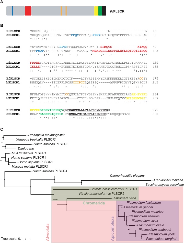
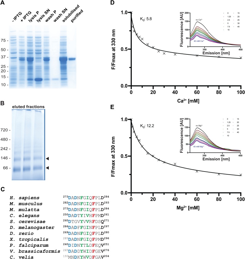
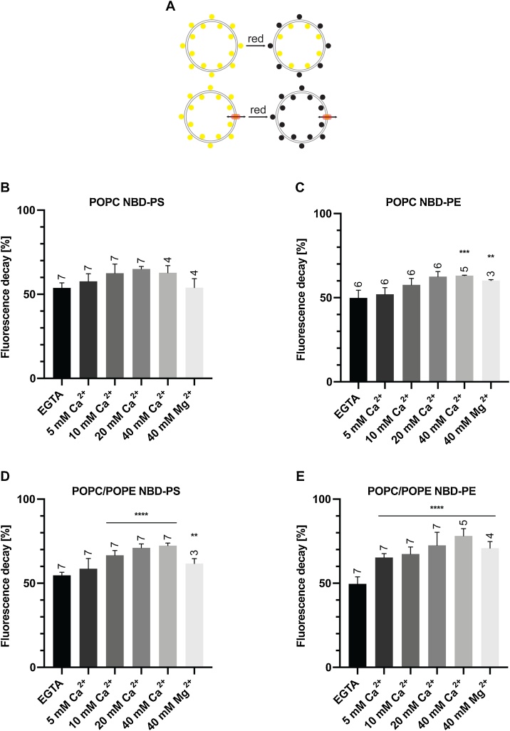
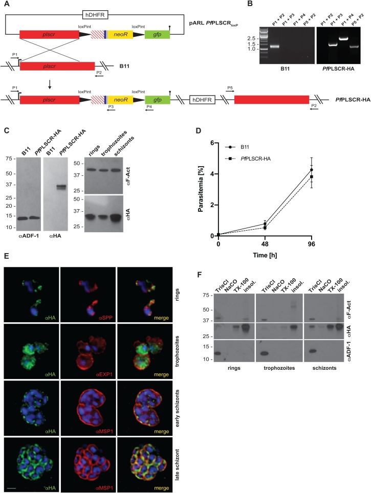
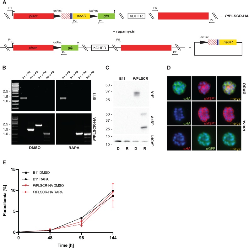
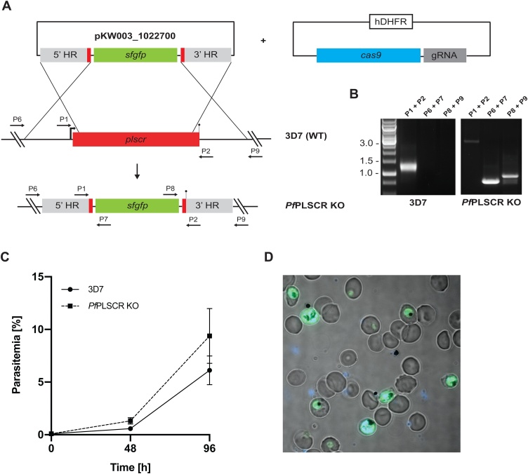
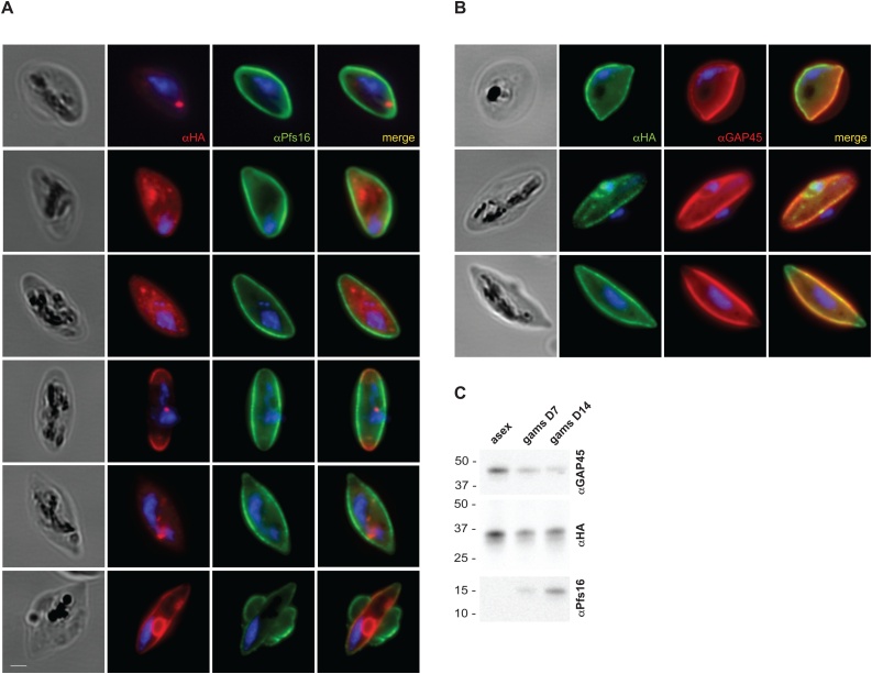
Recent studies highlight the emerging role of lipids as important messengers in malaria parasite biology. In an attempt to identify interacting proteins and regulators of these dynamic and versatile molecules, we hypothesised the involvement of phospholipid translocases and their substrates in the infection of the host erythrocyte by the malaria parasite Plasmodium spp. Here, using a data base searching approach of the Plasmodium Genomics Resources (www.plasmodb.org), we have identified a putative phospholipid (PL) scramblase in P. falciparum (PfPLSCR) that is conserved across the genus and in closely related unicellular algae. By reconstituting recombinant PfPLSCR into liposomes, we demonstrate metal ion dependent PL translocase activity and substrate preference, confirming PfPLSCR as a bona fide scramblase. We show that PfPLSCR is expressed during asexual and sexual parasite development, localising to different membranous compartments of the parasite throughout the intra-erythrocytic life cycle. Two different gene knockout approaches, however, suggest that PfPLSCR is not essential for erythrocyte invasion and asexual parasite development, pointing towards a possible role in other stages of the parasite life cycle.

Keywords: Gametocytes; Invasion; Liposomes; Malaria; Phospholipid scramblase; Plasmodium falciparum.


Conflict of interest statement

The authors report no declarations of interest.

Figures

Fig. 1
Fig. 1
Protein features and conservation of the putative PL scramblase in P. falciparum. A) A schematic of PfPLSCR indicating the PXXP/Y motifs (blue), putative DNA binding region (red), predicted palmitoylation sites (orange) and bipartite nuclear leader sequence (yellow), as well as a putative Ca2+ binding motif (green) and the C-terminal transmembrane helix (black and underlined). B) Alignment of PfPLSCR (PlasmoDB ID: PF3D7_1022700) with the human ortholog hPLSCR1 (UniProtKB ID: O15162). The functional domains, colour-coded as above, are positionally conserved in PfPLSCR. Similar residues are marked by colons (:) and identical residues are indicated by stars (*). Protein sequences were aligned with Clustal Omega [77]. The cut-off score was set to 4.0 for the prediction of palmitoylation sites [49]. C) Phylogenetic tree reconstruction from a selection of PLSCR orthologues. The tree was generated by using the Simple Phylogeny Web services [78] and the tree file was imported into iTOL [79] for illustrative purposes. The following EupathDB gene identifiers were used: PF3D7_1022700 (P. falciparum), PGABG01_1020700 (P. gaboni), PmUG01_10033900 (P. malariae), PKH_060680 (P. knowlesi), PVX_111580 (P. vivax), PocGH01_06015800 (P. ovale), PCHAS_050700 (P. chabaudi), PY17X_0508000 (P. yoelii), PBANKA_050690 (P. berghei), Vbra_21596 (Vitrella brassicaformis PLSCR1), Vbra_2039 (Vitrella brassicaformis PLSCR2) and Cvel_12647 (Chromera velia). The following UniProtKB accession numbers were used: O15162 (H. sapiens PLSCR1), Q9NRY7 (H. sapiens PLSCR2), Q9NRY6 (H. sapiens PLSCR3), Q9NRQ2 (H. sapiens PLSCR4), Q9JJ00 (Mus musculus PLSCR1), H9ZA71 (Macaca mulatta PLSCR1), Q21318 (Caenorhabditis elegans), P47140 (Saccharomyces cerevisae), Q8IQD8 (Drosophila melanogaster), Q6NY24 (Danio rerio) and B5DDV8 (Xenopus tropicalis PLSCR1).
Fig. 2
Fig. 2
PfPLSCR undergoes conformational changes in the presence of metal ions. A) SDS-PAGE of the expression and solubilisation of His::PfPLSCR. The purified recombinant protein runs at the predicted molecular weight of ∼ 37 kDa. Pellet and supernatant fractions are labelled as ‘P’ and ‘SN’, respectively. B) BN-PAGE of eluted fractions from the main peak reveals two protein species between the 66 kDa and 146 kDa protein markers (black arrow heads), indicating a possible dimerisation/oligomerisation of His::PfPLSCR. C) Alignment of the putative Ca2+ binding regions from different PLSCR1 orthologues. Residues in positions 1, 3, 5, 7, 9 and 12 of the EF-hand-like motif in hPLSCR1 have been proposed to octahedrally coordinate the calcium ion [24] and are shown in bold. Highly conserved residues are highlighted in blue, hydrophobic residues (positions 5 and 7) in green and the strictly conserved phenylalanine residues (position 9) are shown red. D & E) Representative binding curves and calculated affinities (for the shown experiments) of recombinant PfPLSCR for Ca2+ and Mg2+ ions, respectively. The concentration-dependent decrease of fluorescence at λ 330 nm is plotted as F/Fmax with F = fluorescence measured in the presence of cations and Fmax= fluorescence under EGTA conditions. A selection of emission spectra is shown in the upper right corner.
Fig. 3
Fig. 3
PL scramblase activity of recombinant PfPLSCR in symmetrically labelled vesicles. A) Schematic of the ‘scramblase assay’. B & D) Transbilayer movement of NBD-PS and C & E) NBD-PE in proteoliposomes of different lipid compositions. Scramblase activity (mean ± SD) is presented as the percentage of fluorescence decrease at 500 s. Number of independent experiments is indicated above the error bars. Statistical significance of differences between activities measured in the presence of EGTA and divalent cations was calculated using unpaired t-tests (****, p < 0.0001; ***, p = 0.0001; **, p ≤ 0.005).
Fig. 4
Fig. 4
PfPLSCR-HA is expressed throughout the asexual life cycle and localises to membranous structures in the parasite. A) Schematic of the strategy to generate the conditional KO line PfPLSCR-HA via SLI mediated single-crossover homologous recombination. The targeting plasmid contains the Pfplscr homology region (red), followed by two synthetic loxPint modules flanking the recodonised Pfplscr sequence (red stripes) with a triple-HA tag (blue), T2A skip peptide (gray) and neomycin resistance cassette (yellow). The GFP coding sequence (green) is followed by a stop codon (black bar). Numbered arrows indicate primers used for genotyping. B) Diagnostic PCR confirms successful integration into the Pfplscr locus by amplification of three distinctive fragments at the expected size (1585, 2455 and 1182 bp) and loss of the wild-type band (1396 bp) in the PfPLSCR-HA parasite line. C) Western blot analysis gives rise to a ∼ 37 kDa protein band as predicted for PfPLSCR-HA. The endogenously tagged protein is expressed throughout the intra-erythrocytic life cycle as indicated by the different parasite stages (hours post invasion): rings (4 ± 2 h), trophozoites (20 ± 4 h) and schizonts (44 ± 4 h). Anti-ADF (actin depolymerising factor) and anti-F-Actin antibodies are used as loading controls. D) Growth curves of parental B11 and PfPLSCR-HA expressing parasites over two cycles. Parasitemias (as quantified by flow cytometry) were averaged from at least three biological replicates using blood from different donors and are presented as mean ± SD. E) Immunofluorescence analysis of PfPLSCR-HA expressing parasites. The upper panel depicts two ring-stage parasites and co-labelling with anti-SPP (signal peptide peptidase) antibodies as an ER marker. Anti-EXP1 (exported protein 1) delineates the PVM surrounding three trophozoites in a multiply infected red blood cell. Lower two panels show deconvolved co-labelling with antibodies against MSP1 (merozoite surface protein 1) in late blood stage parasites. PfPLSCR-HA co-localises with MSP1 at the plasma membrane in late schizonts. Parasite nuclei are stained with DAPI (blue). Scale bar = 1 μM. F)PfPLSCR-HA remains strongly membrane-associated throughout the intra-erythrocytic life cycle as suggested by sequential extraction of the protein and its presence in the TX-100 insoluble fraction. The blot was cut and probed with anti-ADF as a control for soluble ADF protein and anti-F-Actin as a control for partially membrane-bound parasite actin.
Fig. 5
Fig. 5
PL scrambling is not essential for red blood cell invasion and intra-erythrocytic parasite development. A) Schematic of rapamycin induced and DiCre-mediated excision of the floxed sequence in PfPLSCR-HA. B) Diagnostic PCR confirms successful excision of the locus compared to DMSO-treated PfPLSCR-HA parasites. C) The C-terminally truncated PfPLSCR protein is expressed as a GFP fusion protein at the expected molecular weight of ∼ 53 kDa in rapamycin-treated parasites. The ∼ 27 kDa band is characteristic for GFP only. Anti-ADF antibodies are used as a loading control. D) Plasma membrane association is lost in parasites expressing the truncated PfPLSCR-GFP fusion protein, which distributes throughout the parasite cytosol. E) Growth curves of rapamycin and DMSO-treated parasites over three cycles. Parasitemias were averaged from at least three biological replicates using blood from different donors and are presented as mean ± SD.
Fig. 6
Fig. 6
A full gene KO in 3D7 parasites supports the redundancy of PfPLSCR in asexual parasite biology. A) Schematic of the CRISPR-Cas9 mediated KO strategy. Expression of sfGFP is driven under a constitutive BiP promoter. Numbered arrows indicate primers used for genotyping. B) Diagnostic PCR confirms successful replacement of the Pfplscr locus by the sfGFP expression cassette. C) Growth curves of wild-type 3D7 and PfPLSCR KO parasites over two cycles. Parasitemias were averaged from at least three biological replicates using blood from different donors and are presented as mean ± SD. D) Live-cell image of PfPLSCR KO parasites expressing sfGFP throughout asexual parasite development. Parasite nuclei are stained with DAPI (blue).
Fig. 7
Fig. 7
PfPLSCR is expressed in gametocytes. A) Immunofluorescence analysis of stage III-IV gametocytes reveals different localisation patterns of the triple-HA tagged protein. Anti-Pfs16 is used as gametocyte specific antibody marking the PVM. Parasite nuclei are stained with DAPI. Scale bar = 1 μM. B)PfPLSCR-HA co-localises with the IMC marker GAP45, which delineates the parasite periphery. C) Western blot analysis confirms PfPLSCR-HA expression in gametocytes.

Similar articles

Cited by

References

    1. Vial H.J., Eldin P., Tielens A.G.M., van Hellemond J.J. Phospholipids in parasitic protozoa. Mol. Biochem. Parasitol. 2003;126:143–154. - PubMed
    1. Sherling E.S., van Ooij C. Host cell remodeling by pathogens: the exomembrane system in Plasmodium-infected erythrocytes. FEMS Microbiol. Rev. 2016;40:701–721. - PMC - PubMed
    1. Gulati S., Ekland E.H., Ruggles K.V., Chan R.B., Jayabalasingham B., Zhou B., Mantel P.Y., Lee M.C., Spottiswoode N., Coburn-Flynn O. Profiling the essential nature of lipid metabolism in asexual blood and gametocyte stages of plasmodium falciparum. Cell Host Microbe. 2015;18:371–381. - PMC - PubMed
    1. McNamara C.W., Lee M.C., Lim C.S., Lim C.S.H., Roland J., Simon O., Yeung B.K., Chatterjee A.K., McCormack S.L., Manary M.J. Targeting plasmodium PI(4)K to eliminate malaria. Nature. 2013;504:248–253. - PMC - PubMed
    1. Vaid A., Ranjan R., Smythe W.A., Hoppe H.C. Sharma P: PfPI3K, a phosphatidylinositol-3 kinase from Plasmodium falciparum, is exported to the host erythrocyte and is involved in hemoglobin trafficking. Blood. 2010;115:2500–2507. - PMC - PubMed

Publication types

MeSH terms

LinkOut - more resources