. 2021 Apr 21:12:652719.
doi: 10.3389/fmicb.2021.652719. eCollection 2021.

Sumoylation of the Carboxy-Terminal of Human Cytomegalovirus DNA Polymerase Processivity Factor UL44 Attenuates Viral DNA Replication

Affiliations

Sumoylation of the Carboxy-Terminal of Human Cytomegalovirus DNA Polymerase Processivity Factor UL44 Attenuates Viral DNA Replication

Jun Chen et al. Front Microbiol. .

Abstract

Controlled regulation of genomic DNA synthesis is a universally conserved process for all herpesviruses, including human cytomegalovirus (HCMV), and plays a key role in viral pathogenesis, such as persistent infections. HCMV DNA polymerase processivity factor UL44 plays an essential role in viral DNA replication. To better understand the biology of UL44, we performed a yeast two-hybrid screen for host proteins that could interact with UL44. The most frequently isolated result was the SUMO-conjugating enzyme UBC9, a protein involved in the sumoylation pathway. The UBC9-UL44 interaction was confirmed by in vitro His-tag pull-down and in vivo co-immunoprecipitation assays. Using deletion mutants of UL44, we mapped two small regions of UL44, aa 11-16, and 260-269, which might be critical for the interaction with UBC9. We then demonstrated that UL44 was a target for sumoylation by in vitro and in vivo sumoylation assays, as well as in HCMV-infected cells. We further confirmed that 410lysine located within a ψKxE consensus motif on UL44 carboxy-terminal was the major sumoylation site of UL44. Interestingly, although 410lysine had no effects on subcellular localization or protein stability of UL44, the removal of 410lysine sumoylation site enhanced both viral DNA synthesis in transfection-replication assays and viral progeny production in infected cells for HCMV, suggesting sumoylation can attenuate HCMV replication through targeting UL44. Our results suggest that sumoylation plays a key role in regulating UL44 functions and viral replication, and reveal the crucial role of the carboxy-terminal of UL44, for which little function has been known before.

Keywords: DNA replication; UBC9; UL44; human cytomegalovirus; sumoylation.


Conflict of interest statement

The authors declare that the research was conducted in the absence of any commercial or financial relationships that could be construed as a potential conflict of interest.

Figures

FIGURE 1
FIGURE 1
HCMV UL44 interacts with UBC9 in vitro and in vivo. (A) His-tagged UL44 pulled down Myc-tagged UBC9 in vitro. His-UL44 were bound to immobilized cobalt chelate resin then incubated with cell lysates expressing Myc-UBC9. The bound protein complexes were analyzed by western blot using anti-His and anti-Myc antibodies. Representative of three independent experiments. (B) Co-IP of transiently expressed UL44 and UBC9. 293T cells were cotransfected with HA-tagged UL44 and Myc-tagged UBC9 or NB80 as negative control. Cell lysates were incubated with immobilized anti-HA beads to isolate UL44 and its associated proteins. Results were analyzed by western blot using anti-HA and anti-Myc antibodies. Representative of three independent experiments.
FIGURE 2
FIGURE 2
Mapping of the potential UBC9-interacting domains in UL44. (A) Schematic view and structural features of UL44 deletion mutants and their interaction with UBC9 as detected by yeast two-hybrid and β-galactosidase assays. +, positive interaction; –, no interaction. The first and last β-strands are indicated by red box. (B) Effect of different mutation on the interaction of UL44 with UBC9. 293T cells were cotransfected with Myc-tagged UBC9 and HA-tagged UL44 (full length or deletion mutants as indicated), then lysates were incubated with immobilized anti-Myc beads to isolate UBC9 and its associated proteins. (C) Stereoscopic ribbon representation of UL44 (aa 1–290) by YASARA view. UL44 aa 11–16 and 260–269, corresponding to the first and last β-strands, were colored yellow, was likely critical for the interaction of UL44 with UBC9. α-helices and other β-strands were colored blue and red, respectively.
FIGURE 3
FIGURE 3
UL44 is a target for sumoylation. (A) UL44 covalently conjugated with SUMO-1. 293T cells were transiently cotransfected with HA-UL44, Myc-SUMO-1 and UBC9 as indicated. Forty eight hours later, cells were resuspended and boiled in SDS sample buffer, the supernatants were analyzed by western blot using anti-HA and anti-Myc antibodies. β-actin served as loading control. (B) UBC9 dominant-negative mutant abolished the sumoylation of UL44. 293T cells were cotransfected with HA-UL44, Myc-SUMO-1, UBC9, or UBC9-DN as indicated, and then processed the same as in (A). (C) UL44 covalently conjugated with SUMO-1 in vitro. Purified His-tagged UL44, SUMO-activating E1 enzyme, UBC9, UBC9 dominant-negative mutant, and SUMO-1 were mixed in reaction buffer and incubated at 37°C for 2 h. The mixtures were separated by SDS-PAGE and UL44 was detected with anti-His antibody. (D) UL44 co-IPed with SUMO-1. Lysates from 293T cells expressing HA-UL44 and Myc-SUMO-1 were immunoprecipitated by anti-HA, the results were analyzed by western blot using anti-HA and anti-Myc antibodies. For all panels, representative of three independent experiments.
FIGURE 4
FIGURE 4
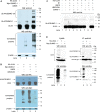
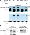
410lysine was the major sumoylation site of UL44 in both transfected and infected cells. (A) 293T cells were transiently cotransfected with HA-UL44 (wild-type or K410A mutant), Myc-SUMO-1 and UBC9 as indicated. Forty eight hours later, cells were resuspended and boiled in SDS sample buffer, the supernatants were analyzed by western blot using anti-HA and anti-Myc antibodies. (B) HFF cells were infected with wild-type HCMV (WT) or HCMV containing UL44 K410A mutant (K410A) at an MOI of 1. At 48 h post-transfection, total cell lysates were either directly analyzed by SDS-PAGE, or immunoprecipitated with covalently conjugated anti-UL44 beads, then probed for UL44 or sumoylated proteins using anti-UL44 or anti-SUMO1 antibodies, respectively. For all panels, representative of three independent experiments.
FIGURE 5
FIGURE 5
410lysine had no effects on subcellular localization or protein stability of UL44. (A) Hela cells were transfected with constructs encoding fusion proteins EGFP-UL44WT, EGFP-UL44-K410A, EGFP-NLSUL44WT, EGFP-NLSUL44–K410A, and UBC9 or SUMO-1 alone or together as indicated in figure, stained with Hoechst 33258 to visualize cell nuclei. Confocal microscopy showed mutation of 410lysine had no effect on the nuclear localization of UL44. (B) HFFs were infected with wild-type HCMV (WT) or HCMV carrying UL44-K410A mutant (K410A) for 48 h. Cells were then pulsed in methionine-free DMEM with methionine analog AHA. After pulse labeling, cells were either immediately harvested (0 h), or chased in regular DMEM for 6 h (6 h). Cell lysates containing AHA-incorporated proteins were then immunoprecipitated with anti-UL44 antibody and further cross-linked with biotin. Biotin-labeled UL44 were resolved and visualized in SDS-PAGE. Pulse-chase test showed mutation of 410lysine had no effects on protein stability of UL44.
FIGURE 6
FIGURE 6
Mutation of UL44 410lysine enhanced oriLyt-dependent DNA synthesis in transfection-replication assays. (A) HFF cells were transiently cotransfected with pCMV-HA-UL44 (wild-type or K410A mutant) and all other plasmids as indicated in Table 2, or without UL54 expression plasmid as control. SUMO-1 and UBC9 expression plasmids were also cotransfected in certain groups as indicated. Total cellular DNA was harvested 5 days post-transfection and was cleaved with DpnI, and then was used as qPCR template to determine the copies of oriLyt-containing plasmid pGL3-14. Each sample was done in triplicate. Each set of assays was repeated 3 times and a representative one was shown. (B) Vero cells were transiently cotransfected with HA-UL44 (wild-type or K410A mutant) and all other plasmids as indicated in Table 2, or without UL54 expression plasmid as control. SUMO-1 and UBC9 expression plasmids were also cotransfected in certain groups as indicated. Luciferase activity was measured 5 days after transfection. Firefly luciferase activity was normalized to Renilla luciferase activity for each sample. To confirm the conjugation of UL44 with SUMO-1, partial of cell lysates were directly probed with anti-HA antibody to visualize UL44. Analyses were repeated 3 times and the results were the average of triplicate experiments. Error bars = SD. * denotes p < 0.05, and *** denotes p < 0.001 by two-tailed unpaired Student’s t-test between indicated two groups.
FIGURE 7
FIGURE 7
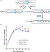
Mutation of 410lysine enhanced the viral replication of HCMV in HFFs. (A) Schematic for BAC construction and generation of wild-type, mutant and revertant HCMV virus. (B) The replication and spread abilities of HCMV were determined by multi-step virus growth curves. HFF cells were infected by appropriate virus (UL44WT-Zeo, UL44-K410A-Zeo, and WT-Rev-Kana) at a moi of 0.1 and collected at the indicated time points. Released virus at each time point was titrated on HFFs by serial dilutions and plaques counting to generate growth curves. Titration of each time point was done in triplicate and titer values were means from triplicate experiments. Error bars = SD. Differences among three viruses at each time point were assessed by One-way ANOVA with Tukey’s post hoc test. * denotes p < 0.05, and ** denotes p < 0.01 by One-way ANOVA with Tukey’s multiple comparison test with UL44WT-Zeo. # denotes p < 0.05, and ## denotes p < 0.01 by One-way ANOVA with Tukey’s multiple comparison test with WT-Rev-Kana.
FIGURE 8
FIGURE 8
UL44 homologs share a highly conserved C terminus. Nuclear localization signals are indicated by red box. Putative ψKxE consensus sumoylation motifs corresponding to HCMV 410lysine are indicated in boldface type and by red box. The single-letter amino acid code is used and * means the end of a protein.

Similar articles

Cited by

References

    1. Adamson A. L. (2005). Effects of SUMO-1 upon Epstein-Barr virus BZLF1 function and BMRF1 expression. Biochem. Biophys. Res. Commun. 336 22–28. 10.1016/j.bbrc.2005.08.036 - DOI - PubMed
    1. Ahn J. H., Xu Y., Jang W. J., Matunis M. J., Hayward G. S. (2001). Evaluation of interactions of human cytomegalovirus immediate-early IE2 regulatory protein with small ubiquitin-like modifiers and their conjugation enzyme Ubc9. J. Virol. 75 3859–3872. 10.1128/jvi.75.8.3859-3872.2001 - DOI - PMC - PubMed
    1. Alvisi G., Jans D. A., Guo J., Pinna L. A., Ripalti A. (2005). A protein kinase CK2 site flanking the nuclear targeting signal enhances nuclear transport of human cytomegalovirus ppUL44. Traffic 6 1002–1013. 10.1111/j.1600-0854.2005.00331.x - DOI - PubMed
    1. Appleton B. A., Brooks J., Loregian A., Filman D. J., Coen D. M., Hogle J. M. (2006). Crystal structure of the cytomegalovirus DNA polymerase subunit UL44 inΔ3, with the C terminus from the catalytic subunit. Differences in structure and function relative to unliganded UL44. J. Biol. Chem. 281 5224–5232. 10.1074/jbc.m506900200 - DOI - PubMed
    1. Appleton B. A., Loregian A., Filman D. J., Coen D. M., Hogle J. M. (2004). The cytomegalovirus DNA polymerase subunit UL44 forms a C clamp-shaped dimer. Mol. Cell 15 233–244. 10.1016/j.molcel.2004.06.018 - DOI - PubMed

LinkOut - more resources