. 2021 May 6;11(1):9660.
doi: 10.1038/s41598-021-88841-0.

Mitochondrial calcium uniporter is essential for hearing and hair cell preservation in congenic FVB/NJ mice

Affiliations

Mitochondrial calcium uniporter is essential for hearing and hair cell preservation in congenic FVB/NJ mice

Mayakannan Manikandan et al. Sci Rep. .

Abstract

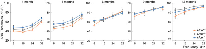
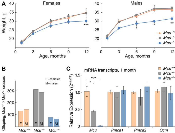
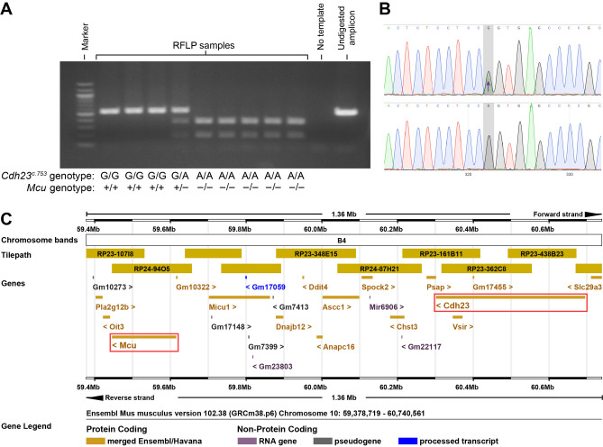
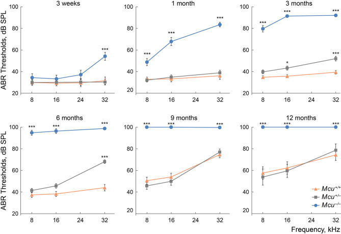
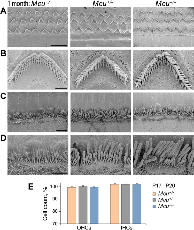
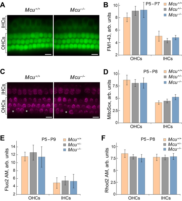
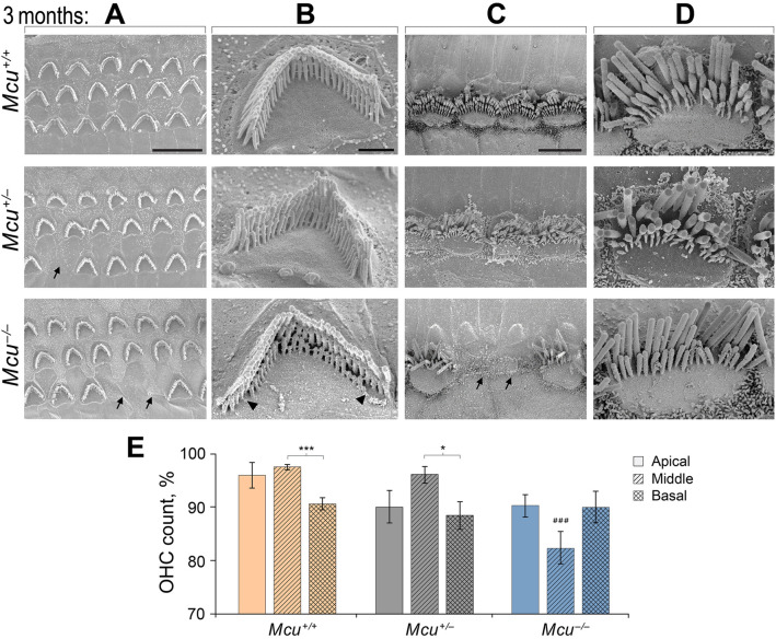
Mitochondrial Ca2+ regulates a wide range of cell processes, including morphogenesis, metabolism, excitotoxicity, and survival. In cochlear hair cells, the activation of mechano-electrical transduction and voltage-gated Ca2+ channels result in a large influx of Ca2+. The intracellular rise in Ca2+ is partly balanced by the mitochondria which rapidly uptakes Ca2+ via a highly selective channel comprised of the main pore-forming subunit, the mitochondrial Ca2+ uniporter (MCU), and associated regulatory proteins. MCU thus contributes to Ca2+ buffering, ensuring cytosolic homeostasis, and is posited to have a critical role in hair cell function and hearing. To test this hypothesis, Ca2+ homeostasis in hair cells and cochlear function were investigated in FVB/NJ mice carrying the knockout allele of Mcu (Mcu+/- or Mcu-/-). The Mcu knockout allele, which originated in C57BL/6 strain cosegregated along with Cdh23ahl allele to the FVB/NJ strain, due to the close proximity of these genes. Neither Mcu+/- nor Mcu-/- genotypes affected cochlear development, morphology, or Ca2+ homeostasis of auditory hair cells in the first two postnatal weeks. However, Mcu-/- mice displayed high-frequency hearing impairment as early as 3 weeks postnatal, which then progressed to profound hearing loss at all frequencies in about 6 months. In Mcu+/- mice, significantly elevated ABR thresholds were observed at 6 months and 9 months of age only at 32 kHz frequency. In three-month-old Mcu-/- mice, up to 18% of the outer hair cells and occasionally some inner hair cells were missing in the mid-cochlear region. In conclusion, mitochondrial Ca2+ uniporter is not required for the development of cochlea in mice, but is essential for hearing and hair cell preservation in congenic FVB/NJ mice.


Conflict of interest statement

The authors declare no competing interests.

Figures

Figure 1
Figure 1
Mice on the mixed B6-CD1 background are predisposed to rapidly progressing hearing loss at all frequencies. Auditory brainstem responses (ABRs) were recorded from the Mcu+/+, Mcu+/−, and Mcu−/− mice on the mixed B6-CD1 background at various time points. Mcu−/− mice show a trend toward slightly elevated ABR thresholds up to three months of age. Although analyses did not show any statistically significant differences among Mcu+/+, Mcu+/− and Mcu−/− mice, as assessed using two way ANOVA followed by Bonferroni multiple comparison test. Not every mouse was tested at each time point. Number of mice (Mcu+/+, Mcu+/−, Mcu−/−): 1 month (23, 32, 11); 3 months (17, 30, 12); 6 months (12, 28, 19); 9 months (10, 25, 19); 12 months (9, 33, 14).
Figure 2
Figure 2
Mcu−/− mice on the FVB/NJ background are smaller in size. (A) Mcu−/− mice have smaller body sizes and reduced body weight in comparison to Mcu+/− and Mcu+/+ littermates. Number of female mice (Mcu+/+, Mcu+/−, Mcu−/−): 1 month (12, 11, 11); 3 months (12, 21, 11); 6 months (7, 15, 9); 9 months (10, 17, 13); 12 months (5, 7, 5). Number of male mice (Mcu+/+, Mcu+/−, Mcu−/−): 1 month (12, 15, 6); 3 months (15, 17, 6); 6 months (8, 22, 9); 9 months (5, 12, 7); 12 months (3, 16, 3). (B) Percentage of female (n = 39) and male (n = 37) offspring obtained from Mcu+/− × Mcu+/− crosses (8 litters). (C) Relative quantitation of the mRNA levels of Mcu, Pmca1, Pmca2, and Ocm in the cochlea of FVB/NJ mice of Mcu−/− and Mcu+/− genotypes as compared to those of Mcu+/+ littermates. Cochleae obtained from 6 animals each of Mcu+/+, Mcu+/−, Mcu−/− genotypes at 1 month of age were used for studying gene expression. *p < 0.05, **p < 0.01, ***p < 0.001, **** p < 0.0001, Two way ANOVA followed by Bonferroni multiple comparison tests.
Figure 3
Figure 3
PCR and restriction fragment length polymorphism (RFLP) based genotyping of Cdh23c.753A/G in FVB/NJ Mcu−/− mice. (A) A representative electropherogram showing the PCR amplicons resolved in a 1.5% agarose gel following digestion with BsrI. The Cdh23c.753G allele renders the binding site for BsrI inaccessible to digest the PCR amplicon of 534 bp, while the Cdh23c.753A allele creates a BsrI binding site resulting in fragments of 350 bp and 184 bp. (B) DNA sequence chromatogram validates the PCR–RFLP method to genotype Cdh23c.753A/G polymorphism. The top panel shows a heterozygous Cdh23c.753A/G allele previously identified by restriction with BsrI. The bottom panel shows a homozygous for Cdh23c.753G sample unaffected by BsrI. (C) Ensembl-based analysis shows genes in a 1.36 Mb region of mouse Chr.10qB4. The genes Mcu and Cdh23 (red boxes) are closely located with an intergenic distance of ~ 686 kb.
Figure 4
Figure 4
Hearing loss in Mcu−/− mice on FVB/NJ background. Early-onset high-frequency hearing loss rapidly progresses to profound deafness with age in FVB/NJ Mcu−/− mice. Auditory brainstem responses were recorded from the Mcu−/−, Mcu+/−, and Mcu+/+ mice of FVB/NJ background, at various time points. Also, note the modest high-frequency hearing loss starting from 3 months of age in Mcu+/− mice. *p < 0.05, ***p < 0.001, two-way ANOVA followed by Bonferroni multiple comparison test. Not every mouse was tested at each time point. Same mice were used as in Fig. 2A plus 8 Mcu+/+, 21 Mcu+/−, and 11 Mcu−/− 3-week-old mice.
Figure 5
Figure 5
Cochlear hair cells in young FVB/NJ Mcu+/+, Mcu+/−, and Mcu−/− mice. (AD) Representative scanning electron micrographs of cochlear hair cells from 1-month-old FVB/NJ Mcu+/+, Mcu+/−, and Mcu−/− mice. (A) The typical three-row arrangement of OHCs, (B) single OHC stereocilia at high magnification, (C) the single row of IHCs, and (D) single IHC stereocilia at high magnification. (E) The number of hair cells in the mid-basal cochlear turn at P17–20. Kruskal–Wallis analysis with Dunn’s multiple comparison test did not show any statistically significant differences between groups. The number of mice is 9 (Mcu+/+), 21 (Mcu+/−), and 9 (Mcu−/−). Scale bars are (in µm): 10 (A); 1 (B); 5 (C); 2 (D).
Figure 6
Figure 6
Mcu is not required for the development of hair cells. Results obtained in vitro demonstrate that mechanotransduction, Ca2+ levels, and mitochondria superoxide activity are not altered in the cochlear hair cells of Mcu−/− mice. (A) Representative images show FM1-43 uptake in hair cells of Mcu+/+, Mcu+/− or Mcu−/− mice (mid-basal turn). (B) The fluorescence intensity of FM1-43 in hair cells is not significantly different between the groups, indicating the normal functioning of MET channels in Mcu−/− mice. (C) Representative images showing the MitoSOX indicator levels in hair cells of Mcu+/+or Mcu−/−. Asterisks show Mcu+/+ and Mcu−/− OHCs, which were excluded from statistical analysis due to no detectable superoxide activity. (D) The fluorescence intensity of MitoSOX in hair cells of Mcu−/− mice is similar to that of the Mcu+/+ mice indicating that there is no difference in oxidative activity caused by cellular superoxide buildup. (E,F) The fluorescence intensity of intracellular (Fluo-2 AM) and mitochondrial (Rhod-2 AM) Ca2+ indicators in hair cells of Mcu+/+, Mcu+/− and Mcu−/− mice were not significantly different. Data represented herein are from mice of age P5–P9. Two-way ANOVA analysis did not show any statistically significant differences between Mcu+/+, Mcu+/− or Mcu−/− groups. The number of mice for all groups was greater than 5. Scale bars in (A) and (C) are 12 µm.
Figure 7
Figure 7
Loss of hair cells in adult Mcu−/− mouse cochlea. (AD) Representative scanning electron micrographs of cochlear hair cells acquired from Mcu+/+ (top row), Mcu+/− (middle row), and Mcu−/− FVB/NJ mice (bottom panel) at P88–P92 time point. (A) The typical three-row arrangement of OHCs, (B) single OHC stereocilia at high magnification, (C) the single row of IHCs, and (D) single IHC stereocilia at high magnification are shown. Note the absence of the third (shortest) row of stereocilia in Mcu−/− mice. The arrows in (A) and (C) indicate missing sensory hair cells and the arrowheads in B indicate the degenerating shortest row of stereocilia in OHCs. (E) The SEM images were categorized based on their cochlear region (apical, middle, and basal turn), and the percentage of missing OHCs were plotted (mean ± SEM). ###p < 0.001 in comparison to the middle region of Mcu+/+ and Mcu+/− mice, *p < 0.05, ***p < 0.001, Kruskal–Wallis with Dunn’s multiple comparison tests. The number of mice is 12 (Mcu+/+), 13 (Mcu+/−), and 18 (Mcu−/−). Scale bars are (in µm): 10 (A); 1 (B); 5 (C); 2 (D).

Similar articles

Cited by

References

    1. Bowl MR, Dawson SJ. Age-related hearing loss. Cold Spring Harb. Perspect. Med. 2019 doi: 10.1101/cshperspect.a033217. - DOI - PMC - PubMed
    1. Wong AC, Ryan AF. Mechanisms of sensorineural cell damage, death and survival in the cochlea. Front. Aging Neurosci. 2015;7:58. doi: 10.3389/fnagi.2015.00058. - DOI - PMC - PubMed
    1. Bottger EC, Schacht J. The mitochondrion: A perpetrator of acquired hearing loss. Hear. Res. 2013;303:12–19. doi: 10.1016/j.heares.2013.01.006. - DOI - PMC - PubMed
    1. Lesus J, et al. Why study inner ear hair cell mitochondria? HNO. 2019;67:429–433. doi: 10.1007/s00106-019-0662-2. - DOI - PMC - PubMed
    1. DiMauro S, Schon EA. Mitochondrial DNA mutations in human disease. Am. J. Med. Genet. 2001;106:18–26. doi: 10.1002/ajmg.1392. - DOI - PubMed

Publication types

LinkOut - more resources