Review
. 2021 Jun 7;220(6):e202102001.
doi: 10.1083/jcb.202102001. Epub 2021 May 5.

Lysosome biogenesis: Regulation and functions

Affiliations
Review

Lysosome biogenesis: Regulation and functions

Chonglin Yang et al. J Cell Biol. .

Abstract

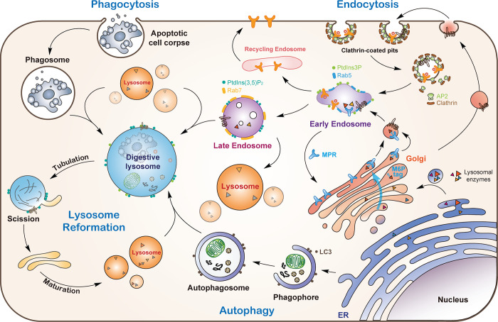
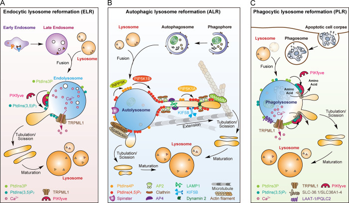
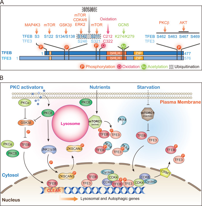
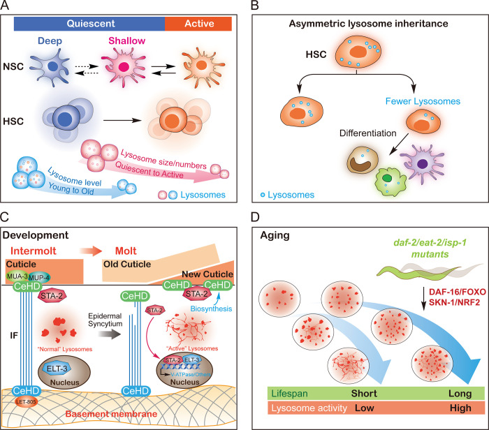
Lysosomes are degradation centers and signaling hubs in cells and play important roles in cellular homeostasis, development, and aging. Changes in lysosome function are essential to support cellular adaptation to multiple signals and stimuli. Therefore, lysosome biogenesis and activity are regulated by a wide variety of intra- and extracellular cues. Here, we summarize current knowledge of the regulatory mechanisms of lysosome biogenesis, including synthesis of lysosomal proteins and their delivery via the endosome-lysosome pathway, reformation of lysosomes from degradative vesicles, and transcriptional regulation of lysosomal genes. We survey the regulation of lysosome biogenesis in response to nutrient and nonnutrient signals, the cell cycle, stem cell quiescence, and cell fate determination. Finally, we discuss lysosome biogenesis and functions in the context of organismal development and aging.


Figures

Figure 1.
Figure 1.
Lysosomes receive proteins and cargos from multiple pathways. Lysosomal hydrolases are synthesized and modified by linkage with oligosaccharides in the ER and transported to the Golgi apparatus. Following recognition of the mannose residues in the oligosaccharide chain by MPR, the hydrolase–MPR complexes are delivered to early endosomes. Newly synthesized lysosomal membrane proteins are either sorted at the TGN and delivered to endosomes (direct pathway) or first delivered to the plasma membrane and then endocytosed to reach early endosomes (indirect pathway). Receptors not destined for lysosomes are recycled back to the plasma membrane or Golgi. Early endosomes undergo a conversion to late endosomes, which then fuse with lysosomes. Phagocytosed cargos are enclosed in phagosomes, which undergo a maturation process and fuse with lysosomes. Autophagic cargos are delivered to lysosomes by fusion of autophagosomes with lysosomes. Lysosomes reform from digestive lysosomes (endo-, phago-, and autolysosomes) by tubulation and scission to form protolysosomes, which mature into functional lysosomes.
Figure 2.
Figure 2.
Graphic summary of lysosome reformation. (A) ELR is regulated by PIKfyve, TRPML1, and Ca2+. PIKfyve converts PtdIns3P to PtdIns(3,5)P2, which activates TRPML1 to regulate lysosomal Ca2+ efflux required for lysosomal tubulation. (B) ALR is achieved by PtdIns(4,5)P2-, clathrin-, and AP2-mediated membrane budding on autolysosomes; KIF5B-driven elongation of membrane tubules along microtubules; dynamin 2–dependent protolysosome scission; and finally protolysosome maturation. AP4 enriches lysosomal membrane proteins for tubulation. WHAMM promotes lysosome tubulation by binding to PtdIns(4,5)P2. The sugar transporter Spinster is also involved in ALR. The generation of PtdIns(4,5)P2 from PtdIns4P is controlled by PtdIns4P 5-kinase 1B on autolysosome membranes and PtdIns4P 5-kinase 1A on protolysosomal tubules. The balance between PtdIns4P and PtdIns(4,5)P2 is also regulated by inositol polyphosphate-5-phosphatase K. Spastizin and spatacsin form a complex to promote tubule initiation on the autolysosome. (C) PLR is regulated by PIKfyve, TRPML1, and amino acid transporters (e.g., SLC-36.1/SLC36A1-4 and LAAT-1/PQLC2). PIKfyve probably regulates the activity of these lysosomal transporters through PtdIns(3,5)P2 to promote lysosome tubulation from phagolysosomes.
Figure 3.
Figure 3.
TFEB/TFE3-dependent lysosome biogenesis. (A) Regulatory modifications of TFEB/TFE3. TFEB/TFE3 are phosphorylated (orange arrows), acetylated (green arrow), and ubiquitinated (gray arrow) by the indicated enzymes. Direct oxidation by reactive oxygen species can also occur (red arrow). (B) mTOR-dependent and PKC-dependent lysosome biogenesis pathways. Under nutrient-rich conditions, TFEB/TFE3 is phosphorylated by lysosome-localized mTOR. Phosphorylated TFEB/TFE3 bind to 14-3-3 proteins and are sequestered in the cytoplasm. With starvation, mTOR is inactivated and unable to phosphorylate TFEB/TFE3, leading to their nuclear translocation and activation. Activation of PKC leads to phosphorylation and inactivation of GSK3β, which then fails to phosphorylate TFEB, thereby inducing nuclear translocation of TFEB. Activated PKC induces JNK2/p38 activation, which phosphorylates ZKSCAN3 and leads to its cytoplasmic translocation and de-repression of lysosomal genes. Nuclear TFEB/TFE3 are rephosphorylated by CDK4/6 (and probably also by mTOR and GSK3β) and relocate back to the cytoplasm in a CRM1-dependent manner.
Figure 4.
Figure 4.
Role of lysosome biogenesis and function in development and aging. (A) The correlation between lysosomes and the different states of NSCs or HSCs. qNSCs and qHSCs have larger and more abundant lysosomes with lower degradation activity (indicated in pink). qNSCs exhibit an age-dependent decrease in lysosome levels (indicated in blue). (B) The correlation between lysosomes and the asymmetric division and differentiation of HSCs. Lysosomes are asymmetrically distributed to daughter cells, and the daughter cells with low levels of lysosomes are metabolically active and induced to differentiate. (C) STA-2– and ELT-3–dependent lysosome biogenesis in ECM remodeling during C. elegans molt. During molt, the cuticle-epidermis attachments are damaged and detected by STA-2. STA-2 translocates to the nucleus and functions together with ELT-3 to activate the expression of lysosomal V-ATPase genes. This accelerates lysosome maturation at molt and facilitates the ECM remodeling required for larval development. (D) The correlation between lysosome biogenesis and lifespan in C. elegans. In aging C. elegans, lysosomes show reduced vesicular morphology; increased tubular morphology; increased mean and total volume; and decreased acidity, motility, and degradation activity. In long-lived mutants from three different longevity pathways, lysosomal gene expression is up-regulated, which requires DAF-16/FOXO and SKN-1/NRF2. Lysosome morphology and activity are well maintained during aging.

Similar articles

  • Lysosomes at the Crossroads of Cell Metabolism, Cell Cycle, and Stemness.
    Nowosad A, Besson A. Nowosad A, et al. Int J Mol Sci. 2022 Feb 18;23(4):2290. doi: 10.3390/ijms23042290. Int J Mol Sci. 2022. PMID: 35216401 Free PMC article. Review.
  • Lysosomes as dynamic regulators of cell and organismal homeostasis.
    Ballabio A, Bonifacino JS. Ballabio A, et al. Nat Rev Mol Cell Biol. 2020 Feb;21(2):101-118. doi: 10.1038/s41580-019-0185-4. Epub 2019 Nov 25. Nat Rev Mol Cell Biol. 2020. PMID: 31768005 Review.
  • Molecular Mechanisms of Lysosome and Nucleus Communication.
    Zhao Q, Gao SM, Wang MC. Zhao Q, et al. Trends Biochem Sci. 2020 Nov;45(11):978-991. doi: 10.1016/j.tibs.2020.06.004. Epub 2020 Jul 2. Trends Biochem Sci. 2020. PMID: 32624271 Free PMC article. Review.
  • Current methods to analyze lysosome morphology, positioning, motility and function.
    Barral DC, Staiano L, Guimas Almeida C, Cutler DF, Eden ER, Futter CE, Galione A, Marques ARA, Medina DL, Napolitano G, Settembre C, Vieira OV, Aerts JMFG, Atakpa-Adaji P, Bruno G, Capuozzo A, De Leonibus E, Di Malta C, Escrevente C, Esposito A, Grumati P, Hall MJ, Teodoro RO, Lopes SS, Luzio JP, Monfregola J, Montefusco S, Platt FM, Polishchuck R, De Risi M, Sambri I, Soldati C, Seabra MC. Barral DC, et al. Traffic. 2022 May;23(5):238-269. doi: 10.1111/tra.12839. Epub 2022 Apr 24. Traffic. 2022. PMID: 35343629 Free PMC article. Review.
  • Lysosomal size matters.
    de Araujo MEG, Liebscher G, Hess MW, Huber LA. de Araujo MEG, et al. Traffic. 2020 Jan;21(1):60-75. doi: 10.1111/tra.12714. Epub 2019 Dec 6. Traffic. 2020. PMID: 31808235 Free PMC article. Review.

Cited by

References

    1. Andrews, N.W., Almeida P.E., and Corrotte M.. 2014. Damage control: Cellular mechanisms of plasma membrane repair. Trends Cell Biol. 24:734–742. 10.1016/j.tcb.2014.07.008 - DOI - PMC - PubMed
    1. Angarola, B., and Ferguson S.M.. 2019. Weak membrane interactions allow Rheb to activate mTORC1 signaling without major lysosome enrichment. Mol. Biol. Cell. 30:2750–2760. 10.1091/mbc.E19-03-0146 - DOI - PMC - PubMed
    1. Annunziata, I., van de Vlekkert D., Wolf E., Finkelstein D., Neale G., Machado E., Mosca R., Campos Y., Tillman H., Roussel M.F., et al. . 2019. MYC competes with MiT/TFE in regulating lysosomal biogenesis and autophagy through an epigenetic rheostat. Nat. Commun. 10:3623. 10.1038/s41467-019-11568-0 - DOI - PMC - PubMed
    1. Appelqvist, H., Wäster P., Kågedal K., and Öllinger K.. 2013. The lysosome: From waste bag to potential therapeutic target. J. Mol. Cell Biol. 5:214–226. 10.1093/jmcb/mjt022 - DOI - PubMed
    1. Bagshaw, R.D., Mahuran D.J., and Callahan J.W.. 2005. A proteomic analysis of lysosomal integral membrane proteins reveals the diverse composition of the organelle. Mol. Cell. Proteomics. 4:133–143. 10.1074/mcp.M400128-MCP200 - DOI - PubMed

Publication types