Review
. 2021 Apr 15:8:664241.
doi: 10.3389/fmolb.2021.664241. eCollection 2021.

The Dynamic SecYEG Translocon

Affiliations
Review

The Dynamic SecYEG Translocon

Julia Oswald et al. Front Mol Biosci. .

Abstract

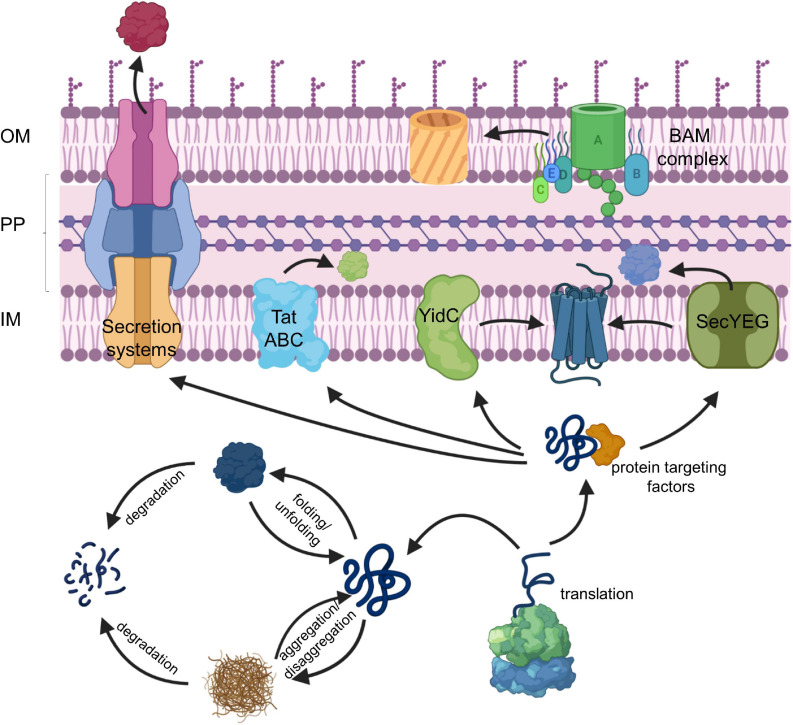
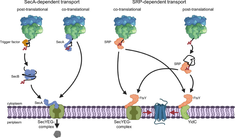
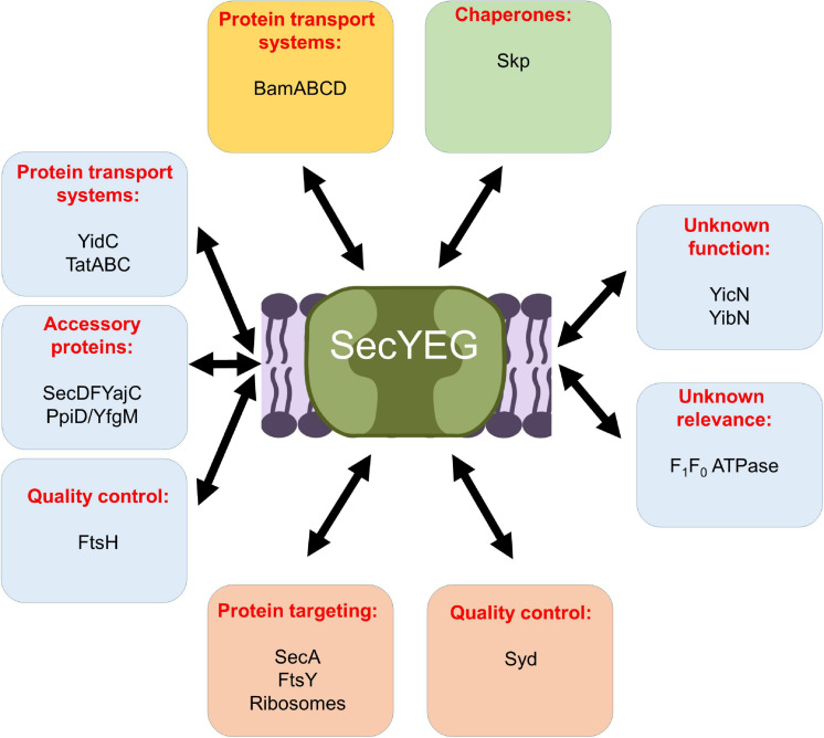
The spatial and temporal coordination of protein transport is an essential cornerstone of the bacterial adaptation to different environmental conditions. By adjusting the protein composition of extra-cytosolic compartments, like the inner and outer membranes or the periplasmic space, protein transport mechanisms help shaping protein homeostasis in response to various metabolic cues. The universally conserved SecYEG translocon acts at the center of bacterial protein transport and mediates the translocation of newly synthesized proteins into and across the cytoplasmic membrane. The ability of the SecYEG translocon to transport an enormous variety of different substrates is in part determined by its ability to interact with multiple targeting factors, chaperones and accessory proteins. These interactions are crucial for the assisted passage of newly synthesized proteins from the cytosol into the different bacterial compartments. In this review, we summarize the current knowledge about SecYEG-mediated protein transport, primarily in the model organism Escherichia coli, and describe the dynamic interaction of the SecYEG translocon with its multiple partner proteins. We furthermore highlight how protein transport is regulated and explore recent developments in using the SecYEG translocon as an antimicrobial target.

Keywords: FtsY; PpiD; SecA; SecYEG translocon; YidC; protein transport; signal recognition particle; stress response.


Conflict of interest statement

The authors declare that the research was conducted in the absence of any commercial or financial relationships that could be construed as a potential conflict of interest.

Figures

FIGURE 1
FIGURE 1
The proteostasis network in bacteria. For details see text. Secretion systems refer to the type I–IX protein secretion systems that have been identified in bacteria, although some of these secretion systems are only found in some species (Christie, 2019). IM, inner membrane; PP, periplasm; OM, outer membrane.
FIGURE 2
FIGURE 2
SecA- and SRP-dependent protein targeting in bacteria. The SecA- and SRP-dependent protein targeting pathways constitute the two main protein targeting pathways in bacteria and both can operate in a co- or post-translational mode. However, post-translational targeting of secretory proteins by SecA and co-translational targeting of membrane proteins by SRP are the preferred modes. Substrates of the post-translational SecA pathway are kept in a translocation competent state by chaperones, like the ribosome-bound TF or the cytosolic SecB. SecA serves as receptor for signal sequences (shown in red) of secretory proteins and is bound to the SecYEG translocon, which serves as main protein transport channel in bacteria. Repetitive ATP hydrolysis cycles by SecA allows for the translocation of the polypeptide across the SecY channel. SecA can also associate with the ribosome and target potential substrates co-translationally to the SecYEG translocon. The subsequent ATP-dependent translocation likely occurs then post-translationally, i.e., after the substrate is released from the ribosome. SRP binds with high affinity to translating ribosomes and traps the signal anchor sequence of a membrane protein when it emerges from the ribosomal peptide tunnel. SRP then delivers the translating ribosome (ribosome-associated nascent chain, RNC) to the SRP receptor FtsY. FtsY serves as SecYEG-bound receptor for nascent membrane proteins and engages similar binding sites as SecA on the SecYEG translocon. After SRP-FtsY contact, the translating ribosome docks onto the SecYEG translocon and ongoing translation inserts the protein into the lipid phase. FtsY can also associate with the YidC insertase and SRP can deliver less complex membrane proteins co-translationally to the YidC insertase for insertion. Small membrane proteins (<50 amino acids) and likely tail-anchored membrane proteins are post-translationally bound by SRP and targeted to SecYEG or YidC only after they have been released from the ribosome. This post-translational insertion by SRP is likely initiated by a so far largely uncharacterized mRNA-targeting step (Steinberg et al., 2020), which is not depicted in this cartoon.
FIGURE 3
FIGURE 3
Structures of SecA and SecA bound to the ribosome. (A) Structure of B. subtilis SecA (PDB 5EUL), showing its multiple domains. The two nucleotide-binding domains NBD1 and NBD2 are shown in cyan and in olive, respectively. The peptide-binding domain (PBD) is shown in raspberry-red, the helical wing domain (HWD) and the helical scaffold domain (HSD) in gray and the two-helix finger (2HF) in red. (B) Structure of E. coli SecA bound to a translating ribosome (PDB 6S0K). The 50S ribosomal subunit is shown in light-blue and the nascent RodZ chain in green. Ribosomal proteins that are in contact with SecA [uL23 (blue), uL29 (pink), and uL24 (green)] and the different domains of SecA are labeled and shown in the same color-code as in (A).
FIGURE 4
FIGURE 4
Structures of the SRP-FtsY-complex and the SRP-ribosome complex. (A) Structure of the E. coli SRP-FtsY complex (PDB 2XXA) (Ataide et al., 2011). Ffh, the protein component of the bacterial SRP is shown in yellow and the 4.5S RNA in dark-blue. The domains of Ffh are indicated. The NG-domain of FtsY is shown in green; the structure of the N-terminal A-domain of FtsY has not been solved yet and is shown as green box. (B) Structure of an SRP-RNC complex (PDB 5GAH). The 50S ribosomal subunit is shown in light-blue and the ribosomal proteins that provide the contact site for SRP are indicated, uL23 (blue), uL29 (pink), uL24 (green), and bL32 (light-green). Ffh is shown in yellow and the 4.5S RNA in dark-blue. The nascent PhoA chain is shown in dark red.
FIGURE 5
FIGURE 5
Structure of the quaternary RNC-SRP-FtsY-SecYEG complex. Structure of the quaternary complex (PDB 5NCO), depicting an early state of co-translational protein insertion. The subunits SecY, SecE and SecG of the SecYEG translocon are indicated by green, orange and blue color, respectively. The color code of the FtsY-SRP complex is as in Figure 4 and the nascent PhoA is shown in dark-red. Please note that in this structure, the SecYEG translocon is only tentatively fitted and would have to tilt by ∼20° to be accommodated within the membrane (Jomaa et al., 2017).
FIGURE 6
FIGURE 6
Structure of the SecYEG translocon in its resting state and active state. (A) Structure of T. thermophilus SecYEG in the resting state (PDB 5AWW) and the active state (PDB 5CH4). SecY is shown in green, SecE in orange and SecG in blue. The SecY transmembrane domains that constitute the lateral gate are shown in light green and the plug in magenta. The upper structures depict the front views of the SecYEG translocon and the lower structures the top view from the cytosol, respectively. (B) Schematic front view and view from the cytosol of the SecYEG translocon.
FIGURE 7
FIGURE 7
Structure of the substrate-engaged SecA-SecYEG complex and model for SecA-dependent translocation across the SecYEG-translocon. (A) Structure of the SecA-SecYEG complex from B. subtilis (PDB 5EUL). SecY and SecE are shown in green and orange, respectively, and the translocating peptide in yellow. The different domains of SecA are indicated. 2HF corresponds to the two-helix finger. (B) Upon ATP binding to SecA, the 2-helix-finger (2HF) inserts into the SecY channel and pushes the polypeptide into the channel. The signal sequence is depicted in red. For preventing back-sliding, the polypeptide binding domain (PBD) of SecA rotates toward the nucleotide-binding domain (NBD2) and forms a clamp that traps the polypeptide. This step likely occurs before or simultaneously with ATP-hydrolysis. Closing the clamp also leads to the retraction of the 2HF. After phosphate release, the clamp opens again and the polypeptide can slide deeper into the channel but in principle also backward. In vivo, backsliding at this stage could be prevented by contacts of the polypeptide to periplasmic chaperones, like Skp (Schäfer et al., 1999) or the PpiD/YfgM complex (Götzke et al., 2014; Jauss et al., 2019). In addition, the membrane potential is likely important for maintaining directionality of translocation (Driessen and Nouwen, 2008; Knyazev et al., 2018). Figure was modified after (Catipovic et al., 2019).
FIGURE 8
FIGURE 8
Model of membrane protein insertion via the SecYEG translocon. In the resting state of the SecYEG-translocon, the lateral gate, composed of transmembrane domains (TMs) 2/3 on one side (orange) and TMs 7/8 (blue) on the other side, is closed. Binding of the translating ribosome to the cytosolically exposed loop connecting TM 6 and 7 of SecY (C5-loop, not shown), causes the lateral gate to slightly open, which is then primed for the approaching nascent chain. The emerging nascent membrane protein (red) disrupts contacts between TM 2 and TM 7 on the cytosolic side of the membrane further, while TM 7 moves closer toward TM 3 on the periplasmic side. This creates a V-shaped crevice during the early state of insertion. This state is likely further stabilized by the two N-terminal TMs of SecE (not shown). Ongoing chain elongation positions the hydrophobic core of the signal peptide (red zylinder) at the lateral gate, where it occupies approx. the same position as TM 2 in the resting SecYEG channel, before it is released into the membrane.
FIGURE 9
FIGURE 9
Schematic view on the protein interaction network of the E. coli SecYEG complex. Interactions within the inner membrane are shown in blue boxes, those that take place at the cytosolic phase of the inner membrane in orange boxes, those at the periplasmic side of the inner membrane in a green box, and those with the outer membrane are boxed in yellow. For details see text.
FIGURE 10
FIGURE 10
Structures of YidC, SecDF and a model of the holo-translocon. (A) Structure of YidC from E. coli (PDB 6AL2). The conserved transmembrane domains (TMs) 2 to 6 of YidC are indicated (TM2, light blue; TM3, yellow; TM4, orange; TM5, light pink; TM6, red), while the structure of TM1 is still unknown. The short amphipathic helix EH1 is depicted in dark blue, the periplasmic loop P1 in dark green and the cytoplasmic loop C1 in light green. (B) Structure of the SecDF complex from Thermus thermophilus (PDB 5YHF). SecDF consists of 12 TMs, six each in SecD (TM1-6, pink) and SecF (TM7-12, green), and three periplasmic domains, termed P1-head (dark blue), P1-base (light blue) and P4 (yellow). (C) Modell of the holo-translocon based on the cryo-EM structure from E. coli (PDB 5MG3). SecY is shown in green, SecE in orange and SecG in blue, its ancillary subunits SecD in pink, SecF in green and YidC in yellow.
FIGURE 11
FIGURE 11
Cellular response to impaired protein transport. Depletion of SRP/FtsY or SecYEG induce a multifaceted response. This includes membrane stabilization via the induction of the phage-shock response (PspABC complex), the inhibition of translation via the induction of the ribosome-modulation factor (RMF) upon FtsY depletion and the induction of the σ32-response via the accumulation of misfolded proteins. Increased chaperone and protease production reduce the cellular concentration of misfolded proteins and provide a negative feedback loop for declining the σ32 response. Chaperones inhibit σ32 directly and the membrane bound protease FtsH degrades σ32. Membrane targeting of σ32 for degradation by FtsH is dependent on SRP/FtsY and SecYEG. Thus, upon SRP/FtsY or SecYEG depletion/saturation, elevated σ32 levels persist. FtsH also degrades misfolded/aggregated membrane proteins and SecY that is not in complex with its partner protein SecE. Ffh, the protein subunit of SRP is also a substrate of the Lon protease; in particular when Ffh is in excess over the 4.5S RNA, the RNA subunit of the bacterial SRP. “+” indicates increased production, “–” indicates reduced production, inhibition or degradation.
FIGURE 12
FIGURE 12
(p)ppGpp-dependent regulation of translation and protein transport in bacteria. The alarmones ppGpp and pppGpp are synthesized upon amino acid starvation by the ribosome-associated protein RelA or by the cytosolic protein SpoT upon carbon or fatty acid starvation. Allosteric regulation of RNA polymerase by (p)ppGpp reduces ribosome biogenesis and increases the stability of the ribonuclease MazF, which degrades multiple mRNAs. This includes the mRNA encoding for Ffh, the protein component of the bacterial SRP, or the ppiD mRNA, encoding for an accessory subunit of the SecYEG translocon. (p)ppGpp also increases the activity of FtsH, which can degrade SecY and YfgM. YfgM forms a complex with PpiD that associates with the SecYEG translocon. Whether SecY is specifically degraded by FtsH upon (p)ppGpp accumulation is not shown yet. (p)ppGpp also acts as competitive inhibitor of GTP-binding proteins like translation factors (IF2 and EF-G) or ribosome biogenesis proteins (ObgE). This leads to reduced ribosome biogenesis and reduced translation upon stress. Although not yet experimentally shown, it appears likely that increasing (p)ppGpp concentrations also inhibit the two GTPases SRP and FtsY, which would fine-tune the protein targeting machinery to the reduced translation rates.
FIGURE 13
FIGURE 13
Inhibitors of bacterial protein translocation. (A) Inhibitors of the ATPase SecA. (B) Inhibitors of the SecYEG-translocon. Ipomeassin F, decatransin, eeyarastatin 1 and eeyarastatin 24 also act on the homologous Sec61 complex in eukaryotes. Chemical structures were retrieved from the Sigma Aldrich web resource (https://www.sigmaaldrich.com/) or adapted from (Li et al., 2008; Van Puyenbroeck and Vermeire, 2018; Zong et al., 2019).

Similar articles

Cited by

References

    1. Abell B. M., Pool M. R., Schlenker O., Sinning I., High S. (2004). Signal recognition particle mediates post-translational targeting in eukaryotes. EMBO J. 23 2755–2764. - PMC - PubMed
    1. Ahmad S., Wang B., Walker M. D., Tran H. R., Stogios P. J., Savchenko A., et al. (2019). An interbacterial toxin inhibits target cell growth by synthesizing (p)ppApp. Nature 575 674–678. - PMC - PubMed
    1. Akiyama Y., Kihara A., Tokuda H., Ito K. (1996). FtsH (HflB) is an ATP-dependent protease selectively acting on SecY and some other membrane proteins. J. Biol. Chem. 271 31196–31201. - PubMed
    1. Akopian D., Dalal K., Shen K., Duong F., Shan S. O. (2013a). SecYEG activates GTPases to drive the completion of cotranslational protein targeting. J. Cell Biol. 200 397–405. 10.1083/jcb.201208045 - DOI - PMC - PubMed
    1. Akopian D., Shen K., Zhang X., Shan S. O. (2013b). Signal recognition particle: an essential protein-targeting machine. Annu. Rev. Biochem. 82 693–721. - PMC - PubMed

LinkOut - more resources