

Glucocorticoid-induced leucine zipper regulates liver fibrosis by suppressing CCL2-mediated leukocyte recruitment
- PMID: 33927191
- PMCID: PMC8085011
- DOI: 10.1038/s41419-021-03704-w
Glucocorticoid-induced leucine zipper regulates liver fibrosis by suppressing CCL2-mediated leukocyte recruitment
Abstract
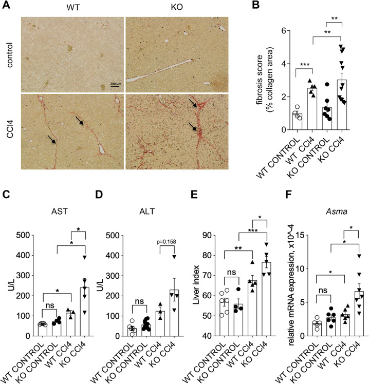
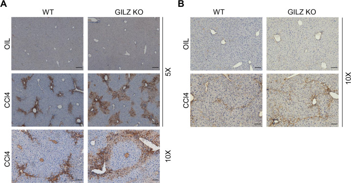
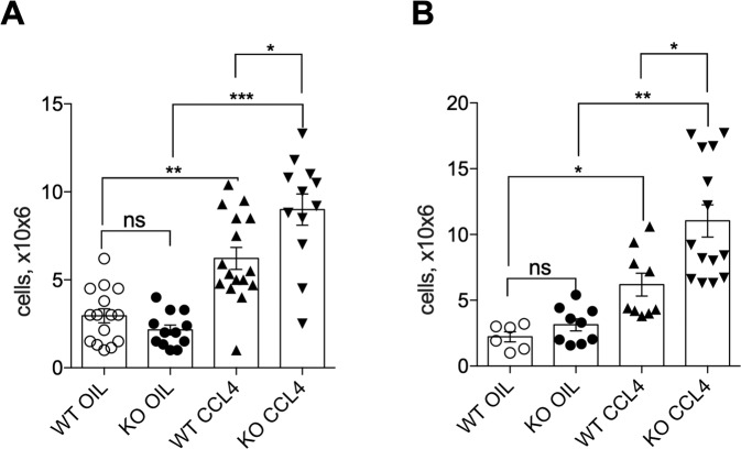
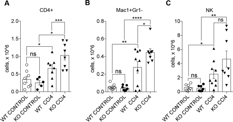
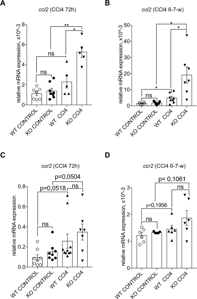
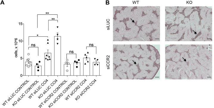
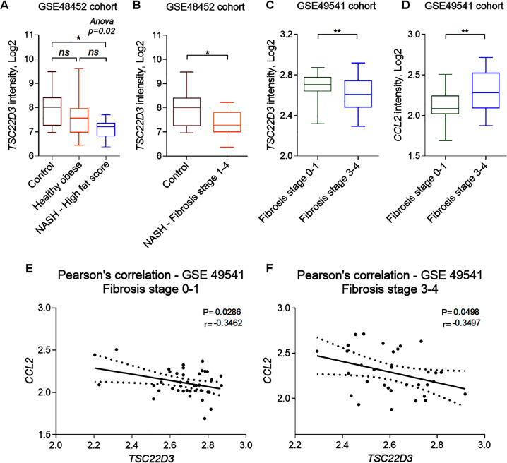
Liver fibrosis (LF) is a dangerous clinical condition with no available treatment. Inflammation plays a critical role in LF progression. Glucocorticoid-induced leucine zipper (GILZ, encoded in mice by the Tsc22d3 gene) mimics many of the anti-inflammatory effects of glucocorticoids, but its role in LF has not been directly addressed. Here, we found that GILZ deficiency in mice was associated with elevated CCL2 production and pro-inflammatory leukocyte infiltration at the early LF stage, resulting in enhanced LF development. RNA interference-mediated in vivo silencing of the CCL2 receptor CCR2 abolished the increased leukocyte recruitment and the associated hepatic stellate cell activation in the livers of GILZ knockout mice. To highlight the clinical relevance of these findings, we found that TSC22D3 mRNA expression was significantly downregulated and was inversely correlated with that of CCL2 in the liver samples of patients with LF. Altogether, these data demonstrate a protective role of GILZ in LF and uncover the mechanism, which can be targeted therapeutically. Therefore, modulating GILZ expression and its downstream targets represents a novel avenue for pharmacological intervention for treating LF and possibly other liver inflammatory disorders.
Conflict of interest statement
The authors declare no competing interests.
Figures
Similar articles
-
Essential role of suppressor of cytokine signaling 1 (SOCS1) in hepatocytes and macrophages in the regulation of liver fibrosis.Cytokine. 2019 Dec;124:154501. doi: 10.1016/j.cyto.2018.07.032. Epub 2018 Aug 8. Cytokine. 2019. PMID: 30097285
-
Downregulation of the glucocorticoid-induced leucine zipper (GILZ) promotes vascular inflammation.Atherosclerosis. 2014 Jun;234(2):391-400. doi: 10.1016/j.atherosclerosis.2014.03.028. Epub 2014 Apr 5. Atherosclerosis. 2014. PMID: 24747114
-
Glucocorticoid-Induced Leucine Zipper Suppresses ICAM-1 and MCP-1 Expression by Dephosphorylation of NF-κB p65 in Retinal Endothelial Cells.Invest Ophthalmol Vis Sci. 2017 Jan 1;58(1):631-641. doi: 10.1167/iovs.16-20933. Invest Ophthalmol Vis Sci. 2017. PMID: 28129426
-
Glucocorticoids and Glucocorticoid-Induced-Leucine-Zipper (GILZ) in Psoriasis.Front Immunol. 2019 Sep 13;10:2220. doi: 10.3389/fimmu.2019.02220. eCollection 2019. Front Immunol. 2019. PMID: 31572404 Free PMC article. Review.
-
Glucocorticoid-Induced Leucine Zipper: A Novel Anti-inflammatory Molecule.Front Pharmacol. 2019 Mar 27;10:308. doi: 10.3389/fphar.2019.00308. eCollection 2019. Front Pharmacol. 2019. PMID: 30971930 Free PMC article. Review.
Cited by
-
High-fat diet in early life triggers both reversible and persistent epigenetic changes in the medaka fish (Oryzias latipes).BMC Genomics. 2023 Aug 21;24(1):472. doi: 10.1186/s12864-023-09557-1. BMC Genomics. 2023. PMID: 37605229 Free PMC article.
-
Anti-Inflammatory Effects of Synthetic Peptides Based on Glucocorticoid-Induced Leucine Zipper (GILZ) Protein for the Treatment of Inflammatory Bowel Diseases (IBDs).Cells. 2023 Sep 16;12(18):2294. doi: 10.3390/cells12182294. Cells. 2023. PMID: 37759516 Free PMC article.
-
Channel Expansion in the Ligand-Binding Domain of the Glucocorticoid Receptor Contributes to the Activity of Highly Potent Glucocorticoid Analogues.Molecules. 2024 Mar 29;29(7):1546. doi: 10.3390/molecules29071546. Molecules. 2024. PMID: 38611825 Free PMC article.
-
GILZ as a Regulator of Cell Fate and Inflammation.Cells. 2021 Dec 30;11(1):122. doi: 10.3390/cells11010122. Cells. 2021. PMID: 35011684 Free PMC article. Review.
-
Single-cell transcriptomic analysis reveals heterogeneous features of myeloid-derived suppressor cells in newborns.Front Immunol. 2024 Jun 11;15:1367230. doi: 10.3389/fimmu.2024.1367230. eCollection 2024. Front Immunol. 2024. PMID: 38919617 Free PMC article.
References
Publication types
MeSH terms
Substances
LinkOut - more resources
Full Text Sources
Other Literature Sources
Medical
Molecular Biology Databases
