. 2021 Apr 26;22(9):4510.
doi: 10.3390/ijms22094510.

Complex Mechanisms of Antimony Genotoxicity in Budding Yeast Involves Replication and Topoisomerase I-Associated DNA Lesions, Telomere Dysfunction and Inhibition of DNA Repair

Affiliations

Complex Mechanisms of Antimony Genotoxicity in Budding Yeast Involves Replication and Topoisomerase I-Associated DNA Lesions, Telomere Dysfunction and Inhibition of DNA Repair

Ireneusz Litwin et al. Int J Mol Sci. .

Abstract

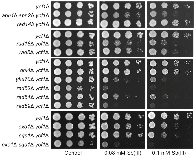
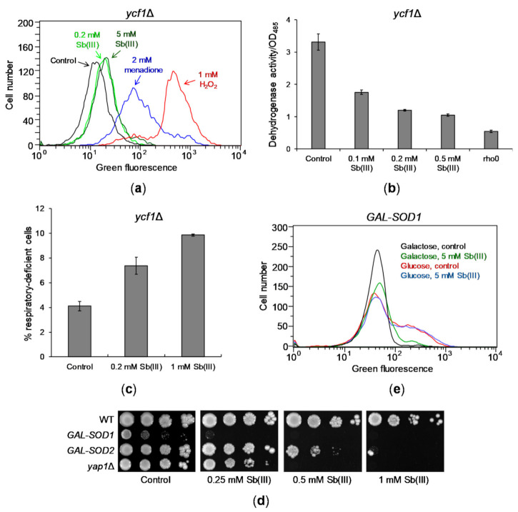
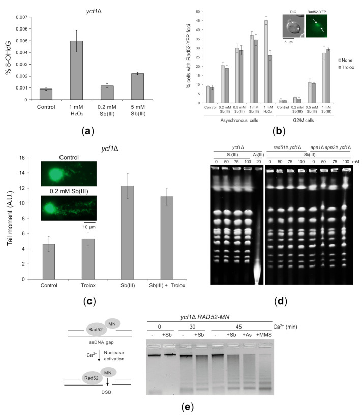
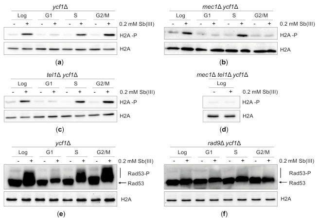
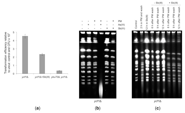
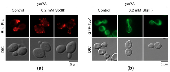
Antimony is a toxic metalloid with poorly understood mechanisms of toxicity and uncertain carcinogenic properties. By using a combination of genetic, biochemical and DNA damage assays, we investigated the genotoxic potential of trivalent antimony in the model organism Saccharomyces cerevisiae. We found that low doses of Sb(III) generate various forms of DNA damage including replication and topoisomerase I-dependent DNA lesions as well as oxidative stress and replication-independent DNA breaks accompanied by activation of DNA damage checkpoints and formation of recombination repair centers. At higher concentrations of Sb(III), moderately increased oxidative DNA damage is also observed. Consistently, base excision, DNA damage tolerance and homologous recombination repair pathways contribute to Sb(III) tolerance. In addition, we provided evidence suggesting that Sb(III) causes telomere dysfunction. Finally, we showed that Sb(III) negatively effects repair of double-strand DNA breaks and distorts actin and microtubule cytoskeleton. In sum, our results indicate that Sb(III) exhibits a significant genotoxic activity in budding yeast.

Keywords: DNA damage; DNA repair; antimony; cell cycle checkpoints; genotoxicity.


Conflict of interest statement

The authors declare no conflict of interest.

Figures

Figure 1
Figure 1
Role of DNA repair pathways in Sb(III) tolerance. Cultures of indicated mutants were serially diluted and plated on rich media in the presence or absence of Sb(III). Plates were incubated at 30 °C for 2 days and then photographed.
Figure 2
Figure 2
Sb(III) generates mild oxidative stress and mitochondrial dysfunction. (a) Sb(III) is a weak inducer of ROS production in yeast cells. The ycf1Δ mutant was exposed to indicated concentrations of Sb(III), menadione and H2O2 in the presence of ROS probe DHR123 for 2 h and subjected to the flow cytometry analysis. Increased levels of green fluorescence reflects formation of R123 as a result of DHR123 oxidation by ROS. Black line, untreated culture; light green line, 0.2 mM Sb(III); dark green line, 5 mM Sb(III); blue line, 2 mM menadione; red line, 1 mM H2O2. (b) Sb(III) impairs mitochondrial functions as measured by downregulation of dehydrogenase activity. Reduction of colorless artificial substrate triphenyltetrazolium chloride (TTC) to a pink product was used to demonstrate the activity of dehydrogenase enzymes. The rho0 mutant devoid of mitochondrial DNA was used as a positive control of respiratory defective cells. (c) Sb(III) elevates the rate of mitochondrial DNA loss. The ycf1Δ mutant was treated with indicated concentrations of Sb(III) for 3 days. The percentage of respiratory-deficient cells was determined by their inability to grow on medium with non-fermentable source of carbon. (b,c) Each bar represents the mean of four independent experiments (each with at least of 3 replicates) with standard deviations (SD). (d) The effect of SOD1 and SOD2 repression on Sb(III) tolerance. Indicated yeast strains were grown on galactose rich media to maintain expression of SOD1 and SOD2 and then washed before plating on rich glucose media (to repress GAL promoter) in the presence or absence of Sb(III). Plates were incubated at 30 °C for 3 days and then photographed. (e) Sb(III) does not increase production of ROS in the absence of superoxide dismutase Cu/Zn Sod1. The GAL-SOD1 strain was grown on galactose (gal) or glucose (glu) and exposed to 5 mM Sb(III) for 2 h in the presence of DHR123 and analyzed by flow cytometry.
Figure 3
Figure 3
Sb(III)-induced DNA damage in yeast cells. (a) Sb(III) induces low levels of oxidative DNA damage. The ycf1Δ mutant was treated with indicated concentrations of Sb(III) or H2O2 for 2 h followed by DNA extraction and quantification of 8-hydroxy-2′-deoxyguanosine (8-OHdG) using ELISA kit. Error bars represent the mean value ± SD (n = 3). (b) Sb(III) triggers formation of Rad52-YFP nuclear foci. Asynchronous and G2/M-arrested cells were exposed to indicated concentrations of Sb(III) or H2O2 for 2 h in the absence or presence of 1 mM Trolox. Live cells were analyzed by fluorescence microscopy to visualize Rad52-YFP foci. Error bars represent the mean value ± SD (n = 3). Representative image of Rad52-YFP foci is shown. DIC, differential interphase contrast. Scale bar: 5 μm. (c) Analysis of Sb(III)-induced DNA damage by the comet assay. Exponentially growing ycf1Δ cells were treated with 0.2 mM Sb(III) for 2 h or mock-treated in the absence or presence of 1 mM Trolox. The tail moment was calculated based on the analysis of 200 DNA comets from three independent experiments, with at least 50 comets per experiment (mean ± SD). Representative images of DNA comets are shown. A.U., arbitrary units. Scale bar: 10 μm. (d) PFGE analysis of yeast chromosomes isolated from indicated strains exposed to various concentrations of Sb(III) or As(III) for 6 h or left untreated. (e) ChEC analysis revealed the presence of replicative DNA lesions induced by Sb(III). The ycf1Δ RAD52-MN strain was synchronized in G1 and released in S phase in the presence of 0.2 mM Sb(III), 0.2 mM As(III) or 0.05% MMS for 2 h. Before total DNA extraction, cells were permeabilized and treated with 2 mM CaCl2 to initiate DNA cleavage by the Rad52-MN fusion protein. Quantifications of DNA digestion are shown in Supplementary Figure S1.
Figure 4
Figure 4
Sb(III) activates DNA damage response in budding yeast. (ad) Sb(III) induces Mec1/Tel1-dependent histone H2A phosphorylation at S129 (H2A-P) in S and G2/M but not in G1 phase of the cell cycle. Cells were grown logarithmically (Log), synchronized and kept arrested in G1 with α-factor (G1), synchronized in G1 and released from the α-factor block to allow DNA replication (S) or synchronized and kept arrested in G2/M with nocodazole (G2/M) followed by treatment with 0.2 mM Sb(III) for 2 h or mock-treatment. Next, total protein extracts were prepared and analyzed by western blot using anti-phospho H2A (S129) and anti-H2A antibodies as a loading control. The lethality of mec1Δ mutation was suppressed by deletion of the SML1 gene encoding a ribonucleotide reductase inhibitor. (e,f) Sb(III) triggers Rad9-dependent hyperphosphorylation of Rad53 (Rad53-P). The ycf1Δ and rad9Δ ycf1Δ cultures were prepared and treated as described above. Western blot analysis was performed with anti-Rad53 and anti-H2A antibodies as a loading control (source data are shown in Supplementary Figure S2).
Figure 5
Figure 5
The effect of Sb(III) on cell cycle progression and the role of DNA damage checkpoint pathway in Sb(III) tolerance. (a) Sb(III) does not trigger checkpoint-dependent G1 delay. The ycf1Δ and rad9Δ ycf1Δ strains were synchronized in G1 with α-factor, washed and released in the presence or absence of 0.2 mM Sb(III). Percentage of cells arrested in G1 was determined by the α-factor-nocodazole trap assay. (b) Sb(III) slows S phase progression, partially in a Rad9-dependent manner. Cells were prepared and treated as described in (a). DNA content was determined by flow cytometry. 1C, DNA content. (c) Sb(III)-induced G2/M delay partially depends on Rad9. Cells were arrested at G2/M boundary with nocodazole and then released in the presence or absence of 0.2 mM Sb(III). Percentage of binucleate (post-mitotic) cells was determined by fluorescence microscopy. (d) Sensitivity of DNA damage checkpoint mutants to Sb(III). Cultures of indicated mutants were serially diluted and plated on rich media in the presence or absence of Sb(III). Plates were incubated at 30 °C for 2 days and then photographed.
Figure 6
Figure 6
The yKu complex suppresses Sb(III)-induced DNA damage checkpoint activation in G1. (a,b) Deletion of YKU70 results in Mec1/Tel1/Rad9-dependent G1 checkpoint activation in the presence of Sb(III). Indicated strains were synchronized in G1 with α-factor and then exposed to 0.2 mM Sb(III) for 2 h or left untreated. Next, total protein extracts were prepared and analyzed by western blot using anti-phospho H2A (S129), anti-Rad53 and anti-H2A antibodies as a loading control (source data are shown in Supplementary Figure S3). The lethality of mec1Δ mutation was suppressed by deletion of the SML1 gene. (c) Sb(III) triggers formation of Rfa1-YFP nuclear foci in G1 cells lacking YKU70. G1-arrested ycf1Δ and yku70Δ ycf1Δ cells were exposed to indicated concentrations of Sb(III) for 2 h or left untreated. Live cells were analyzed by fluorescence microscopy to visualize Rfa1-YFP foci. Error bars represent the mean value ± SD (n = 3). Representative image of Rfa1-YFP foci is shown. DIC, differential interphase contrast. Scale bar: 5 μm. (d) Sb(III) induces Rad9-dependent G1 delay in the absence of YKU70. The ycf1Δ and rad9Δ ycf1Δ strains were synchronized in G1 with α-factor, washed and released fresh media in the presence or absence of 0.2 mM Sb(III). Percentage of cells arrested in G1 was determined by the α-factor-nocodazole trap assay.
Figure 7
Figure 7
Sb(III) sensitivity of telomere maintenance mutants is suppressed by either checkpoint inactivation or preventing resection of chromosome ends. (a) Genetic interaction between tel1Δ and yku70Δ in the presence of Sb(III). (b) Deletion of RAD9 checkpoint gene suppresses sensitivity of yku70Δ to Sb(III). (c) The cdc13-1 mutation renders yeast cells hypersensitive to Sb(III) at both permissive (23 °C) and semi-permissive (25 °C) temperature. (d) Sb(III) sensitivity of cdc13-1 ycf1Δ cells is reversed by concomitant deletion of EXO1 or PIF1. (ad) Exponentially growing cultures were serially diluted and spotted onto YPD plates with or without Sb(III).
Figure 8
Figure 8
The effect of Sb(III) on telomere homeostasis. (a) Sb(III) does not alter telomere length in yeast cells. Cells of indicated strains were cultivated for six days in the presence or absence of Sb(III). Afterwards, genomic DNA was isolated, digested with XhoI and analyzed by Southern blot using a probe detecting Y’ telomere repeats. (b) Sb(III) inhibits telomere fusions in cells lacking Uls1. Indicated strains were cultivated in the presence or absence of 0.2 mM Sb(III) for 6 days followed by genomic DNA preparation and PCR analysis of fusions between X and Y’-only telomeres using a pair of primers specific for X and Y’ subtelomeric regions. (c,d) Cdc13 association with telomeric DNA is increased in the presence of Sb(III). The telomere VI-R DNA binding of cMyc-tagged Cdc13 in ycf1Δ cells treated or not treated with 0.2 mM Sb(III) for 2 h was analyzed by ChIP performed with anti-Myc antibodies followed by qPCR. ARS306 locus was used as a control for the Cdc13-free internal chromosome region. The % input value represents the enrichment of Cdc13-cMyc protein at the specific locus and is normalized to the ACT1 reference gene. Error bars are standard deviations from two independent experiments. DNA content analysis of asynchronous cultures used for ChIP-qPCR is embedded in panel C.
Figure 9
Figure 9
Sb(III) triggers Top1-dependent DNA lesions. (a) The lack of Top1 slightly improves the growth of cells with the tel1Δ mutation in the presence of Sb(III). Exponentially growing cell cultures were serially diluted and spotted onto YPD plates with or without indicated compounds and incubated at 30 °C for two days. (b) Sb(III)-induced DNA breaks partially depends on the activity of Top1. Exponentially growing cells were treated with 0.2 mM Sb(III) for 2 h or mock-treated and analyzed by the comet assay as described in Figure 3C. A.U., arbitrary units. (c) Deletion of TOP1 decreases levels of Sb(III)-induced replicative DNA lesions. The presence of ssDNA-containing DNA lesions were assessed by ChEC as described in Figure 3E.
Figure 10
Figure 10
The repair of DBSs is negatively affected by Sb(III). (a) Sb(III) decreases the ability of yeast cells to repair cohesive DSBs by NHEJ. Indicated strains were transformed in parallel with PstI-cleaved YCplac111 and supercoiled YCplac111. In addition, ycf1Δ cells were exposed to 0.2 mM Sb(III) for two hours, washed and used for transformation with the same set of plasmids. Transformation efficiency was expressed as the ratio of colonies obtained with the linear plasmid divided by colonies obtained with the uncut plasmid normalized to the number of viable cells used for transformation. Each bar represents the mean of four independent experiments with SD. (b) Sb(III) does not enhance genotoxicity of phleomycin (PM). The ycf1Δ mutant was exposed to 10 μg/mL PM, 5 mM As(III) or 5 mM Sb(III) for 6 h followed by isolation of genomic DNA and PGFE analysis. (c) Sb(III) inhibits the repair of PM-induced DSBs. The ycf1Δ mutant was exposed to 35 μg/mL PM for 2 h, washed and released in the presence or absence of 0.2 mM Sb(III) for up to 12 h. Cells were also left untreated or cultivated in the presence of 0.2 mM Sb(III) for 12 h. At indicated time-points, genomic DNA was prepared and analyzed by PGFE.
Figure 11
Figure 11
Sb(III) targets actin and microtubule cytoskeleton. (a) Exponentially growing ycf1Δ cells were exposed to 0.2 mM Sb(III) for 2 h or left untreated and then chemically fixed for actin staining with rhodamine-phalloidin. (b) The ycf1Δ mutant expressing chromosomally encoded α-tubulin fused to GFP (GFP-Tub1) was grown to log phase and then treated with 0.2 mM Sb(III) for 2 h or mock-treated. Next, viable cells were analyzed by fluorescence microscopy to visualize microtubules. Scale bars: 5 μm.

Similar articles

Cited by

References

    1. Filella M., Belzile N., Chen Y.W. Antimony in the environment: A review focused on natural waters: I. Occurrence. Earth Sci. Rev. 2002;57:125–176. doi: 10.1016/S0012-8252(01)00070-8. - DOI
    1. Cooper R.G., Harrison A.P. The exposure to and health effects of antimony. Indian J. Occup. Environ. Med. 2009;13:3–10. doi: 10.4103/0019-5278.50716. - DOI - PMC - PubMed
    1. Wang X., He M., Xie J., Xi J., Lu X. Heavy metal pollution of the world largest antimony mine-affected agricultural soils in Hunan province (China) J. Soils Sediments. 2010;10:827–837. doi: 10.1007/s11368-010-0196-4. - DOI
    1. Filella M., Hennebert P., Okkenhaug G., Turner A. Occurrence and fate of antimony in plastics. J. Hazard. Mater. 2020;390:121764. doi: 10.1016/j.jhazmat.2019.121764. - DOI - PubMed
    1. Haldimann M., Blanc A., Dudler V. Exposure to antimony from polyethylene terephthalate (PET) trays used in ready-to-eat meals. Food Addit. Contam. 2007;24:860–868. doi: 10.1080/02652030701297511. - DOI - PubMed

MeSH terms

LinkOut - more resources