. 2021 Apr 19;10(4):942.
doi: 10.3390/cells10040942.

MRCKα Is Dispensable for Breast Cancer Development in the MMTV-PyMT Model

Affiliations

MRCKα Is Dispensable for Breast Cancer Development in the MMTV-PyMT Model

Mei Qi Kwa et al. Cells. .

Abstract

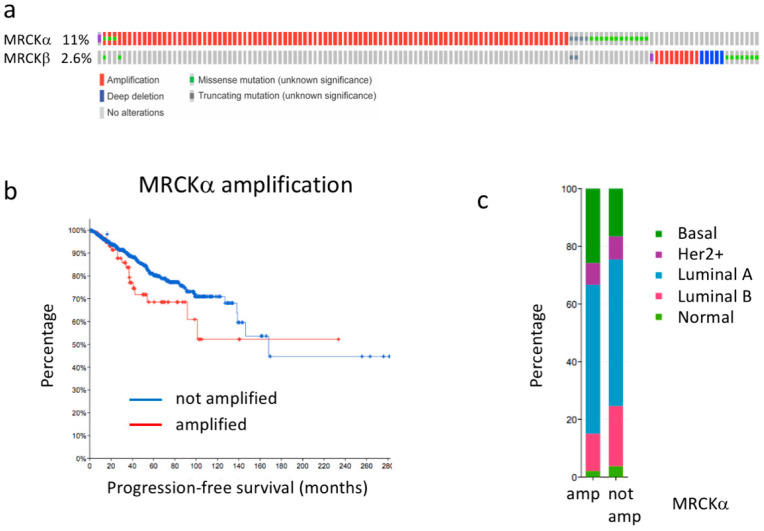
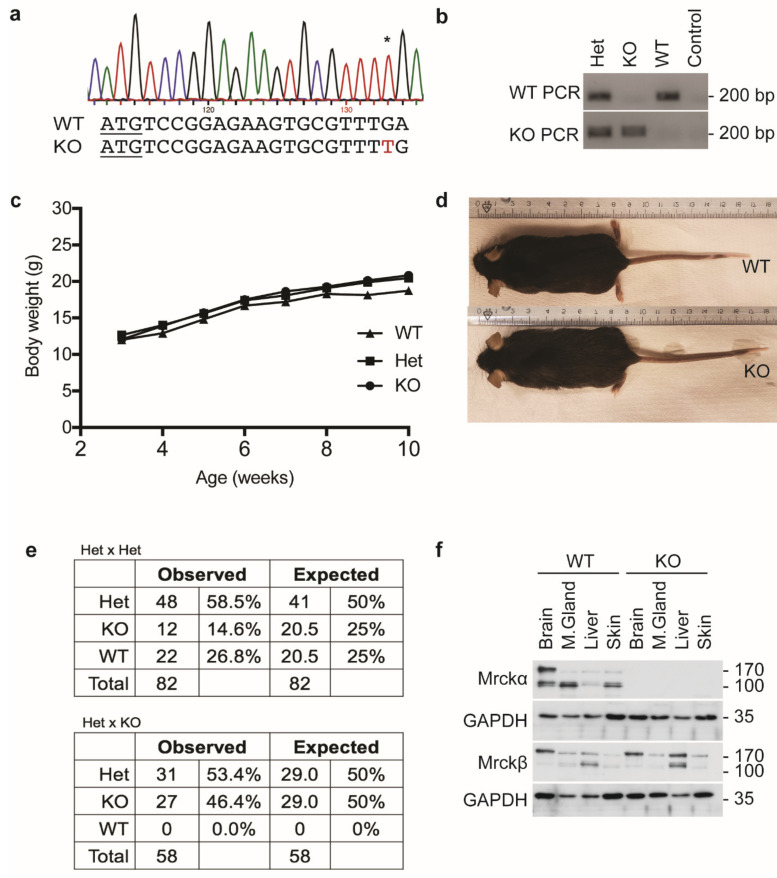
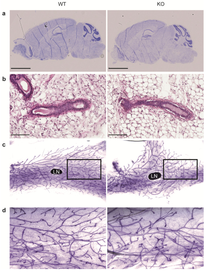
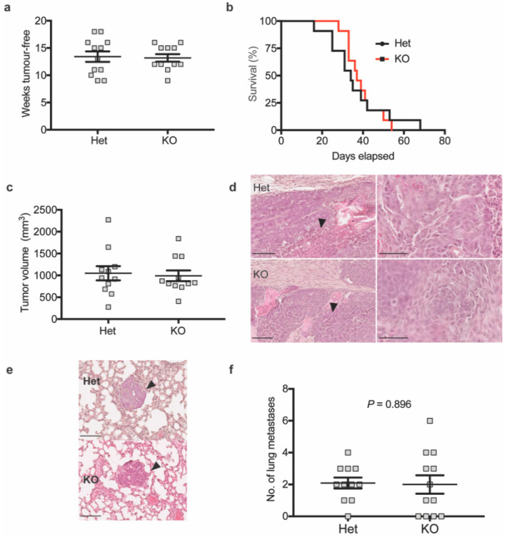
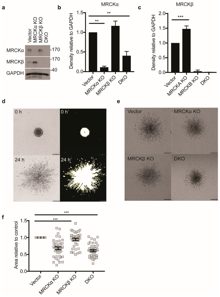
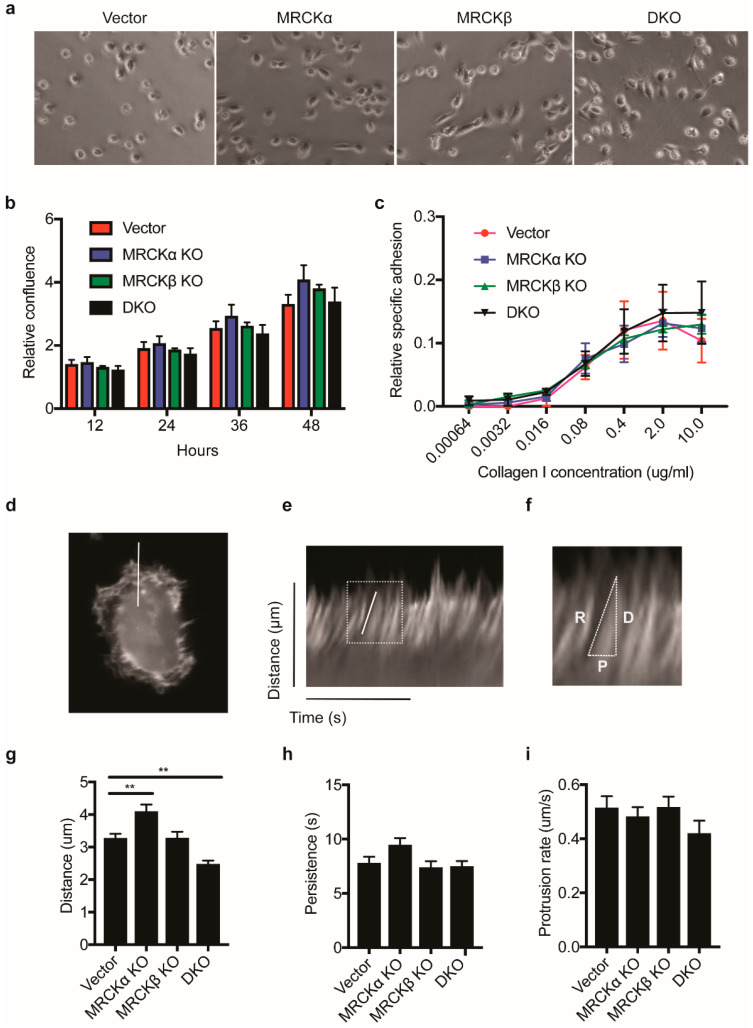
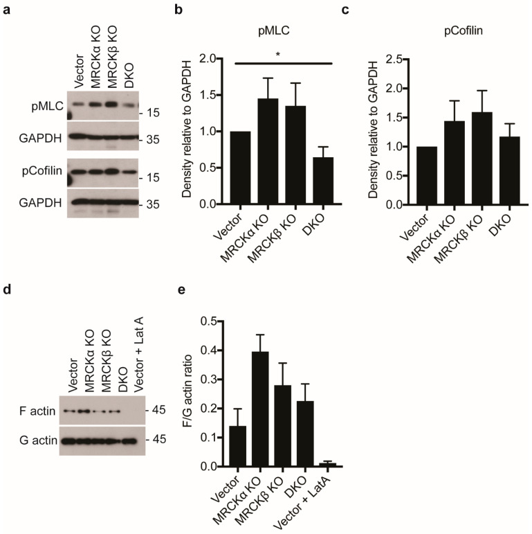
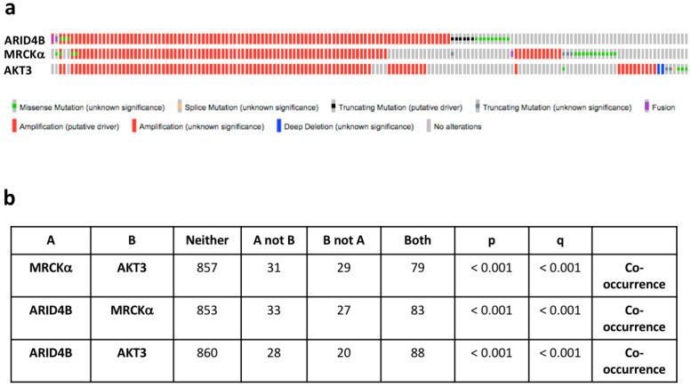
MRCKα is a ubiquitously expressed serine/threonine kinase involved in cell contraction and F-actin turnover, which is highly amplified in human breast cancer and part of a gene expression signature for bad prognosis. Nothing is known about the in vivo function of MRCKα. To explore MRCKα function in development and in breast cancer, we generated mice lacking a functional MRCKα gene. Mice were born close to the Mendelian ratio and showed no obvious phenotype including a normal mammary gland formation. Assessing breast cancer development using the transgenic MMTV-PyMT mouse model, loss of MRCKα did not affect tumor onset, tumor growth and metastasis formation. Deleting MRCKα and its related family member MRCKβ in two triple-negative breast cancer cell lines resulted in reduced invasion of MDA-MB-231 cells, but did not affect migration of 4T1 cells. Further genomic analysis of human breast cancers revealed that MRCKα is frequently co-amplified with the oncogenes ARID4B and AKT3 which might contribute to the prognostic value of MRCKα expression. Collectively, these data suggest that MRCKα might be a prognostic marker for breast cancer, but probably of limited functional importance.

Keywords: MRCK; breast cancer; invasion.


Conflict of interest statement

The authors declare no conflict of interest. The funders had no role in the design of the study; in the collection, analyses, or interpretation of data; in the writing of the manuscript, or in the decision to publish the results.

Figures

Figure 1
Figure 1
MRCKα gene is amplified in breast cancer. (a) Genetic alterations of the MRCKα and MRCKβ genes in breast cancer. (b) Progression free survival of breast cancer patients with amplified and non-amplified MRCKα gene (p = 0.087). (c) Breast cancer subtype distribution of patients with amplified (amp) and not amplified (not amp) MRCKα gene. All graphs were generated in cBioPortal.org using the TCGA PanCancer Atlas data set for invasive breast carcinoma (accessed on 20 March 2021).
Figure 2
Figure 2
MRCKα ko mice show no obvious phenotype. (a) Genotype profile of ko mice showing the p.Leu7fs*35 mutation. * indicates the inserted nucleotide in the chromatogram. (b) Representative results from genotyping PCR using primers targeting the mutation specifically. (c) Body weight measurements of post-weaning female wild-type, heterozygous and ko mice. (d) Representative images of wild-type and ko mice. (e) Mendelian ratio of the observed and expected frequencies of wild-type, heterozygous and ko mice in Het × Het mating (n = 82) and Het × KO (n = 58) mating pairs. (f) Immunoblot analysis of MRCKα and MRCKβ expression in wild-type and ko mice.
Figure 3
Figure 3
Mrckα knockout does not affect brain and mammary gland development. (a) Representative Nissl staining of sagittal brain sections from adult wild-type and ko mice. Scale bars: 2.5 mm. (b) Representative H&E staining of the 4th inguinal mammary glands from 7-weeks old virgin female wild-type and KO mice. Scale bars: 100 μm. (c) Representative carmine alum-stained mammary gland whole mounts. LN denotes the inguinal lymph node. A black box was drawn to indicate the area of magnification of the image, as shown in (d).
Figure 4
Figure 4
Mrckα knockout does not affect primary mammary tumour development and metastasis in MMTV-PyMT mice. (a) Weeks tumour-free (b) survival analysis (p = 0.8243, log-rank test) and (c) tumor volume at end-point of MMTV-PyMT heterozygous (Het) vs. KO mice (n ≥ 10/10). (d) Representative H&E staining of MMTV-PyMT heterozygous or Mrckα KO mice primary tumours showing extensive necrosis (black arrows). Images on the right represent the same images at higher magnification showing the abnormal cancer cell nuclei. Scale bars: 100 μm. (e) Representative H&E staining of lung metastases of MMTV-PyMT heterozygous vs. KO mice. Scale bars: 100 μm. (f) Quantitation of lung metastases by counting of foci from the left lung lobe from MMTV-PyMT heterozygous and KO mice upon reaching end point. (n: 10/10; mean ± SEM, two-tailed unpaired t-test).
Figure 5
Figure 5
MRCKα and MRCKβ knockout affect 3D invasion. (a) Representative immunoblot showing successful MRCKα and MRCKβ, single and double knockout (DKO) in MDA-MB-231 cells. Quantitation of (b) MRCKα and (c) MRCKβ expression in lysates relative to GAPDH (n = 3, mean ± SEM, one-way ANOVA; ** p ≤ 0.01; *** p ≤ 0.001). (d) Strategy for quantifying area of invasion of spheroids by thresholding using ImageJ. Briefly, area of spheroids immediately after embedding in type I collagen was subtracted from the total area of the same spheroids 24 h post-embedding, to obtain the area of invasion (e) 3D matrix invasion of vector control, MRCKα KO, MRCKβ KO, or DKO MDA-MB-231 heterospheroids co-cultured with CAFs and embedded in type I collagen. Scale bar: 200 μm. (f) Area of collagen invasion relative to vector control (n = 3, totaling > than 100 cells per group; mean ± SEM, one-way ANOVA; *** p ≤ 0.001).
Figure 6
Figure 6
MRCK KO has a modest effect on F-actin polymerization. (a) Representative phase contrast images showing the morphologies of control, MRCKα KO, MRCKβ KO, or DKO MDA-MB-231 cells (b) Proliferation rate of MRCKα KO, MRCKβ KO and DKO MDA-MB-231 cells as measured by relative confluency over time. (c) Relative adhesion of MRCKα KO, MRCKβ KO and DKO MDA-MB-231 cells to increasing concentration of type I collagen. Data is obtained for 3 biological replicates. (di) Kymograph analysis of actin protrusion distance, rate and persistence in MRCK KO cells (d) Representative still image of an MDA-MB-231 cell expressing LifeAct-GFP used in live imaging. A white line was drawn perpendicular to the cell body to indicate the position where kymograph was obtained. (e) Example of a kymograph generated using the reslice function on ImageJ program, showing the distance and time in the y and x axis, respectively. A white box is used to highlight an actin protrusion and the image magnified in (f). (f) Example of a measurement of actin protrusion distance, D, persistence, P and protrusion rate, R. (g) Kymograph measurements obtained for protrusion distance, ** p ≤ 0.01 (h) persistence and (i) protrusion rate.
Figure 7
Figure 7
MRCK KO does not effectively reduce myosin and cofilin phosphorylation in MDA-MB-231 cells (a) Representative immunoblot showing myosin light chain (pMLC) and phospho-cofilin in MDA-MB-231 cells with MRCKα KO, MRCKβ KO, or DKO. (b,c) Quantitation of indicated proteins from lysates obtained from the knockout cells (n: 6/6/6/6; mean ± SEM, one-way ANOVA; * p ≤ 0.05). (d) Representative immunoblot showing levels of F-actin and G-actin in control, MRCKα KO, MRCKβ KO and DKO MDA-MB-231 cells. As a control, cells were also treated with 1 μM latrunculin A (Lat A) for 15 min. (e) Quantification of F/G-actin ratio (n: 6/6/6/6; mean ± SEM, not significant with one-way ANOVA, * p ≤ 0.05 with two-tailed t-test for Vector vs. MRCKa KO).
Figure 8
Figure 8
MRCKα and MRCKβ are dispensable in 4T1 cells matrix invasion. (a) Representative immunoblot showing successful knockout of MRCKα and MRCKβ in 4T1 cells. DKO was achieved by using one of two different CRISPR gRNAs for MRCKα (“MRCKα 1 + 2”) and one gRNA for MRCKβ (b) Representative phase contrast images of control, MRCKα, MRCKβ and DKO 4T1 cell morphologies in the presence and absence of Y-27632 (10 μM). (c) 3D matrix invasion of MRCK KO 4T1 spheroids in type I collagen, in the presence and absence of Y-27632 (10 μM). Black boxes highlight the invasion front of the control spheroids, as shown magnified in the right panel. (d) Quantification of the area of invasion of the 4T1 spheroids from (c) relative to 24 h control (n: 3; mean ± SEM, one-way ANOVA; * p ≤ 0.05, *** p ≤ 0.001). (e) Representative immunoblot and (f,g) quantification of the expression of the indicated proteins. (n: 5; mean ± SEM, one-way ANOVA).
Figure 9
Figure 9
MRCKα is co-amplified with ARID4B and AKT3. (a) Oncoprints of genomic alterations of MRCKα, ARID4B and AKT3 in human breast cancers. (b) Analysis of co-occurrence of MRCKα with the oncogenes ARID4B and AKT3. All analyses were generated in cBioportal (TCGA PanCancer Atlas, breast cancer samples, accessed on 20 February 2021).

Similar articles

Cited by

References

    1. Lawson C.D., Ridley A.J. Rho GTPase signaling complexes in cell migration and inva-sion. J. Cell Biol. 2018;217:447–457. doi: 10.1083/jcb.201612069. - DOI - PMC - PubMed
    1. Leung T., Chen X.Q., Tan I., Manser E., Lim L. Myotonic dystrophy kinase-related Cdc42-binding kinase acts as a Cdc42 effector in promoting cytoskeletal reorganization. Mol. Cell Biol. 1998;18:130–140. doi: 10.1128/MCB.18.1.130. - DOI - PMC - PubMed
    1. Schwarz J., Proff J., Hävemeier A., Ladwein M., Rottner K., Barlag B., Pich A., Tatge H., Just I., Gerhard R. Serine-71 phosphorylation of Rac1 modulates downstream signaling. PLoS ONE. 2012;7:e44358. - PMC - PubMed
    1. Unbekandt M., Belshaw S., Bower J., Clarke M., Cordes J., Crighton D., Croft D.R., Drysdale M.J., Garnett M.J., Gill K., et al. Discovery of Potent and Selective MRCK Inhibitors with Therapeutic Effect on Skin Cancer. Cancer Res. 2018;78:2096–2114. doi: 10.1158/0008-5472.CAN-17-2870. - DOI - PMC - PubMed
    1. Tan I., Seow K.T., Lim L., Leung T. Intermolecular and intramolecular interactions regulate catalytic activity of myotonic dystrophy kinase-related Cdc42-binding kinase alpha. Mol. Cell Biol. 2001;21:2767–2778. doi: 10.1128/MCB.21.8.2767-2778.2001. - DOI - PMC - PubMed

Publication types

MeSH terms

Grants and funding