Subcellular Localization and Vesicular Structures of Anthocyanin Pigmentation by Fluorescence Imaging of Black Rice (Oryza sativa L.) Stigma Protoplast
- PMID: 33918111
- PMCID: PMC8066712
- DOI: 10.3390/plants10040685
Subcellular Localization and Vesicular Structures of Anthocyanin Pigmentation by Fluorescence Imaging of Black Rice (Oryza sativa L.) Stigma Protoplast
Abstract
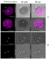
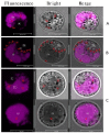
Anthocyanins belong to the group of flavonoid compounds broadly distributed in plant species responsible for attractive colors. In black rice (Oryza sativa L.), they are present in the stems, leaves, stigmas, and caryopsis. However, there is still no scientific evidence supporting the existence of compartmentalization and trafficking of anthocyanin inside the cells. In the current study, we took advantage of autofluorescence with anthocyanin's unique excitation/emission properties to elucidate the subcellular localization of anthocyanin and report on the in planta characterization of anthocyanin prevacuolar vesicles (APV) and anthocyanic vacuolar inclusion (AVI) structure. Protoplasts were isolated from the stigma of black and brown rice and imaging using a confocal microscope. Our result showed the fluorescence displaying magenta color in purple stigma and no fluorescence in white stigma when excitation was provided by a helium-neon 552 nm and emission long pass 610-670 nm laser. The fluorescence was distributed throughout the cell, mainly in the central vacuole. Fluorescent images revealed two pools of anthocyanin inside the cells. The diffuse pools were largely found inside the vacuole lumen, while the body structures could be observed mostly inside the cytoplasm (APV) and slightly inside the vacuole (AVI) with different shapes, sizes, and color intensity. Based on their sizes, AVI could be grouped into small (Ф < 0.5 um), middle (Ф between 0.5 and 1 um), and large size (Ф > 1 um). Together, these results provided evidence about the sequestration and trafficking of anthocyanin from the cytoplasm to the central vacuole and the existence of different transport mechanisms of anthocyanin. Our results suggest that stigma cells are an excellent system for in vivo studying of anthocyanin in rice and provide a good foundation for understanding anthocyanin metabolism in plants, sequestration, and trafficking in black rice.
Keywords: anthocyanin; anthocyanin vacuolar inclusion; autofluorescence; confocal microscopy; protoplast; stigma; subcellular localization.
Conflict of interest statement
The authors declare no conflict of interest.
Figures


Similar articles
-
Computational and Transcriptomic Analysis Unraveled OsMATE34 as a Putative Anthocyanin Transporter in Black Rice (Oryza sativa L.) Caryopsis.Genes (Basel). 2021 Apr 16;12(4):583. doi: 10.3390/genes12040583. Genes (Basel). 2021. PMID: 33923742 Free PMC article.
-
Recent Insights into Anthocyanin Pigmentation, Synthesis, Trafficking, and Regulatory Mechanisms in Rice (Oryza sativa L.) Caryopsis.Biomolecules. 2021 Mar 7;11(3):394. doi: 10.3390/biom11030394. Biomolecules. 2021. PMID: 33800105 Free PMC article. Review.
-
New insight into the structures and formation of anthocyanic vacuolar inclusions in flower petals.BMC Plant Biol. 2006 Dec 17;6:29. doi: 10.1186/1471-2229-6-29. BMC Plant Biol. 2006. PMID: 17173704 Free PMC article.
-
Determinant Factors and Regulatory Systems for Anthocyanin Biosynthesis in Rice Apiculi and Stigmas.Rice (N Y). 2021 Apr 21;14(1):37. doi: 10.1186/s12284-021-00480-1. Rice (N Y). 2021. PMID: 33881644 Free PMC article.
-
Different localization patterns of anthocyanin species in the pericarp of black rice revealed by imaging mass spectrometry.PLoS One. 2012;7(2):e31285. doi: 10.1371/journal.pone.0031285. Epub 2012 Feb 17. PLoS One. 2012. PMID: 22363605 Free PMC article.
Cited by
-
Anthocyanic Vacuolar Inclusions: From Biosynthesis to Storage and Possible Applications.Front Chem. 2022 Jun 28;10:913324. doi: 10.3389/fchem.2022.913324. eCollection 2022. Front Chem. 2022. PMID: 35836677 Free PMC article. Review.
-
Identification and Spatial Distribution of Bioactive Compounds in Seeds Vigna unguiculata (L.) Walp. by Laser Microscopy and Tandem Mass Spectrometry.Plants (Basel). 2022 Aug 18;11(16):2147. doi: 10.3390/plants11162147. Plants (Basel). 2022. PMID: 36015450 Free PMC article.
-
The Content of Anthocyanins in Cowpea (Vigna unguiculata (L.) Walp.) Seeds and Contribution of the MYB Gene Cluster to Their Coloration Pattern.Plants (Basel). 2023 Oct 20;12(20):3624. doi: 10.3390/plants12203624. Plants (Basel). 2023. PMID: 37896090 Free PMC article.
-
A high-efficiency PEG-Ca2+-mediated transient transformation system for broccoli protoplasts.Front Plant Sci. 2022 Dec 12;13:1081321. doi: 10.3389/fpls.2022.1081321. eCollection 2022. Front Plant Sci. 2022. PMID: 36578340 Free PMC article.
-
Detection of QTLs Regulating Six Agronomic Traits of Rice Based on Chromosome Segment Substitution Lines of Common Wild Rice (Oryza rufipogon Griff.) and Mapping of qPH1.1 and qLMC6.1.Biomolecules. 2022 Dec 11;12(12):1850. doi: 10.3390/biom12121850. Biomolecules. 2022. PMID: 36551278 Free PMC article.
References
-
- Hemamalini S., Umamaheswari D.S., Lavanya D.R., Umamaheswara R.D.C. Exploring the therapeutic potential and nutritional properties of ‘karuppu kavuni’ variety rice of Tamil nadu. Int. J. Pharma Bio Sci. 2018;9:88–96. doi: 10.22376/ijpbs.2018.9.1.p88-96. - DOI
-
- Yazhen S., Wenju W., Panpan Z., Yuanyuan Y., Panpan D., Wusen Z., Yanling W. Flavonoids—A Coloring Model for Cheering up Life. IntechOpen; London, UK: 2019. Anthocyanins: Novel Antioxidants in Diseases Prevention and Human Health. - DOI
Grants and funding
LinkOut - more resources
Full Text Sources
Other Literature Sources

