Review
. 2021 Apr 8;13(8):1786.
doi: 10.3390/cancers13081786.

Roles of Inflammasomes in Epstein-Barr Virus-Associated Nasopharyngeal Cancer

Affiliations
Review

Roles of Inflammasomes in Epstein-Barr Virus-Associated Nasopharyngeal Cancer

Chin King Looi et al. Cancers (Basel). .

Abstract

Epstein-Barr virus (EBV) infection is recognised as one of the causative agents in most nasopharyngeal carcinoma (NPC) cases. Expression of EBV viral antigens can induce host's antiviral immune response by activating the inflammasomes to produce pro-inflammatory cytokines, such as interleukin-1β (IL-1β) and IL-18. These cytokines are known to be detrimental to a wide range of virus-infected cells, in which they can activate an inflammatory cell death program, called pyroptosis. However, aberrant inflammasome activation and production of its downstream cytokines lead to chronic inflammation that may contribute to various diseases, including NPC. In this review, we summarise the roles of inflammasomes during viral infection, how EBV evades inflammasome-mediated immune response, and progress into tumourigenesis. The contrasting roles of inflammasomes in cancer, as well as the current therapeutic approaches used in targeting inflammasomes, are also discussed in this review. While the inflammasomes appear to have dual roles in carcinogenesis, there are still many questions that remain unanswered. In particular, the exact molecular mechanism responsible for the regulation of the inflammasomes during carcinogenesis of EBV-associated NPC has not been explored thoroughly. Furthermore, the current practical application of inflammasome inhibitors is limited to specific tumour types, hence, further studies are warranted to discover the potential of targeting the inflammasomes for the treatment of NPC.

Keywords: Epstein–Barr virus; cancer; immune response; inflammasome; inflammation; nasopharyngeal carcinoma; viral evasion.


Conflict of interest statement

The authors declare no conflict of interest. Where authors are identified as personnel of the International Agency for Research on Cancer/World Health Organisation, the authors alone are responsible for the views expressed in this article and they do not necessarily represent the decisions, policy, or views of the International Agency for Research on Cancer/World Health Organisation.

Figures

Figure 1
Figure 1
Human NLR subfamilies and their members. The canonical structure of the NLR consists of a tripartite structure, including a N-terminal domain, a central NACHT domain, and C-terminal LRRs. A total of 22 human NLRs have been identified to date and they can be classified into four subfamilies based on their N-terminal effector domains. NLRA subfamily consists of one NLR member–CIITA, which possesses AT at its N-terminal region, while NLRB subfamily comprises NAIP that have BIR domains. NLRC subfamily members are characterised with the presence of CARD domain; NLRP subfamily members are PYD-containing NLRs and they are well-known for the regulation of NF-κB pathway to modulate the release of pro-inflammatory cytokines through the formation of the oligomeric inflammatory complex termed the “inflammasome”. (NLR, NOD-like receptor; NACHT, also known as NOD (nucleotide binding and oligomerisation domain); LRRs, leucine-rich repeats; AT, acidic transactivation domain; PYD, pyrin domain; CARD, caspase activation and recruitment domain; BIR, baculoviral inhibition of apoptosis protein repeat; NF-κB, nuclear factor kappa B; FIND, function-to-find domain; X, unknown).
Figure 2
Figure 2
An overview of canonical inflammasome activation during Epstein–Barr virus (EBV) infection. The viral constituents can serve as a PAMP and be recognised by PRRs such as TLR and NLR. As such, TLR/NLR can recognise EBV during infection. The interaction of viral components with TLRs or NLRs results in the transcription and expression of the NLRP3 inflammasome, pro-IL-1β and pro-IL-18 via NF-κB activation (signal 1). An additional stimulus (signal 2) induced by cytosolic danger signals (referred to as DAMPs), such as lysosomal damage, mitochondrial ROS, potassium efflux and calcium ion influx, is usually required for the production and subsequent extracellular release of IL-1β and IL-18. Moreover, the binding of ATP released from autophagic dying cells to P2X7R induces P2X7R activation and subsequent potassium ion efflux and calcium ion influx. Together, these signals promote assembly of the NLRP3 inflammasome to ASC which then recruits pro-caspase-1 via its CARD. This leads to the oligomerisation and activation of the inflammasome complex. The inflammasome complex triggers the cleavage of pro-caspase-1 into caspase-1, and subsequent maturation of pro-inflammatory cytokines. Activated caspase-1 induces pyroptosis of infected cells via cleavage of GSDMD. On the other hand, IL-1β and IL-18 are important mediators of innate and adaptive immune response. (PAMP, pathogen-associated molecular pattern; TLRs, toll-like receptors; NLRs, NOD-like receptors; IL-1β, interleukin-1β; IL-18, interleukin-18; NF-κB, nuclear factor kappa B; DAMPs, damage-associated molecular patterns; ROS, reactive oxygen species; ATP, adenosine triphosphate; P2X7R, P2X7 purinergic receptor; NLRP3, NOD-like receptor pyrin domain containing 3; ASC, apoptosis-associated speck-like protein; CARD, caspase activation and recruitment domain; GSDMD, gasdermin D).
Figure 3
Figure 3
IKKs act as the negative regulator of inflammasome activation. IKK kinase complex which is made up of two kinases (IKKα and IKKβ) and a regulatory subunit (IKKγ), is the key regulator of NF-κB signalling. The phosphorylation of the inhibitor of NF-κB proteins, particularly the IκBα by IKKs, resulting in the ubiquitination and proteasomal degradation of IκBα. The activated NF-κB is then translocated into the nucleus to regulate target gene expression, which contributes to inflammasome activation, maturation and production of IL-1β and IL-18. The ROS released from damaged mitochondria serve as an important inflammasome activating signal, however, IKKs can induce autophagy clearance of damaged mitochondria by inducing the expression and accumulation of autophagy receptor, p62/SQSTM1. In addition, degradation of inflammasome components through ubiquitination triggers expression of p62/SQSTM1, and hence, negatively regulates inflammasome activation by promoting mitophagy. (IKK, inhibitory kappa B kinases; NF-κB, nuclear factor kappa B; IκBα, inhibitory subunit of nuclear factor kappa B alpha; ROS, reactive oxygen species; p62/SQSTM1, p62/sequestosome-1).
Figure 4
Figure 4
Overview of the role of EBV genes in regulating NF-κB activation. LMP1 induces a constitutive activation of NF-κB via interaction of CTAR/TES with ubiquitinated TRAFs, followed by phosphorylation of the IκBα to sustain latent infection. Meanwhile, NF-κB upregulates LMP1 expression, creating an amplification loop that displays constitutive NF-κB activity during EBV latency. Dysregulation of NF-κB activity may lead to unfavourable immune response such as recruitment of Tregs via upregulation of CCL20 expression. This in turn diminishes cytotoxicity effect of CTLs and results in the suppression of immune surveillance. In addition, constitutive NF-κB activity contributes to the malignant progression of EBV-associated NPC through the upregulation of genes involved in proliferation, anti-apoptosis, and maintaining latent infection. An upregulation of EBV BART is also associated with constitutive activation of NF-κB signalling in NPC cells. On the other hand, EBV may inhibit the activation of NF-κB and the subsequent pro-inflammatory cytokines production. For instance, the expression of BPLF1, an EBV deubiquitinase can deubiquitinate TRAF to antagonise NF-κB activity as well as inflammasome activation; miR-BHRF1-2-5p as EBV miRNA, can directly target the 3′UTR of the IL-1R, and thereby, inhibiting IL-1 signalling. (EBV, Epstein–Barr virus; LMP1, latent membrane protein 1; NF-κB, nuclear factor kappa B; CTAR/TES, C-terminal activating region/transformation effector site; TRAFs, tumour necrosis factor receptor-associated factors; Tregs, regulatory T cells; CCL20, C-C motif chemokine ligand 20; CTLs, cytotoxic T lymphocytes; NPC, nasopharyngeal cancer; miRNAs, microRNAs; UTR, untranslated region; IL-1R, interleukin-1 receptor; IL-1, interleukin-1).
Figure 5
Figure 5
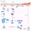
Overview of EBV-induced inflammation and NPC carcinogenesis. EBV in saliva passes through pharynx epithelium to infect naive B cells in the underlying lymphoid tissue via interaction of the viral glycoprotein gp350/220 with CD21 expressed on the B cells. This in turn drives the formation of proliferating lymphoblasts. Once EBV enters the cell, the viral capsid dissolves and viral genome incorporated into the host nucleus. The B cell blasts can differentiate into memory compartment as resting, long-lived, memory B cells with no viral gene expression. Furthermore, EBV encodes vIL-10 to escape host anti-viral responses and to establish latency by enhancing viability and differentiation of infected B cells. The latently infected memory B cells circulate in the periphery and return to the lymphoid tissue, where they can be signalled by cognate antigen to differentiate into plasma cells and activate viral replication program to produce more infectious virus. The lysis of infected cells leads the release of virion to initiate a new round of naïve B cell infection or infect the epithelial cells. Additionally, the plasma membrane break induces the efflux of potassium ion that activates inflammasome. Sustained inflammasome activation may promote viral replication and persistent infection. During a persistent infection, infected cells secrete excessive amount of pro-inflammatory cytokines/chemokines via NF-κB and STAT3 signalling, in order to establish a chronic inflammatory microenvironment that persistently damage the surrounding tissue. Over time, the chronic inflammation may trigger the initiation and development of cancer. In immunocompetent individuals, effective CTLs can recognise infected cells through the presentation of EBV antigen via MHC Class I and eliminate them by apoptosis. However, immune incompetent individual may be at risk of developing EBV-associated tumour during the chronic inflammation. (EBV, Epstein–Barr virus; NF-κB, nuclear factor kappa B; STAT3; signal transducer and activator of transcription 3; CTLs, cytotoxic T lymphocytes; MHC, major histocompatibility complex; vIL-10, viral IL-10).

Similar articles

Cited by

References

    1. Plummer M., de Martel C., Vignat J., Ferlay J., Bray F., Franceschi S. Global burden of cancers attributable to infections in 2012: A synthetic analysis. Lancet Glob. Health. 2016;4:e609–e616. doi: 10.1016/S2214-109X(16)30143-7. - DOI - PubMed
    1. IARC Working Group on the Evaluation of Carcinogenic Risks to Humans Biological agents. Volume 100 B. A review of human carcinogens. IARC Monogr. Eval. Carcinog. Risks Hum. 2012;100:1–441. - PMC - PubMed
    1. Massini G., Siemer D., Hohaus S. EBV in Hodgkin Lymphoma. Mediterr. J. Hematol. Infect. Dis. 2009;1:e2009013. doi: 10.4084/MJHID.2009.013. - DOI - PMC - PubMed
    1. Brady G., Macarthur G.J., Farrell P.J. Epstein-Barr virus and Burkitt lymphoma. Postgrad. Med J. 2008;84:372–377. doi: 10.1136/jcp.2007.047977. - DOI - PubMed
    1. Xiong F., Deng S., Huang H.B., Li X.Y., Zhang W.L., Liao Q.J., Ma J., Li X.L., Xiong W., Li G.Y., et al. Effects and mechanisms of innate immune molecules on inhibiting nasopharyngeal carcinoma. Chin. Med J. 2019;132:749–752. doi: 10.1097/CM9.0000000000000132. - DOI - PMC - PubMed

LinkOut - more resources