Review
. 2022 Jan;47(1):85-110.
doi: 10.1007/s11064-021-03331-z. Epub 2021 Apr 27.

Reconstitution of GABA, Glycine and Glutamate Transporters

Affiliations
Review

Reconstitution of GABA, Glycine and Glutamate Transporters

Niels Christian Danbolt et al. Neurochem Res. 2022 Jan.

Abstract

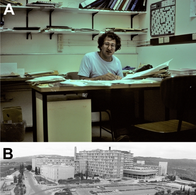
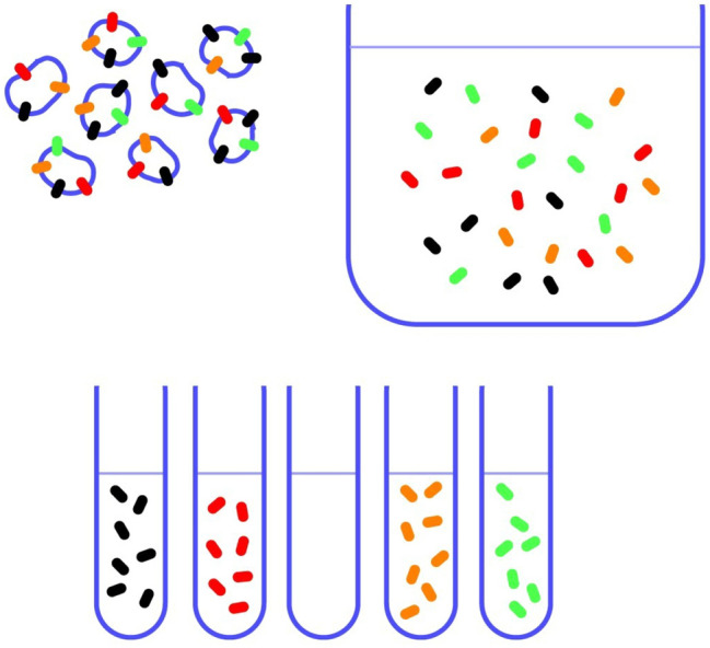
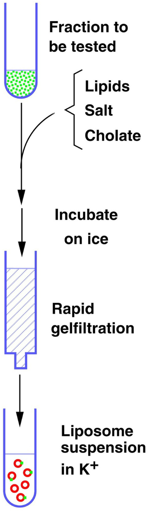
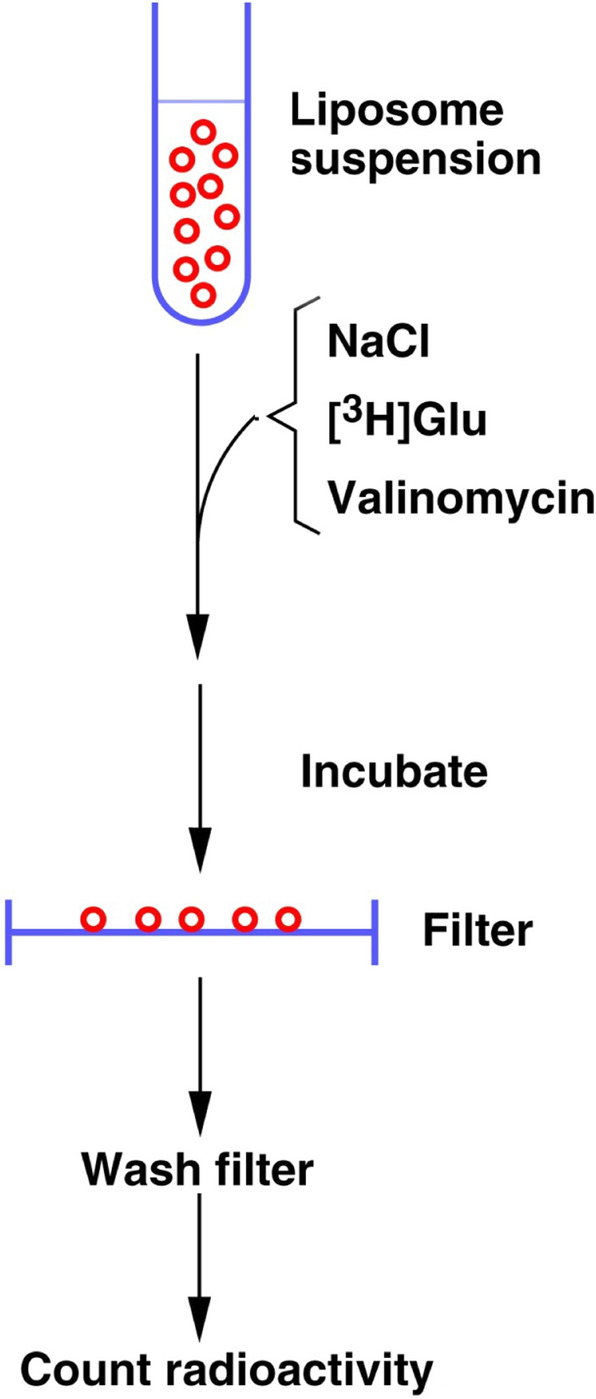
In contrast to water soluble enzymes which can be purified and studied while in solution, studies of solute carrier (transporter) proteins require both that the protein of interest is situated in a phospholipid membrane and that this membrane forms a closed compartment. An additional challenge to the study of transporter proteins has been that the transport depends on the transmembrane electrochemical gradients. Baruch I. Kanner understood this early on and first developed techniques for studying plasma membrane vesicles. This advanced the field in that the experimenter could control the electrochemical gradients. Kanner, however, did not stop there, but started to solubilize the membranes so that the transporter proteins were taken out of their natural environment. In order to study them, Kanner then had to find a way to reconstitute them (reinsert them into phospholipid membranes). The scope of the present review is both to describe the reconstitution method in full detail as that has never been done, and also to reveal the scientific impact that this method has had. Kanner's later work is not reviewed here although that also deserves a review because it too has had a huge impact.

Keywords: GABA transporter; Glutamate transporter; Glycine transporter; Liposomes; Reconstitution; Scientific history.


Conflict of interest statement

The authors declare that they have no conflict of interest.

Figures

Fig. 1
Fig. 1
a Baruch Kanner in his office in 1987 at b the Hadassah Medical School, the Hebrew University, Ein Kerem, Jerusalem, Israel. Copyright: Neurotransporter.org; reproduced with permission
Fig. 2
Fig. 2
A schematic illustration of the purpose of solubilization and the need for a method to detect the transporter of interest after separation. Copyright: Neurotransporter.org; reproduced with permission
Fig. 3
Fig. 3
Schematic illustration of the reconstitution process. Solubilized transporter proteins are mixed with the reconstitution mixture containing lipids, salt and cholate. After incubation on ice, cholate is removed by gel filtration and the liposomes form spontaneously. The fluid in which they are suspended becomes trapped inside and therefore become the internal medium. Copyright: Neurotransporter.org; reproduced with permission
Fig. 4
Fig. 4
Illustrating the principle of gel filtration in removing detergents during reconstitution. A mixture (200 µl) of a low molecular mass dye (phenol red) and a high molecular mass compound (blue-dextran 2000) were added to 1 ml spin columns which are packed with Sephadex G-50 in internal medium. Copyright: Neurotransporter.org; reproduced with permission
Fig. 5
Fig. 5
Schematic illustration of the uptake assay. The liposome suspension (e.g. 20 µl) is diluted into an external medium (e.g. 500 µl) containing radioactively labeled substrate and incubated. The reaction is stopped by dilution in ice-cold medium without substrate and quickly filtered to isolate the liposomes. After quick rinsing of the filters with more of the ice-cold medium, the radioactivity retained in the trapped liposomes is measured. Copyright: Neurotransporter.org; reproduced with permission
Fig. 6
Fig. 6
The environment in Kanner’s lab and in the department as a whole was very friendly and very creative. Further, “out of the box” thinking was stimulated by a multicultural encounter between people from different backgrounds and countries. a Annie Bendahan is here photographed while sonicating lipids for reconstitution of GABA transporters. She worked with Kanner for 30 years from 1981 and generated or helped generating many of the most important data from the laboratory. b Rodica Radian-Gordon who isolated GABA transporter 1 [14, 20] is here depicted in the laboratory assisted by her husband, Alexander Gordon (Alex). Her career path has been extraordinary. She is now (since 2019) the Israeli Ambassador to Spain after having been Israeli Ambassador to several other countries! c Alex obtained a partial purification of glutamate transporters using lectin (WGA; Wheat germ agglutinin) affinity chromatography [15]. Here he is photographed when running a WGA-column in the cold room. d Niels Christian Danbolt learned from Alex, modified both the WGA-chromatography and the reconstitution procedure and eventually managed to isolate active glutamate transporters using three chromatographic steps [13]. Here he is photographed in the cold room (by Michela Tessari [49]). e Beatriz López-Corcuera visited Kanner’s lab to get help with the isolation of glycine transporters. She also succeeded [21, 35]. f This is the old tabletop centrifuge used for all of the reconstitutions in Kanner’s laboratory. This clearly illustrates that intelligence is more important than fancy equipment! Copyright: Neurotransporter.org; reproduced with permission
Fig. 7
Fig. 7
Astroglial EAAT2 is silent in a synaptosome containing tissue homogenate, but not if reconstituted into artificial cell membranes (proteoliposomes). a Mice homozygote for the floxed EAAT2 (Slc1a2) gene (i.e. B6.Cg-Slc1a2tm1.1Ncd/J; RRID:IMSR_JAX:026619 [59]) were bred with mice harboring one flox-EAAT2 allele and one Cre allele (B6.Cg-Tg(Syn1-cre)671Jxm/J}; RRID:IMSR_JAX:003966 [207]). This breeding yielded both mice with normal EAAT2 expression (wild-type; WT) and mice lacking EAAT2 (nKO) in neurons (for details see [9]). b Western blots (not shown) and immunocytochemistry showed that deletion of the EAAT2 gene in neurons had only minor impact on the total tissue content of EAAT2 protein (tntibody to EAAT2: RRID:AB_2714090; 44). c Forebrains from the nKO mice were homogenized in 0.32 M sucrose and assayed for glutamate uptake. The homogenates from the nKO mice had considerably lower glutamate uptake activity (left). The reason may be that (right) many of the astroglial cell fragments do not properly reseal so that only those indicated (red asterisk) accumulate glutamate. Consequently, all the EAAT2 transporter molecules (black dots) in the other fragments are unable to contribute to the uptake, and loss of the low number of transporters on neuronal membranes (blue lines) results in a major reduction in uptake activity. d When all the transporters are taken out of their original membranes and reconstituted into artificial ones (liposomes, green lines), then all transporters, regardless of whether they originate from astrocytes or neurons, have the same possibility to contribute to the uptake. In that case, loss of the few neuronal EAAT2 transporter molecules do not matter much. (Copyright: Panels B, C and left part of D are reproduced from Figs. 1 and 2a and d in Zhou et al. [9])
Fig. 8
Fig. 8
a Two modes of glutamate translocation [55]. Large open circles represent liposomes, and smaller filled gray circles represent transporter molecules. Stoichiometric transport is indicated with red arrows and uncoupled anion (A) fluxes by blue arrows. Net uptake is a process in which external substrate is taken up in a manner dependent on internal K+. This leads to net removal of substrate from the extracellular fluid. In contrast, (hetero)exchange is a process in which internal unlabeled substrate is exchanged with external labeled substrate in a 1:1 relationship. The latter process does not alter the number of substrate molecules on each side of the membrane but allows molecules to switch location. This process requires Na+, but not K+, and occurs in the absence of transmembrane gradients. Consequently, dissipation of transmembrane ion gradients with the ionophore nigericin (Nig) abolishes net uptake (c), but does not have much effect on exchange (e). In the absence of K+, the transporters are locked in exchange mode. An important consequence is the following: when transportable uptake inhibitors are added to cell cultures, the inhibitors induce glutamate release from the cells (e.g. [25, 55]). Panels b and d: The glutamate uptake activity measured with the reconstituted system is mostly due to EAAT2 when forebrain tissue is used as the source of transporter proteins. b Potassium-loaded proteoliposomes prepared from wild-type (WT) and EAAT2 knockout mice (KO [199]) were diluted into a large volume of a sodium-containing medium with radiolabeled glutamate to measure net uptake. Internal medium: 135 mM KPi (pH7.4) with 1 % (v/v) glycerol. External medium: 135 mM NaPi (pH7.4), 1 % (v/v) glycerol, 3 µM valinomycin, 2 µM Na-glutamate, and 1.4 µCi l-[3 H]glutamate. Liposomes prepared from the EAAT2-deficient animals (KO) had little uptake activities compared with those prepared from WT littermates. d Sodium- and glutamate-loaded proteoliposomes, prepared from WT and KO mice, were diluted into a large volume of a sodium containing medium with radiolabeled glutamate to measure heteroexchange. Internal medium: 120 mM NaPi (pH7.4), 20 mM Na-glutamate, and 1 % (v/v) glycerol. External medium: 135 mM NaPi (pH7.4), 1 % (v/v) glycerol, 2 µM Na-glutamate, and 1.4 µCi l-[3 H]glutamate. c Addition of nigericin (Nig.) to potassium-loaded proteoliposomes abolishes the uptake activities. e Glutamate uptake activities by proteoliposomes loaded with sodium and glutamate are insensitive to nigericin. (Copyright: The figure was reproduced from Fig. 1 in Zhou et al. [19])
Fig. 9
Fig. 9
The effect of intraliposomal potassium on the rate of net uptake. Proteoliposomes were loaded with various internal media: 400 mM K+ (400 mM potassium ions, 250 mM HEPES, 100 mM citric acid, and 1% v/v glycerol), 200 mM K+, 200 mM Li+ (200 mM potassium ions, 200 mM lithium ions, 250 mM HEPES, 100 mM citric acid, and 1% v/v glycerol), 200 mM K+, 200 mM Na+ (200 mM potassium ions, 200 mM sodium ions, 250 mM HEPES, 100 mM citric acid, and 1% v/v glycerol); 200 mM K+, 200 mM ethanolamine (200 mM potassium ions, 200 mM ethanolamine, 250 mM HEPES, 100 mM citric acid, and 1% v/v glycerol). They were added into external medium containing 400 mM sodium ions, 250 mM HEPES, 100 mM citric acid, 1% v/v glycerol, 2 µM Na-glutamate, 50 nM l-[3 H]glutamate, and 3 µM valinomycin. The data represent average ± SEM of one experiment with triplicates. (Copyright: The figure was reproduced from Fig. 7A1 in Zhou et al. [19])
Fig. 10
Fig. 10
Stimulation of net uptake by the negative membrane potential created by the addition of the potassium ionophore valinomycin. Proteoliposomes were loaded with 20 mM K-gluconate, 15 mM KPi (pH7.4), 145 mM KCl and 1% (v/v) glycerol). The reactions were started by the addition of proteoliposomes into reaction buffers with varying concentrations of external glutamate (0.2, 2 or 5 µM unlabeled Na-glutamate, 50 nM l-[3 H]glutamate, 20 mM Na-gluconate, 15 mM NaPi (pH7.4), 145 mM NaCl, and 1% (v/v) glycerol. The addition of 3 µM valinomycin was indicated as a solid bar. The incubation time was 5 s. 3 µM nigericin was added as negative control (*). The figure represents average ± SEM of one representative experiment (n = 3 replicates). (Copyright: The figure was reproduced from Fig. 2B in Zhou et al. [19])
Fig. 11
Fig. 11
The effect of membrane potential built by anions on the rate of net uptake. Proteoliposomes were loaded with various internal media: NO3 (100 mM KNO3, 20 mM KPi pH 7.4, and 1% v/v glycerol), Cl (100 mM KCl, 20 mM KPi pH 7.4, and 1% v/v glycerol), Pi- (100 mM KPi pH7.4, and 1% v/v glycerol). Then they were added into related external media containing 2 µM l-[3 H]glutamate and sodium without valinomycin (blue solid line) or with 3 µM valinomycin (dashed red line). (Copyright: The figure was reproduced from Fig. 7a in Zhou et al. [19])
Fig. 12
Fig. 12
Electroneutral glutamate exchange is voltage dependent. Proteoliposomes were loaded (indicated as “in”) with either: NaPi (120 mM NaPi pH7.4, 20 mM Na-glutamate, and 1% v/v glycerol) or NaNO3 (100 mM NaNO3, 40 mM NaPi pH 7.4, 20 mM Na-glutamate and 1% v/v glycerol), and then diluted into external media containing 2 µM l-[3 H]glutamate and either NaNO3 (100 mM NaNO3, 40 mM NaPi pH 7.4, 20 mM Na-gluconate and 1% v/v glycerol), or NaPi (120 mM NaPi pH7.4, 20 mM Na-gluconate, and 1% v/v glycerol). (Copyright: The figure was reproduced from Fig. 5C in Zhou et al. [19])
Fig. 13
Fig. 13
The effect of intraliposomal glutamate on the exchange rate. Proteoliposomes were loaded with internal media consisting of 15 mM KPi (pH 7.4), 145 mM KCl and the indicated concentrations of unlabeled l-Glu (1 20 mM). The liposomes (20 µl) were diluted into 1500 µl of external medium consisting of 15 mM NaPi (pH 7.4), 145 mM NaCl, 2 µM labeled L[3H]glutamate. Sodium gluconate was added to balance the osmolarity of the internal glutamate. The uptake reaction was stopped by dilution and filtration after the time indicated (0–180 s). Note that external substrate (L[3 H]glutamate, 2 µM) enters faster into liposomes if the liposomes already contain high substrate concentrations. The figure represents unpublished data from the study of the relative rates of net uptake and heteroexchange [19]. Copyright: Neurotransporter.org; reproduced with permission
Fig. 14
Fig. 14
Bell-shaped time course due to uptake followed by reversed uptake. Liposomes are loaded with the standard K+-containing internal medium (0.12 M potassium phosphate with 1 ml/dl glycerol) and the uptake reaction is started by diluting the liposomes in Na+-containing external medium (0.15 M NaCl with 1 ml/dl glycerol, 3 µM valinomycin) with 50 nM l-[3H]glutamate). Glutamate accumulates inside the liposomes (solid squares) until the ion gradients are dissipated. Then glutamate leaks out presumably to reversal of the transporters giving rise to the bell-shaped time course. Addition of nigericin abolishes uptake when added at start (asterisks) and induces reversal when added later (solid triangles). Dihydrokainate inhibits uptake (open squares) but is also able to block reversal because it is a non-transportable substrate as illustrated by the addition of both nigericin and dihydrokainate (open triangles). Data from the same material as in [224]). Copyright: The figure was reproduced from Fig. 4E in Zhou et al. [19]

Similar articles

Cited by

References

    1. Hediger MA, Romero MF, Peng J-B, Rolfs A, Takanaga H, Bruford EA. The ABCs of solute carriers: physiological, pathological and therapeutic implications of human membrane transport proteinsIntroduction. Pflugers Arch. 2004;447:465–468. doi: 10.1007/s00424-003-1192-y. - DOI - PubMed
    1. Cesar-Razquin A, Snijder B, Frappier-Brinton T, Isserlin R, Gyimesi G, Bai X, Reithmeier RA, Hepworth D, Hediger MA, Edwards AM, Superti-Furga G. A Call for Systematic Research on Solute Carriers. Cell. 2015;162:478–487. doi: 10.1016/j.cell.2015.07.022. - DOI - PubMed
    1. Bröer S, Bröer A. Amino acid homeostasis and signalling in mammalian cells and organisms. Biochem J. 2017;474:1935–1963. doi: 10.1042/BCJ20160822. - DOI - PMC - PubMed
    1. Kanner BI, Sharon I. Active transport of L-glutamate by membrane vesicles isolated from rat brain. Biochemistry. 1978;17:3949–3953. doi: 10.1021/bi00612a011. - DOI - PubMed
    1. Kanner BI. Active transport of gamma-aminobutyric acid by membrane vesicles isolated from rat brain. Biochemistry. 1978;17:1207–1211. doi: 10.1021/bi00600a011. - DOI - PubMed

MeSH terms

LinkOut - more resources