

The Neurobiology of Zika Virus: New Models, New Challenges
- PMID: 33897363
- PMCID: PMC8059436
- DOI: 10.3389/fnins.2021.654078
The Neurobiology of Zika Virus: New Models, New Challenges
Abstract
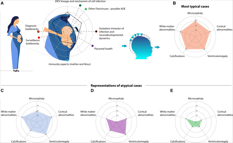
The Zika virus (ZIKV) attracted attention due to one striking characteristic: the ability to cross the placental barrier and infect the fetus, possibly causing severe neurodevelopmental disruptions included in the Congenital Zika Syndrome (CZS). Few years after the epidemic, the CZS incidence has begun to decline. However, how ZIKV causes a diversity of outcomes is far from being understood. This is probably driven by a chain of complex events that relies on the interaction between ZIKV and environmental and physiological variables. In this review, we address open questions that might lead to an ill-defined diagnosis of CZS. This inaccuracy underestimates a large spectrum of apparent normocephalic cases that remain underdiagnosed, comprising several subtle brain abnormalities frequently masked by a normal head circumference. Therefore, new models using neuroimaging and artificial intelligence are needed to improve our understanding of the neurobiology of ZIKV and its true impact in neurodevelopment.
Keywords: Zika virus; artificial intelligence; brain abnormalities; cell death; congenital Zika syndrome; neurodevelopment; neuroimaging.
Copyright © 2021 Moura, Ferreira, Loureiro, de Paiva, Rosa-Ribeiro, Amaro, Soares and Machado.
Conflict of interest statement
The authors declare that the research was conducted in the absence of any commercial or financial relationships that could be construed as a potential conflict of interest.
Figures
Similar articles
-
Lessons Learned at the Epicenter of Brazil's Congenital Zika Epidemic: Evidence From 87 Confirmed Cases.Clin Infect Dis. 2017 May 15;64(10):1302-1308. doi: 10.1093/cid/cix166. Clin Infect Dis. 2017. PMID: 28329257
-
Differential gene expression elicited by ZIKV infection in trophoblasts from congenital Zika syndrome discordant twins.PLoS Negl Trop Dis. 2020 Aug 3;14(8):e0008424. doi: 10.1371/journal.pntd.0008424. eCollection 2020 Aug. PLoS Negl Trop Dis. 2020. PMID: 32745093 Free PMC article.
-
Perinatal characteristics and longer-term outcomes in Brazilian children with confirmed or suspected congenital Zika infection: ZIKAction Paediatric Registry.Dialogues Health. 2023 Jan 20;2:100104. doi: 10.1016/j.dialog.2023.100104. eCollection 2023 Dec. Dialogues Health. 2023. PMID: 38515475 Free PMC article.
-
Pre-Clinical Pregnancy Models for Evaluating Zika Vaccines.Trop Med Infect Dis. 2019 Apr 7;4(2):58. doi: 10.3390/tropicalmed4020058. Trop Med Infect Dis. 2019. PMID: 30959955 Free PMC article. Review.
-
Congenital Zika syndrome: Pitfalls in the placental barrier.Rev Med Virol. 2018 Sep;28(5):e1985. doi: 10.1002/rmv.1985. Epub 2018 May 15. Rev Med Virol. 2018. PMID: 29761581 Review.
Cited by
-
Zika virus induced microcephaly and aberrant hematopoietic cell differentiation modeled in novel neonatal humanized mice.Front Immunol. 2023 Feb 7;14:1060959. doi: 10.3389/fimmu.2023.1060959. eCollection 2023. Front Immunol. 2023. PMID: 36825016 Free PMC article.
-
Zika Virus: A New Therapeutic Candidate for Glioblastoma Treatment.Int J Mol Sci. 2021 Oct 12;22(20):10996. doi: 10.3390/ijms222010996. Int J Mol Sci. 2021. PMID: 34681654 Free PMC article. Review.
-
Pilot deployment of a cloud-based universal medical image repository in a large public health system: A protocol study.PLoS One. 2024 Aug 29;19(8):e0307022. doi: 10.1371/journal.pone.0307022. eCollection 2024. PLoS One. 2024. PMID: 39208265 Free PMC article.
References
-
- Adebanjo T., Godfred-Cato S., Viens L., Fischer M., Staples J. E., Kuhnert-Tallman W., et al. (2017). Update: interim guidance for the diagnosis, evaluation, and management of infants with possible congenital Zika virus infection—United States, October 2017. MMWR Morb. Mortal. Wkly. Rep. 66:1089. - PMC - PubMed
-
- Aguiar R. S., Pohl F., Morais G. L., Nogueira F. C. S., Carvalho J. B., Guida L., et al. (2020). Molecular alterations in the extracellular matrix in the brains of newborns with congenital Zika syndrome. Sci. Signal. 13:eaay6736. - PubMed
Publication types
LinkOut - more resources
Full Text Sources
Other Literature Sources