. 2021 Apr 27;118(17):e2023130118.
doi: 10.1073/pnas.2023130118.

Caveolar peroxynitrite formation impairs endothelial TRPV4 channels and elevates pulmonary arterial pressure in pulmonary hypertension

Affiliations

Caveolar peroxynitrite formation impairs endothelial TRPV4 channels and elevates pulmonary arterial pressure in pulmonary hypertension

Zdravka Daneva et al. Proc Natl Acad Sci U S A. .

Abstract

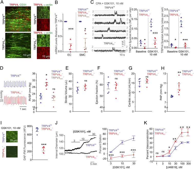
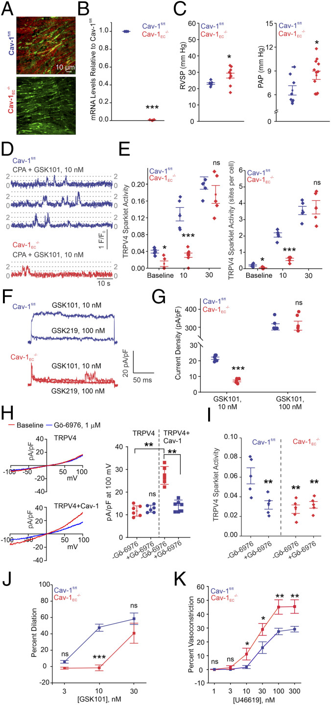
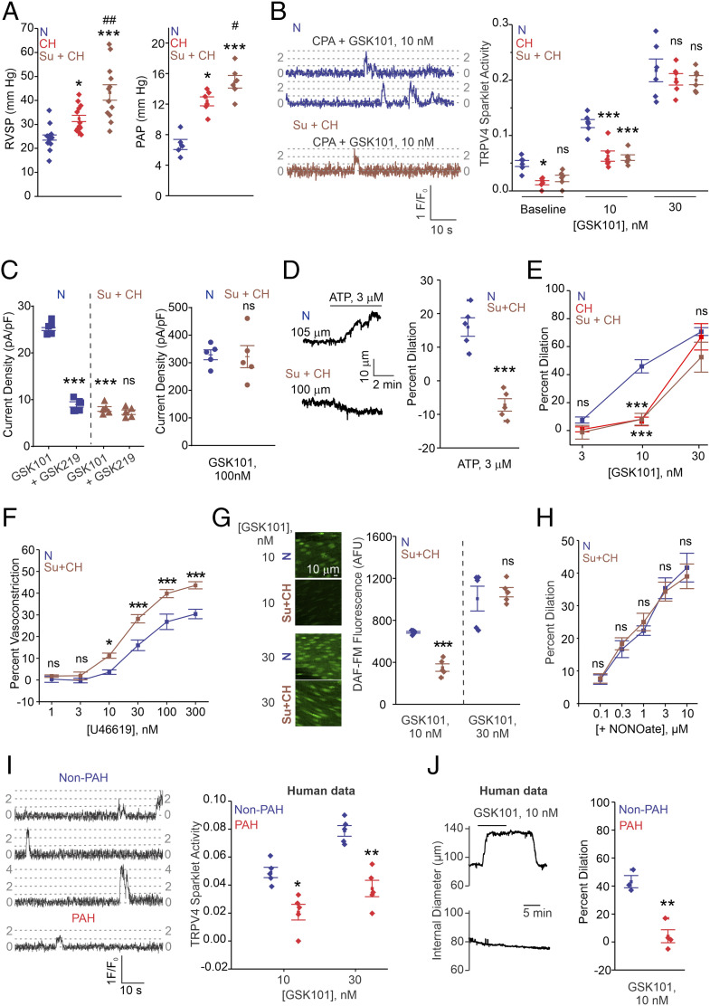
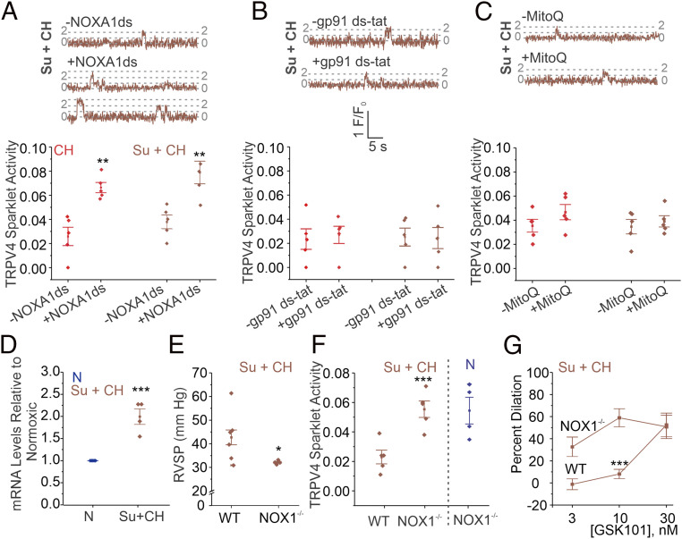
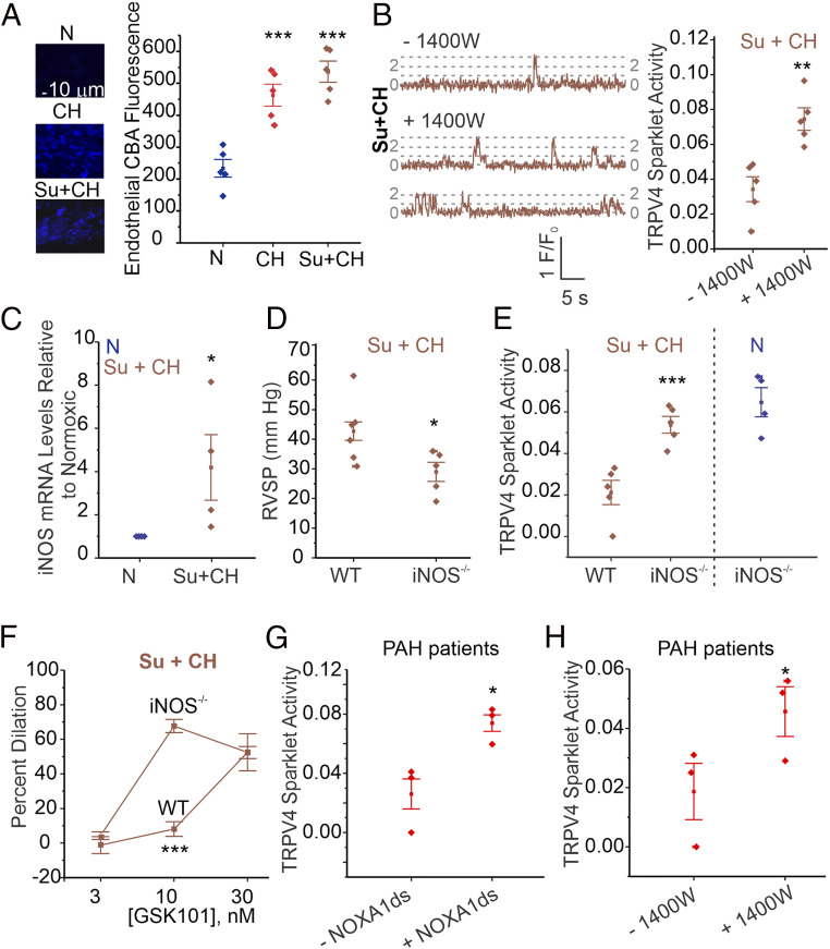
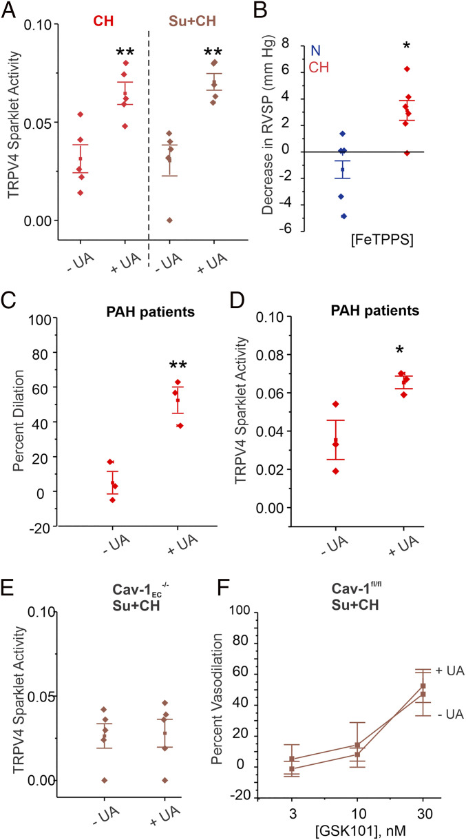
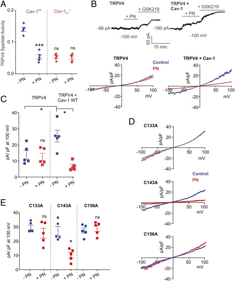
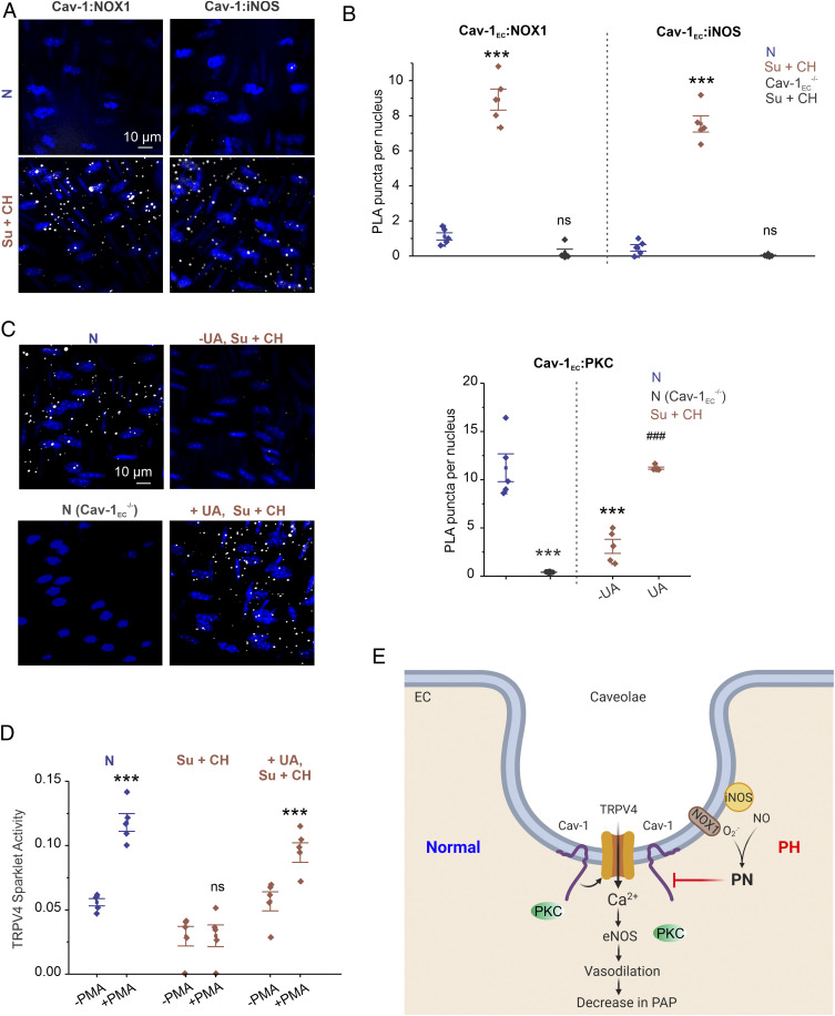
Recent studies have focused on the contribution of capillary endothelial TRPV4 channels to pulmonary pathologies, including lung edema and lung injury. However, in pulmonary hypertension (PH), small pulmonary arteries are the focus of the pathology, and endothelial TRPV4 channels in this crucial anatomy remain unexplored in PH. Here, we provide evidence that TRPV4 channels in endothelial cell caveolae maintain a low pulmonary arterial pressure under normal conditions. Moreover, the activity of caveolar TRPV4 channels is impaired in pulmonary arteries from mouse models of PH and PH patients. In PH, up-regulation of iNOS and NOX1 enzymes at endothelial cell caveolae results in the formation of the oxidant molecule peroxynitrite. Peroxynitrite, in turn, targets the structural protein caveolin-1 to reduce the activity of TRPV4 channels. These results suggest that endothelial caveolin-1-TRPV4 channel signaling lowers pulmonary arterial pressure, and impairment of endothelial caveolin-1-TRPV4 channel signaling contributes to elevated pulmonary arterial pressure in PH. Thus, inhibiting NOX1 or iNOS activity, or lowering endothelial peroxynitrite levels, may represent strategies for restoring vasodilation and pulmonary arterial pressure in PH.

Keywords: TRP channel; caveolin; endothelium; peroxynitrite; pulmonary hypertension.


Conflict of interest statement

The authors declare no competing interest.

Figures

Fig. 1.
Fig. 1.
Inducible TRPV4EC−/− mice show elevated resting PAP. (A) TRPV4EC immunofluorescence images in en face fourth-order PAs from TRPV4fl/fl and TRPV4EC−/− mice. (B) Endothelial TRPV4 mRNA levels (left of the dotted line) relative to those in TRPV4fl/fl mice and SMC TRPV4 mRNA levels (right of the dotted line) relative to those in TRPV4fl/fl mice (n = 4 to 5; *P < 0.05, ***P < 0.001 versus TRPV4fl/fl; one-way ANOVA). (C, Left) TRPV4EC sparklet traces in one EC from a fluo-4–loaded en face PA in response to TRPV4 channel activator GSK101 (10 nmol/L). (Center) TRPV4EC sparklet activity in PAs, expressed as NPO per site (n = 5; *P < 0.05; ***P < 0.001 versus TRPV4fl/fl; two-way ANOVA). N is the number of channels per site, and PO is the open state probability of the channel. Experiments were performed in Fluo-4–loaded fourth-order PAs in the presence of cyclopiazonic acid (CPA; 20 μmol/L) to eliminate Ca2+ release from intracellular stores. (Right) TRPV4EC sparklet activity sites per cell in PAs (n = 5; *P < 0.05; ***P < 0.001 versus TRPV4fl/fl; two-way ANOVA). (D) Representative RVSP (millimeters of mercury) traces (Left) and averaged RVSP values (Right; n = 6; *P < 0.05 versus TRPV4fl/fl; ns indicates no significance; one-way ANOVA). (E) Averaged stroke volume (microliters) in TRPV4fl/fl and TRPV4EC−/− mice. Data represented as mean ± SEM (n = 5). (F) Averaged ejection fraction (%) in TRPV4fl/fl and TRPV4EC−/− mice (n = 5 mice; ns indicates no significance). (G) Cardiac output (milliliters per minute) in TRPV4fl/fl and TRPV4EC−/− mice (n = 5 mice). (H) Averaged PAP (mm Hg) in isolated perfused lungs from TRPV4fl/fl and TRPV4EC−/− mice (n = 6; **P < 0.01; t test). (I) DAF-FM fluorescence analysis of NO production in response to GSK101 (10 nmol/L) in TRPV4fl/fl and TRPV4EC−/− mice, respectively (n = 5; ***P < 0.001; t test). (J) Pressure myography traces (Left) and averaged percent vasodilation (Right) of PAs to GSK101 (3 to 30 nmol/L). Fourth-order PAs were pressurized to 15 mm Hg (n = 5 to 6; ***P < 0.001 versus TRPV4fl/fl; two-way ANOVA). (K) Percent vasoconstriction of PAs in response to the thromboxane A2 receptor agonist U46619 (1 to 300 nmol/L; n = 5; *P < 0.05 [10 nmol/L] and **P < 0.01 [30 to 300 nmol/L] versus TRPV4fl/fl, two-way ANOVA).
Fig. 2.
Fig. 2.
Cav-1EC–TRPV4EC signaling in PAs lowers resting PAP. (A) Cav-1EC immunofluorescence in en face preparations of fourth-order PAs. (B) Cav-1EC mRNA levels in PAs relative to those in Cav-1fl/fl mice (n = 4; ***P < 0.001; t test). (C) Averaged resting RVSP (Left; n = 5 to 9; *P < 0.05; t test) and PAP values (Right; n = 8 to 11; *P < 0.05; t test). (D) TRPV4EC sparklet traces in one EC from a fluo-4–loaded en face PAs in response to TRPV4 channel activator GSK101 (10 nmol/L) in Cav-1fl/fl and Cav-1EC−/− mice. (E, Left) Baseline or GSK101-induced (10 to 30 nmol/L) TRPV4EC sparklet activity, expressed as NPO per site (n = 5; *P < 0.05 versus Cav-1fl/fl [Baseline]; ***P < 0.001 versus Cav-1fl/fl [10 nmol/L]; ns indicates no significance; two-way ANOVA). Experiments were performed in fluo-4–loaded fourth-order PAs in the presence of cyclopiazonic acid (CPA; 20 μmol/L) to eliminate Ca2+ release from intracellular stores. (Right) TRPV4EC sparklet sites per cell in PAs from Cav-1fl/fl or Cav-1EC−/− mice (n = 5; *P < 0.05 versus Cav-1fl/fl [Baseline]; ***P < 0.001 versus Cav-1fl/fl [10 nmol/L]; two-way ANOVA). (F) Representative GSK101 (10 nmol/L)-induced outward TRPV4EC currents in freshly isolated ECs from Cav-1fl/fl or Cav-1EC−/− mice and effect of GSK2193874 (GSK219; 100 nmol/L) in the presence of GSK101 (10 nmol/L). (G) Scatterplot showing TRPV4EC currents in the presence of GSK101 (10 and 100 nmol/L; n = 5; ***P < 0.001 versus Cav-1fl/fl [10 nmol/L]; two-way ANOVA). (H, Left) Representative traces showing TRPV4 currents in the absence or presence of Gö-6976 (PKC-α/β inhibitor; 1 μmol/L) in HEK293 cells transfected with TRPV4 only or cotransfected with TRPV4 plus WT Cav-1, recorded in the whole-cell patch-clamp configuration. (Right) Current density plot of TRPV4 currents at +100 mV in the absence or presence of Gö-6976 (1 μmol/L) in HEK293 cells transfected with TRPV4 or TRPV4 + Cav-1 (n = 5; **P < 0.01 versus TRPV4 [−Gö-6976] and TRPV4 + Cav-1 [−Gö-6976]; two-way ANOVA). (I) Effect of Gö-6976 (1 μmol/L) on TRPV4EC sparklet activity in en face preparations of PAs from Cav-1fl/fl and Cav-1EC−/− mice, expressed as NPO per site (n = 5; **P < 0.01 versus Cav-1fl/fl [−Gö-6976]; two-way ANOVA). (J) Percent dilation of PAs in response to GSK101 (3 to 30 nmol/L; n = 6; *** P < 0.001 versus Cav-1fl/fl [10 nmol/L]; two-way ANOVA). (K) Percent constriction of PAs in response to the thromboxane A2 receptor agonist U46619 (1 to 300 nmol/L; n = 5; *P < 0.05 versus Cav-1fl/fl [10 and 30 nmol/L]; **P < 0.01 versus Cav-1fl/fl [100 and 300 nmol/L]; two-way ANOVA).
Fig. 3.
Fig. 3.
TRPV4EC channel activity and vasodilation are reduced in PAs from mouse models of PH and PAH patients. (A, Left) Averaged RVSP (mmHg) values in mice exposed to N, CH (3 wk; 10% O2), or Su+CH (n = 12; *P < 0.05 versus N; ***P < 0.001 versus N; ##P < 0.01 versus CH; one-way ANOVA). (Right) Averaged PAP (millimeters of mercury) values in mice exposed to N, CH, and Su+CH (n = 6; *P < 0.05 versus N; ***P < 0.001 versus N; #P < 0.05 versus CH; one-way ANOVA). (B, Left) TRPV4EC sparklet traces in one EC from a fluo-4–loaded en face PA from N and Su+CH mice in response to TRPV4 channel activator GSK101 (10 nmol/L). (Right) Baseline or GSK101 (10 and 30 nmol/L)-induced TRPV4EC sparklet activity in N, CH, or Su+CH mice, expressed as NPO per site (n = 5; *P < 0.05 versus N [baseline]; ***P < 0.001 versus N [10 nmol/L]; two-way ANOVA). Experiments were performed in fluo-4–loaded fourth-order PAs in the presence of cyclopiazonic acid (CPA; 20 μmol/L) to eliminate Ca2+ release from intracellular stores. (C, Left) GSK101 (10 nmol/L)-induced outward currents at +100 mV and inhibition by GSK2193874 (GSK219; 100 nmol/L; n = 5; ***P < 0.001 versus N [GSK101]; two-way ANOVA). (Right) GSK101 (100 nmol/L)-induced outward currents at +100 mV in PAs from N and Su+CH mice (n = 5). (D) Pressure myography trace (Left) and percent vasodilation (Right) of PAs in response to ATP (3 μmol/L) in N and Su+CH mice (n = 5; ***P < 0.001 versus N; t test). (E) Percent vasodilation of PAs from N, CH, and Su+CH mice in response to GSK101 (3 to 30 nmol/L; n = 5 to 10; ***P < 0.001 versus N [GSK101; 10 nmol/L]; two-way ANOVA). (F) Percent vasoconstriction to thromboxane A2 receptor agonist U46619 (1 to 300 nmol/L) in PAs from N or Su+CH mice (n = 5; *P < 0.05 [U46619; 10 nmol/L] and ***P < 0.001 [U466; 30 to 300 nmol/L] versus N; two-way ANOVA). (G) DAF-FM fluorescence analysis of NO production in response to GSK101 (10 and 30 nmol/L) in N and Su+CH mice, respectively (n = 5; ***P < 0.001 versus N [GSK101; 10 nmol/L]; two-way ANOVA). (H) Percent vasodilation of PAs from N and Su+CH mice in response to spermine NONOate (NO donor; 0.1 to 10 μmol/L; n = 5). (I) TRPV4EC sparklet traces (Left) and GSK101 (10 and 30 nmol/L)-induced sparklet activity (Right) in PAs from non-PAH and PAH patients (n = 5; *P < 0.05 [GSK101; 10 nmol/L] versus non-PAH; **P < 0.01 [GSK101; 30 nmol/L] versus non-PAH; two-way ANOVA). (J, Left) Representative diameter traces showing GSK101 (10 nmol/L)-induced dilation of PAs from non-PAH individuals and PAH patients, preconstricted with U46619 (50 nmol/L). (Right) Percent vasodilation of PAs from non-PAH and PAH patients in response to GSK101 (10 nmol/L; n = 3 to 4; **P < 0.01; t test).
Fig. 4.
Fig. 4.
Endothelial NOX1 impairs TRPV4EC channel activity in PH. (AC) TRPV4EC sparklet traces in one EC from a fluo-4–loaded en face PA (Top) and TRPV4EC sparklet activity per site (Bottom; NPO per site) in the absence and presence of NOXA1ds (NOX 1 inhibitor; 1 μmol/L; A), gp91ds-tat (NOX2 inhibitor; 1 μmol/L; B), or mitoQ (mitochondrial antioxidant; 1 μmol/L; C) in PAs from C57BL6/J mice exposed to CH (3 wk; 10% O2) or Su+CH (n = 5; **P < 0.01 versus [−NOXA1ds]; two-way ANOVA), expressed as NPO per site. (D) NOX1 mRNA levels in ECs from N and Su+CH mice relative to N mice (n = 5; ***P < 0.001; t test). (E) Averaged resting RVSP (millimeters of mercury) values in WT and NOX1−/− mice exposed to Su+CH (n = 4 to 6; *P < 0.05; t test). (F) GSK101-induced (1 nmol/L) TRPV4EC sparklet activity in PAs from WT and NOX1−/− mice exposed to Su+CH and NOX1−/− mice exposed to N (n = 5; ***P < 0.001 versus WT [Su+CH]; two-way ANOVA). (G) Percent dilation of PAs from WT and NOX1−/− mice exposed to Su+CH in response to GSK101 (3 to 30 nmol/L; n = 5; ***P < 0.001 versus WT [3 and 10 nmol/L]; two-way ANOVA).
Fig. 5.
Fig. 5.
Endothelial iNOS/NOX1-generated PN impairs TRPV4EC channel activity in PH. (A, Left) Representative nonconfocal images for coumarin boronic acid (CBA, PN indicator) fluorescence in ECs from PAs of mice exposed to N, CH (3 wk; 10% O2), or Su+CH. (Right) Scatterplot of CBA fluorescence intensity in en face preparations of fourth-order PAs from mice exposed to N, CH, or Su+CH (n = 5; ***P < 0.001 versus N; one-way ANOVA). Experiments were performed in the presence of PEG-catalase (H2O2 metabolizing enzyme; 500 U/mL) and taurine (hypochlorous acid-lowering agent; 1 mmol/L). (B) TRPV4EC sparklet traces in one EC from fluo-4–loaded en face PAs (Left) and sparklet activity per site (Right; expressed as NPO per site) in the absence and presence of the 1400W (iNOS inhibitor; 1 μmol/L) in PAs from C57BL6/J mice exposed to Su+CH (n = 5; **P < 0.01; t test). (C) iNOS mRNA levels in ECs from PAs of N and Su+CH mice, expressed relative to N mice (n = 5; *P < 0.05; t test). (D) Averaged resting RVSP (millimeters of mercury) values in WT and iNOS−/− mice after exposure to Su+CH (n = 5 to 6; *P < 0.05; t test). (E) TRPV4EC sparklet activity in PAs from WT and iNOS−/− mice exposed to Su+CH and iNOS−/− mice exposed to N (n = 5; * P < 0.05; two-way ANOVA). (F) GSK101 (3–30 nmol/L)-induced vasodilation of PAs from WT and iNOS−/− mice exposed to Su+CH (n = 5; ***P < 0.001 versus WT [10 nmol/L]; two-way ANOVA). (G) Effect of the NOXA1ds (NOX1 inhibitor, 1 μmol/L) on GSK101 (10 nmol/L)-induced TRPV4EC sparklet activity in PAs from PAH patients (n = 3; *P < 0.05; t test). (H) Effect of the 1400W (1 μmol/L) on TRPV4EC sparklet activity in PAs from PAH patients (n = 3; *P < 0.05; t test).
Fig. 6.
Fig. 6.
PN inhibition rescues TRPV4EC channel activity in PH. (A) Effects of the UA (PN scavenger; 200 μmol/L) on TRPV4EC sparklet activity in PAs from mice treated with CH (3 wk; 10% O2) or Su+CH, expressed as NPO per site (n = 5; **P < 0.01 versus [−UA]; one-way ANOVA). (B) Scatter plot showing the FeTPPS (PN decomposer; 30 mg/kg i.p.)-induced decrease in RVSP (millimeters of mercury) in mice exposed to N or CH (n = 5 to 6; *P < 0.05; t test). (C) Effect of UA (200 μmol/L) on GSK101 (10 nmol/L)-induced vasodilation of PAs from PAH patients (n = 3; **P < 0.01; t test). (D) Effect of UA (200 μmol/L) on GSK101 (10 nmol/L)-induced TRPV4EC sparklet activity of PAs from PAH patients (n = 3; *P < 0.05; t test). (E) Effect of the UA (200 μmol/L) on TRPV4EC sparklet activity in PAs from Cav-1EC−/− mice exposed to Su+CH (n = 5). (F) Effect of UA (200 μmol/L) on GSK101 (3 to 30 nmol/L)-induced vasodilation of PAs from Cav-1EC−/− mice exposed to Su+CH (n = 5).
Fig. 7.
Fig. 7.
PN inhibits TRPV4 channel activity by targeting cysteine residues on Cav-1. (A) GSK101 (10 nmol/L)-induced TRPV4EC sparklet activity in PAs from Cav-1fl/fl and Cav-1EC−/− mice in the presence or absence of PN (5 μmol/L), expressed as NPO per site (n = 5; ***P < 0.001 versus Cav-1fl/fl [−PN]; two-way ANOVA). (B, Top) Patch-clamp traces showing continuous recordings of TRPV4 currents at −100 mV under basal conditions, followed by PN (5 μmol/L) and GSK2193874 (GSK219; 100 nmol/L) in HEK293 cells transfected with TRPV4 alone or TRPV4 + Cav-1. Cells were held at −100 mV in the whole-cell patch-clamp configuration. (Bottom) Representative traces showing TRPV4 currents in the absence or presence of PN (5 μmol/L) in HEK293 cells transfected with TRPV4 alone or TRPV4 + Cav-1, recorded in the whole-cell patch-clamp configuration. (C) Current density plot of TRPV4 currents at +100 mV in the absence or presence of PN in HEK293 cells transfected with TRPV4 or TRPV4 + WT Cav-1 (n = 5; *P < 0.05 versus TRPV4 + Cav-1 [−PN]; two-way ANOVA). (D) Representative traces showing TRPV4 currents in the absence or presence of PN in HEK293 cells transfected with TRPV4 plus Cav-1 mutants, recorded in the whole-cell patch-clamp configuration. (E) Current density plot of TRPV4 currents at +100 mV in the absence or presence of PN (5 μmol/L) in HEK293 cells transfected with TRPV4 + Cav-1C133A, TRPV4 + Cav-1C143A, or TRPV4 + Cav-1C156A (n = 5; * P < 0.05 versus [−PN; TRPV4 + Cav-1C143A]; two-way ANOVA).
Fig. 8.
Fig. 8.
Colocalization of endothelial NOX1 and iNOS with Cav-1EC impairs Cav-1EC:PKC interaction in PH. (A) Representative merged images of PLA showing EC nuclei and Cav-1:NOX1 (Left) or Cav-1:iNOS (Right) colocalization in fourth-order PAs from mice treated with N or CH (3 wk; 10% O2) + SU5416. (B) Quantification of Cav-1:NOX1 and Cav-1:iNOS colocalization in PAs of N and Su+CH mice and Cav-1EC−/− mice treated with Su+CH (n = 5; ***P < 0.001 versus N; two-way ANOVA). (C, Left) Representative merged images of PLA showing EC nuclei and Cav-1EC:PKC colocalization in PAs from N mice (Top Left), N Cav-1EC−/− mice (Bottom Left), and Su+CH mice in the absence (Top Right) or presence (Bottom Right) of UA (PN scavenger; 200 μmol/L). (Right) Quantification of Cav-1EC:PKC colocalization in PAs from N mice, N Cav-1EC−/− mice, and Su+CH mice in the absence or presence of UA (200 μmol/L; n = 5; ***P < 0.001 versus N; ###P < 0.001 versus Su+CH [−UA]; two-way ANOVA). (D) TRPV4EC sparklet activity in PAs from mice treated with N, Su+CH, or Su+CH + UA (200 μmol/L) in the absence or presence of Phorbol 12-myristate 13-acetate (PMA, PKC activator; 10 nmol/L), expressed as NPO per site (n = 5; ***P < 0.001 versus N [−PMA]; ***P < 0.001 versus Su+CH + UA [−PMA]). (E) Schematic depiction of the PN-dependent impairment of endothelial function in PH. TRPV4EC channel-dependent vasodilation reduces PAP. Cav-1EC enhances TRPV4EC channel activity via its interaction with PKC. Up-regulation of iNOS and NOX1 enzymes in caveolae results in increased PN formation in PH. PN, in turn, disrupts Cav-1EC:PKC localization, impairs TRPV4EC channel signaling and vasodilation, and increases PAP in PH.

Similar articles

Cited by

References

    1. Rich S., Haworth S. G., Hassoun P. M., Yacoub M. H., Pulmonary hypertension: The unaddressed global health burden. Lancet Respir. Med. 6, 577–579 (2018). - PubMed
    1. Budhiraja R., Tuder R. M., Hassoun P. M., Endothelial dysfunction in pulmonary hypertension. Circulation 109, 159–165 (2004). - PubMed
    1. Ottolini M., et al. ., Mechanisms underlying selective coupling of endothelial Ca2+ signals with eNOS versus IK/SK channels in systemic and pulmonary arteries. J. Physiol. 598, 3577–3596 (2020). - PMC - PubMed
    1. Marziano C., et al. ., Nitric oxide-dependent feedback loop regulates transient receptor potential vanilloid 4 (TRPV4) channel cooperativity and endothelial function in small pulmonary arteries. J. Am. Heart Assoc. 6, e007157 (2017). - PMC - PubMed
    1. Xia Y., et al. ., TRPV4 channel contributes to serotonin-induced pulmonary vasoconstriction and the enhanced vascular reactivity in chronic hypoxic pulmonary hypertension. Am. J. Physiol. Cell Physiol. 305, C704–C715 (2013). - PMC - PubMed

Publication types

MeSH terms