

Anti-tumoral activity of the Pan-HER (Sym013) antibody mixture in gemcitabine-resistant pancreatic cancer models
- PMID: 33876707
- PMCID: PMC8078530
- DOI: 10.1080/19420862.2021.1914883
Anti-tumoral activity of the Pan-HER (Sym013) antibody mixture in gemcitabine-resistant pancreatic cancer models
Abstract
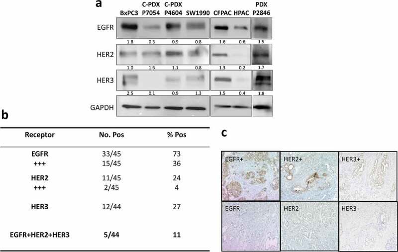
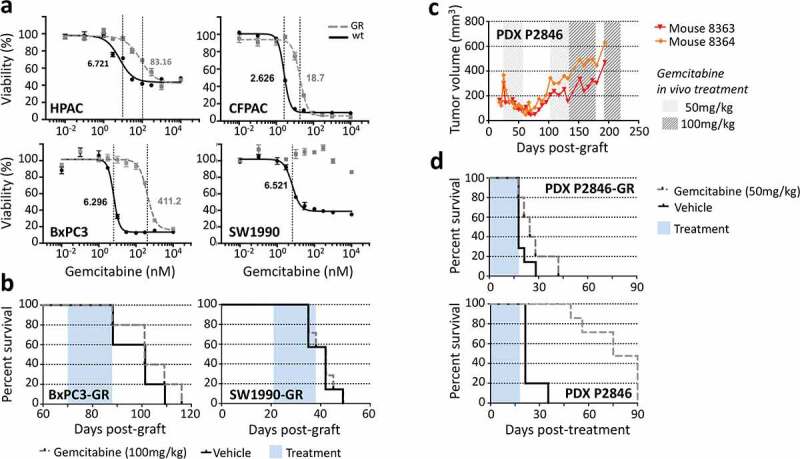
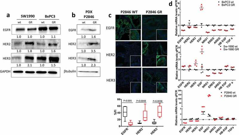
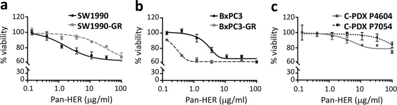
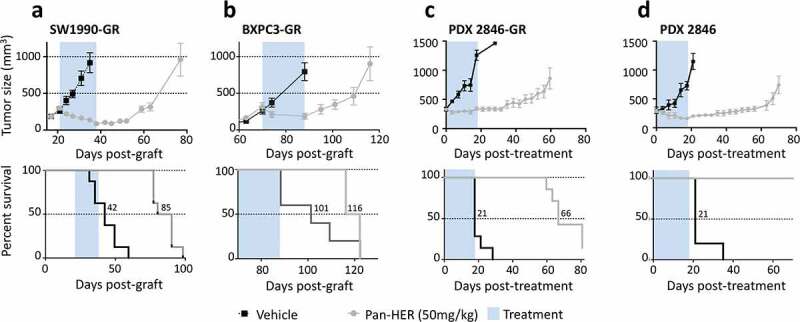
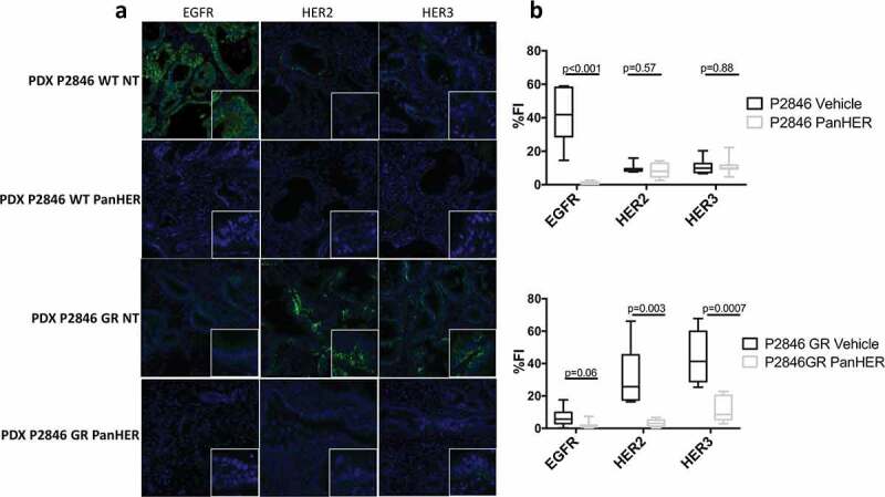
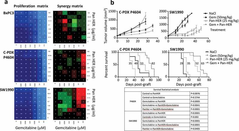
Chemoresistance, particularly to gemcitabine, is a major challenge in pancreatic cancer. The epidermal growth factor receptor (EGFR) and human epidermal growth factor receptors 2 and 3 (HER2, HER3) are expressed in many tumors, and they are relevant therapeutic targets due to their synergistic interaction to promote tumor aggressiveness and therapeutic resistance. Cocktails of antibodies directed against different targets are a promising strategy to overcome these processes. Here, we found by immunohistochemistry that these three receptors were co-expressed in 11% of patients with pancreatic adenocarcinoma. We then developed gemcitabine-resistant pancreatic cancer cell models (SW-1990-GR and BxPC3-GR) and one patient-derived xenograft (PDX2846-GR) by successive exposure to increasing doses of gemcitabine. We showed that expression of EGFR, HER2 and HER3 was increased in these gemcitabine-resistant pancreatic cancer models, and that an antibody mixture against all three receptors inhibited tumor growth in mice and downregulated HER receptors. Finally, we demonstrated that the Pan-HER and gemcitabine combination has an additive effect in vitro and in mice xenografted with the gemcitabine-sensitive or resistant pancreatic models. The mixture of anti-EGFR, HER2 and HER3 antibodies is a good candidate therapeutic approach for gemcitabine-sensitive and -resistant pancreatic cancer.
Keywords: EGFR; HER2; HER3; Pan-Her; chemoresistance; gemcitabine; pancreatic cancer.
Figures
Similar articles
-
An ERBB1-3 Neutralizing Antibody Mixture With High Activity Against Drug-Resistant HER2+ Breast Cancers With ERBB Ligand Overexpression.J Natl Cancer Inst. 2017 Nov 1;109(11):djx065. doi: 10.1093/jnci/djx065. J Natl Cancer Inst. 2017. PMID: 29059433 Free PMC article.
-
In vivo imaging of therapy response to a novel pan-HER antibody mixture using FDG and FLT positron emission tomography.Oncotarget. 2015 Nov 10;6(35):37486-99. doi: 10.18632/oncotarget.6060. Oncotarget. 2015. PMID: 26460961 Free PMC article.
-
Intracellular KRAS-specific antibody enhances the anti-tumor efficacy of gemcitabine in pancreatic cancer by inducing endosomal escape.Cancer Lett. 2021 Jun 1;507:97-111. doi: 10.1016/j.canlet.2021.03.015. Epub 2021 Mar 17. Cancer Lett. 2021. PMID: 33744388
-
The Metastasis Suppressor, N-MYC Downstream-regulated Gene-1 (NDRG1), Down-regulates the ErbB Family of Receptors to Inhibit Downstream Oncogenic Signaling Pathways.J Biol Chem. 2016 Jan 15;291(3):1029-52. doi: 10.1074/jbc.M115.689653. Epub 2015 Nov 3. J Biol Chem. 2016. PMID: 26534963 Free PMC article.
-
Inhibiting signal transducer and activator of transcription-3 increases response to gemcitabine and delays progression of pancreatic cancer.Mol Cancer. 2013 Sep 11;12(1):104. doi: 10.1186/1476-4598-12-104. Mol Cancer. 2013. PMID: 24025152 Free PMC article.
Cited by
-
Design and selection of optimal ErbB-targeting bispecific antibodies in pancreatic cancer.Front Immunol. 2023 Apr 20;14:1168444. doi: 10.3389/fimmu.2023.1168444. eCollection 2023. Front Immunol. 2023. PMID: 37153618 Free PMC article.
-
HER2 Overexpression in Periampullary Tumors According to Anatomical and Histological Classification-A Systematic Review.J Pers Med. 2024 Apr 27;14(5):463. doi: 10.3390/jpm14050463. J Pers Med. 2024. PMID: 38793045 Free PMC article. Review.
-
HER3 in cancer: from the bench to the bedside.J Exp Clin Cancer Res. 2022 Oct 21;41(1):310. doi: 10.1186/s13046-022-02515-x. J Exp Clin Cancer Res. 2022. PMID: 36271429 Free PMC article. Review.
-
HER3-targeted therapy: the mechanism of drug resistance and the development of anticancer drugs.Cancer Drug Resist. 2024 Apr 29;7:14. doi: 10.20517/cdr.2024.11. eCollection 2024. Cancer Drug Resist. 2024. PMID: 38835349 Free PMC article. Review.
-
Generation of bispecific antibodies by structure-guided redesign of IgG constant regions.Front Immunol. 2023 Jan 10;13:1063002. doi: 10.3389/fimmu.2022.1063002. eCollection 2022. Front Immunol. 2023. PMID: 36703993 Free PMC article.
References
-
- Rahib L, Smith BD, Aizenberg R, Rosenzweig AB, Fleshman JM, Matrisian LM.. Projecting cancer incidence and deaths to 2030: the unexpected burden of thyroid.; liver.; and pancreas cancers in the United States. Cancer Res. 2014;74:2913–476. PMID: 24840647. doi:10.1158/0008-5472.CAN-14-0155. - DOI - PubMed
-
- Conroy T, Hammel P, Hebbar M, Ben Abdelghani M, Wei AC, Raoul JL, Choné L, Francois E, Artru P, Biagi JJ, et al. Canadian cancer trials group and the unicancer-GI–PRODIGE group. FOLFIRINOX or gemcitabine as adjuvant therapy for pancreatic cancer. N Engl J Med. 2018;379:2395–406. PMID: 30575490. doi:10.1056/NEJMoa1809775. - DOI - PubMed
-
- Gourgou-Bourgade S, Bascoul-Mollevi C, Desseigne F, Ychou M, Bouché O, Guimbaud R, Bécouarn Y, Adenis A, Raoul JL, Boige V, et al. Impact of FOLFIRINOX compared with gemcitabine on quality of life in patients with metastatic pancreatic cancer: results from the PRODIGE 4/ACCORD 11 randomized trial. J Clin Oncol. 2013;31(1):23–29. PMID: 23213101. doi:10.1200/JCO.2012.44.4869. - DOI - PubMed
Publication types
MeSH terms
Substances
Grants and funding
LinkOut - more resources
Full Text Sources
Other Literature Sources
Medical
Research Materials
Miscellaneous