. 2021 Jun 10:334:106-113.
doi: 10.1016/j.jconrel.2021.04.009. Epub 2021 Apr 16.

Advanced genetic engineering to achieve in vivo targeting of adenovirus utilizing camelid single domain antibody

Affiliations

Advanced genetic engineering to achieve in vivo targeting of adenovirus utilizing camelid single domain antibody

Myungeun Lee et al. J Control Release. .

Abstract

For the developing field of gene therapy the successful address of the basic requirement effective gene delivery has remained a critical barrier. In this regard, the "Holy Grail" vector envisioned by the field's pioneers embodied the ability to achieve efficient and specific in vivo gene delivery. Functional linkage of antibody selectivity with viral vector efficiency represented a logical strategy but has been elusive. Here we have addressed this key issue by developing the technical means to pair antibody-based targeting with adenoviral-mediated gene transfer. Our novel method allows efficient and specific gene delivery. Importantly, our studies validated the achievement of this key vectorology mandate in the context of in vivo gene delivery. Vectors capable of effective in vivo delivery embody the potential to dramatically expand the range of successful gene therapy cures.

Keywords: Adenoviral vectors (Ad); CD276 [B7-H3]; Camelid single domain antibody (sdAb); Gene delivery; Human epithelial ovarian cancer cell (SKOV3.ip1); Ovarian Cancer (OvCa) xenograft mouse model.


Conflict of interest statement

Declaration of Competing Interest

No potential conflicts of interest were disclosed.

Figures

Fig. 1.
Fig. 1.
Schema to accomplish in vivo targeting via the CD276 axis exploiting camelid single domain antibody (sdAb) incorporated into the adenoviral vector capsid. Hexon modification is based on chimerism of the hypervariable region 7 (HVR7) whereby the HVR7 of human adenovirus serotype 3 substitutes for the corresponding domain in human adenovirus serotype 5. This modification is employed to mitigate liver sequestration of in vivo delivered adenovirus vector. To modify vector particle primary binding to target cells a strategy of fiber replacement is employed. A fiber chimera consisting of fiber tail from huAd5 is fused with fibritin from T4 Pol and an anti-CD276 sdAb that is tagged with 6 Histidine (6H).
Fig. 2.
Fig. 2.
Confirmation of capsid incorporation of engineered fiber with anti-CD276 sdAb. A. Gene and fiber map of fiber chimera designed for Ad retargeting. Fiber tail from huAd5 is fused in series with fibritin derived from T4 Pol and anti-CD276 sdAb with COOH terminus 6 Histidine tag (6H). B. Western blot analysis of engineered adenovirus designed for targeting via CD276 axis. Purified virus engineered to incorporate chimeric fiber with anti-CD276 sdAb (5 × 10⁹ v.p.) were subjected to SDS-PAGE followed by western blot analysis using antibodies to Ad5 fiber tai mAb (anti-4D2 Ab) or anti-CD276 sdAb (anti-tetra His Ab), respectively. Analysis was carried out as follows: Lane 1 – Ad5 with incorporated irrelevant control sdAb [Ad5ff.(−)sdAb-CD276.H5/H3]; Lane 2 – Ad5 with incorporated anti-CD276 [Ad5ff.(+)sdAb-CD276.H5/H3]; Lane 3 – Ad5 primary staining was with antifiber tail antibody or anti-sdAb antibody. Secondary staining was with anti-mouse antibodies conjugated to alkaline phosphate.
Fig. 3.
Fig. 3.
Targeted gene delivery in vitro via CD276 axis by adenoviral vector incorporating anti-CD276 sdAb to modified CHO cell lines. A. The virus infection efficiency was evaluated in CHO (Chinese Hamster Ovary) cell lines (CHO-human CD276 over expressed and CHO-mouse CD276 over expressed cell line). These cell lines were kindly provided by Dr. Brad St. Croix]. The cells were infected with indicated virus [Ad5 or Ad5ff. (+) sdAb-CD276.H5/H3] at a multiplicity of infection (MOI) of 100, and EGFP expression were imaged on two days (48 h) after virus infection using fluorescence microscopy. B. Quantification of in vitro gene transfer utilizing luciferase assay. The functionality of the targeted virus was verified by measuring gene transfer efficiency by quantitative analysis of luciferase activity. The sdAb-CD276 targeted virus was evaluated for mediating Ad vector selectivity of the targeted adenoviral vector [Ad5ff. (+) sdAb-CD276.H5/H3] in human CD276 overexpressing CHO cells (CHO-hCD276). The efficiency was verified with different MOIs (MOI; 200, 500, 1000, 2000 or 5000) by measuring of firefly luciferase gene activity (RLU: relative light units) at 72 h post infection.
Fig. 4.
Fig. 4.
Targeted gene delivery to human tumor cells via CD276 axis by tropism modified adenovirus. A. Expression of CD276 in human tumor cell lines. Various human cancer cell lines (including ovarian and breast cancer cells [e.g. SKOV3.ip1 or MDA-MB-231]) were assessed to check the hCD276 gene expression using human CD276 primary antibody by western blotting analysis. B. Gene delivery to human ovarian cancer cell line via targeted and non-targeted adenoviral vectors. Evaluating of the CD276-mediated gene transfer efficiency of the targeted adenoviral vector [Ad5ff. (+) sdAb-CD276.H5/H3] and non-targeted Ad [Ad5ff. (−) sdAb-CD276.H5/H3] in the human ovarian cancer cells (SKOV3.ip1). The relative efficiency was evaluated with different MOIs (MOI; 200, 500 or 1000) by measuring of firefly luciferase gene activity (RLU: relative light units) at 48 h post infection.
Fig. 5.
Fig. 5.
Evaluation of in vivo gene delivery of CD276-targeted adenoviral vectors in murine orthotopic xenograft model of human ovarian cancer. A. Bioluminescence imaging (BLI) of mice given i.p. vector encoding the luciferase gene reporter. Indicated vectors were untargeted Ad5 control [Ad5 (untargeted)], Ad5 with capsid incorporated chimeric fiber with irrelevant sdAb [Ad5ff. (−) sdAb-CD276.H5/H3], or Ad5 with capsid incorporated chimeric fiber with anti-CD276 sdAb [Ad5ff. (+) sdAb-CD276.H5/H3]. B. Quantification of in vivo gene transfer utilizing BLI image analysis. Quantification of total flux was measured from indicated mice body regions of interest (ROIs) via Living Image 2.6 software. Data are expressed as BLI bioluminescence (photons/s/cm2/sr), mean ± SD) (n = 3–4 animals per each group) statistical analysis was via analysis of variance (ANOVA) using GraphPad Prism (La Jolla, CA, USA).
Fig. 6.
Fig. 6.
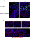
Analysis of targeted versus ectopic in vivo gene delivery of adenoviral vectors in human ovarian cancer model. A. The ectopic gene transduction in vivo with sdAb-CD276 targeted adenoviral vector was analyzed in the tissues (liver and tumor) using histopathological image analysis. OvCa tumor (SKOV3.ip1) bearing mice were intraperitoneally injected with 1 × 10¹¹ vp of viruses (each of untargeted Ad5 or CD276 targeted [Ad5ff. (+) sdAb-CD276.H5/H3]) via i.p injection, and liver and tumor (omentum tissues) were harvested at three days for immunohistochemistry staining (IHC) analysis. Blue: nuclei (stained with Hoechst 33258), Green: GFP signal through untargeted Ad5-EGFP vector, and Red: RFP signal through targeted Ad vector [Ad5ff. (+) sdAb-CD276.H5/H3], the transgene was detected with anti-firefly luciferase (Luc) antibody and used Alexa Fluor 594-conjugated secondary antibodies (in red signal). B. Immunofluorescence analysis of Ad localization in ovarian tumors and other organs (Liver, Spleen and Small Intestine). The images were taken using epifluorescence microscopy (Olympus America, Center Valley, PA). Blue: nuclei (stained with Hoechst 33258) and Red: signal through targeted Ad vector [Ad5ff. (+) sdAb-CD276.H5/H3], the transgene was detected with anti-firefly luciferase (Luc) antibody. All assay were conducted and analyzed in parallel conditions with Fig. 6A. (For interpretation of the references to colour in this figure legend, the reader is referred to the web version of this article.)
Fig. 7.
Fig. 7.
Evaluation of specificity of in vivo gene delivery accomplished by CD276 targeted adenoviral vector. A. The targeting specificity of in vivo gene delivery was verified in the context of vascular ECs. The tissue slices were co-stained with vascular EC markers CD31 and endomucin (Red), combined with luciferase gene (Green) using each of specific primary antibodies for Immunohistochemistry fluorescence analysis. The images were taken using epifluorescence microscopy (Olympus America, Center Valley, PA). The transduced gene expression via CD276 targeting adenoviral vector was verified by reporter gene (firefly luciferase gene), that was evaluated the co-localization signal in the tumor vascular ECs with combined signal. Reporter gene has shown co-localized with vascular ECs in yellow (Merged). Blue: nuclei (stained with Hoechst 33258), Red: vascular ECs (CD31/endomucin), Green: reporter gene (acquisition of firefly luciferase gene with anti-firefly luc Abs), and Yellow: merged (reporter gene expression in vascular ECs). The non-targeting adenoviral vector [Ad5ff. (−) sdAb-CD276] was compare as a control, both adenoviral vectors were shown in liver un-targeting via H5/H3 hexon modification (liver data shown only in targeting Ad [Adff. (+) sdAb-CD276]). B. The targeting specificity of in vivo gene delivery was also confirmed in the context of a human CD276 (hCD276) expressing tumor. The tissue slices were immunofluorescently co-stained using primary antibodies for CD276 (human) [Red] and luciferase reporter protein (Luc) [Green] and are shown along with a merged signal [Yellow]. Note the co-localization in the targeted Ad [Adff. (+) sdAb-CD276]) tissue. All assay were conducted and analyzed in parallel conditions with Fig. 7A. (For interpretation of the references to colour in this figure legend, the reader is referred to the web version of this article.)

Similar articles

Cited by

References

    1. Crystal RG, Adenovirus: the first effective in vivo gene delivery vector, Hum. Gene Ther 25 (2014) 3–11. - PMC - PubMed
    1. Wold WS, Toth K, Adenovirus vectors for gene therapy, vaccination and cancer gene therapy, Curr. Gene Ther 13 (2013) 421–433. - PMC - PubMed
    1. Lukashev AN, Zamyatnin AA Jr., Viral vectors for gene therapy: current state and clinical perspectives, Biochemistry 81 (2016) 700–708. - PubMed
    1. Gao J, Mese K, Bunz O, Ehrhardt A, State-of-the-art human adenovirus vectorology for therapeutic approaches, FEBS Lett. 593 (2019) 3609–3622. - PubMed
    1. Keeler AM, ElMallah MK, Flotte TR, Gene therapy 2017: progress and future directions, Clin. Transl. Sci 10 (2017) 242–248. - PMC - PubMed

Publication types

Substances