Review
. 2021 Jun;40(2):603-624.
doi: 10.1007/s10555-021-09960-8. Epub 2021 Apr 19.

Rethinking the biology of metastatic melanoma: a holistic approach

Affiliations
Review

Rethinking the biology of metastatic melanoma: a holistic approach

Hendrik Hld Vandyck et al. Cancer Metastasis Rev. 2021 Jun.

Abstract

Over the past decades, melanoma-related mortality has remained nearly stable. The main reason is treatment failure of metastatic disease and the inherently linked knowledge gap regarding metastasis formation. In order to elicit invasion, melanoma cells manipulate the tumor microenvironment, gain motility, and adhere to the extracellular matrix and cancer-associated fibroblasts. Melanoma cells thereby express different cell adhesion molecules like laminins, integrins, N-cadherin, and others. Epithelial-mesenchymal transition (EMT) is physiological during embryologic development, but reactivated during malignancy. Despite not being truly epithelial, neural crest-derived malignancies like melanoma share similar biological programs that enable tumorigenesis, invasion, and metastasis. This complex phenomenon is termed phenotype switching and is intertwined with oncometabolism as well as dormancy escape. Additionally, it has been shown that primary melanoma shed exosomes that create a favorable premetastatic niche in the microenvironment of secondary organs and lymph nodes. Although the growing body of literature describes the aforementioned concepts separately, an integrative holistic approach is missing. Using melanoma as a tumor model, this review will shed light on these complex biological principles in an attempt to clarify the mechanistic metastatic pathways that dictate tumor and patient fate.

Keywords: Dormancy; Epithelial-mesenchymal transition; Melanoma; Metastasis; Phenotype switching; Warburg effect.


Conflict of interest statement

The authors declare no competing interests.

Figures

Fig. 1
Fig. 1
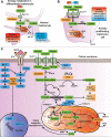
Phenotype switching in melanoma. Proposed model of phenotype switching in melanoma with integrative reciprocity of dormancy, metabolic reprogramming, and role of the TME. Phenotype switching is orchestrated by EMT-TFs and involves MITFlow and MITFhigh interchangeable states that provide context-dependent malignant potential. Note that multiple intermediate states exist and that phenotype switching in melanoma is not a binary process, just as EMT/MET is only partial in carcinogenesis. The recently discovered distinct transcriptional melanoma cell states include: undifferentiated, NCSC, intermediate, SMC, melanocyte-like and hyperdifferentiated/pigmented state. The different melanoma cell states predominantly are the result of epigenetic induction that contribute to plasticity, reversibility and therapy resistance. Adopted from [5, 7, 27]. Abbreviations: EMT, epithelial-mesenchymal transition; MET, mesenchymal epithelial transition; MITF, microphtalmia transcription factor; NCSC, neural crest stem cell; NF-1, neurofibromin 1; RTK, receptor tyrosine kinase; SMC, starved melanoma cell; TF, transcription factor
Fig. 2
Fig. 2
Cell-cell and cell-matrix adhesion of keratinocytes and melanocytes in normal skin versus melanoma. A) Intercellular contacts in epidermis with Ai) epithelial-epithelial and epithelial-basal membrane connections of keratinocytes (orange) and Aii) contacts of epidermal melanocytes (purple) with keratinocytes and basal membrane. Normal epidermal melanocytes interact with adjacent keratinocytes through E-cadherin, desmoglein 1, and gap junctions, which are formed by two connexons. B) Alterated adhesion pathways in melanoma with gain of motility during invasion. The first step in melanoma development is loss of connections between melanocytes with keratinocytes and basal membrane. Melanoma cells escape keratinocyte control and instead interact with Bi) fibroblasts (brown), Bii) other melanoma cells (purple), or Biii) endothelial cells (red), mainly during vertical growth phase that elicits the metastatic potential. Desmoglein 1 and other connections are disrupted while new adhesive and communication properties are conferred. Melanoma cells can express FN1 and the intermediate filament vimentin. They interact with fibroblasts Bi) through N-cadherin, FN1, gap junctions, and with other melanoma cells Bii) through αvβ3-integrin, L1-CAM, MUC18/MCAM, L1-CAM, gap junctions and N-cadherin. Transendothelial migration is mediated by adhesion of melanoma cells with endothelial cells Biii) through N-cadherin, MUC18/MCAM, MCAM ligand, α4β1-integrin, VCAM, αvβ3-integrin, and L1-CAM. Adopted from [–91]. Abbreviations: BM, basement membrane; BRN2 = POU3F2, POU domain, class 3, transcription factor 2; CAF, cancer-associated fibroblast, FN1, fibronectin 1; L1-CAM, L1-cell adhesion molecule; MCAM, melanocyte cell adhesion molecule; OXPHOS, oxidative phosphorylation; SNAIL, Snail family transcriptional repressor 1; SLUG: Snail family transcriptional repressor 2; TWIST1 = TWIST, Twist family bHLH transcription factor 1; VCAM, vascular cell adhesion molecule; ZEB1, zinc finger E-box binding homeobox 1; ZEB2, zinc finger E-box binding homeobox 2
Fig. 3
Fig. 3
Metabolic reprogramming and the Warburg effect in melanoma. A) Metabolism in normal melanocyte with depiction of glycolysis, TCA cycle and OXPHOS. Glucose enters the cytoplasm via GLUT-1 and undergoes glycolysis to pyruvate. After entering the mitochondrium, the enzyme PDH converts pyruvate to Ac-CoA, which is metabolized and burnt in the TCA-cycle and during OXPHOS. Anaerobic glycolysis results in less efficient ATP generation along with lactate production during hypoxia. Lactate can leave the cytoplasm via MCTs. B) The Warburg effect refers to the metabolic switch in highly proliferating cells. Somewhat counterintuitive, these cells prefer less efficient ATP generating glycolysis as main metabolic pathway despite the presence of oxygen (i.e., aerobic glycolysis). C) Warburg effect is exploited by rapidly proliferating melanoma cells when nutrients are abundant. This is mediated by multiple mechanisms that result in increased glucose uptake, accelerated glycolytic flux and decreased mitochondrial respiration. Glycolytic intermediate metabolites can be recycled and synthesized into macromolecules for synthesis of DNA, lipids and cellular proteins that are needed for proliferation. Adopted from [128]. Abbreviations: Ac-CoA, acetyl coenzyme A; ATP, adenosine triphosphate; GLUT, glucose transporter; HIF1α, hypoxia-inducible factor alpha; MCT, monocarboxylate transporter; PDH, pyruvate dehydrogenase; PDK; PDH kinase; ROS, reactive oxygen species; TCA, tricarboxylic acid
Fig. 4
Fig. 4
Metabolic symbiosis as a result of nutrient trade-off between different melanoma cell subsets and adjacent CAFs. Stromal symbiosis is the result of (unidirectional) nutrient trade-off by CAFs and melanoma cells (reverse Warburg effect). This is mediated by aerobic glycolysis in adjacent CAFs that produce metabolites like lactate, pyruvate and ketone bodies. These metabolites are shuttled through MCTs to sustain the anabolism of adjacent melanoma cells. An additional effect of the exploited lactate shuttling is the symbiosis between better and worse oxygenated melanoma cells. Hypoxic melanoma cells produce more lactate that is preferentially taken up and metabolized by better oxygenated melanoma cells. The latter in turn spare the glucose for hypoxic melanoma cells. Adopted from [15, 19]. Abbreviations: Ac-CoA, acetyl coenzyme A; ATP, adenosine triphosphate; CAF, cancer-associated fibroblast; GLUT, glucose transporter; HIF1α, hypoxia-inducible factor 1 alpha; MCT, monocarboxylate transporter; PDH, pyruvate dehydrogenase; PDK, PDH kinase; ROS, reactive oxygen species, TCA, tricarboxylic acid
Fig. 5
Fig. 5
The role of dormancy in melanoma. The majority of disseminated melanoma cells die (A); however, a fraction possesses the potential to adapt to various new environments, followed either by early recurrence (B) with metastasis or induction of dormancy (C), which can yield in late recurrence (D) of metastatic disease. At the primary tumor aggressive subclones can dedifferentiate to mimic vascular channels, delined by melanoma cells that express VE-cadherin in a process named vasculogenic mimicry. After dissemination, invasive melanoma cells can migrate intra- or perivascularly and as single cells or cell clusters. The vascular niche comprises the non-mutually exclusive extravascular and intravascular niche. The former involves the migration of melanoma cells via the abluminal side of pre-existing vascular structures via extravascular migration (EVM). Eventually this leads to metastatic outgrowth at secondary sites through extravascular migratory metastasis (EVMM). Dormancy can be subdivided in cellular dormancy and tumor mass dormancy due to angiostasis (angiogenic dormancy) or immuno surveillance (immunogenic dormancy). Dormant melanoma cells can reside for years at their dormant niches and potentially transdifferentiate to endothelial cells by endothelial transition (EndT), where melanocytic markers are lost and the endothelial marker CD31 is gained. Intrinsic and/or extrinsic factors ultimately induce an outbreak from dormancy, thereby promoting a clinically visible and/or symptomatic metastasic state of the disease. The essential prerequisite for metastasis is that the surrounding new TME allows the adapted or adaptive melanoma cells to survive. Shedded exosomes help to create a cancer-friendly secondary TME which ultimately leads to organotropism. Escape from dormancy is the result of immune evasion, angiogenic switch and/or EndMT and will ultimately lead to a metastatic state of the disease. Finally, in the (macro)metastatic state, metabolic alterations of melanoma cells and their TME such as the (reverse) Warburg effect become significant. Abbreviations: CAF, cancer-associated fibroblast; CTC, circulation tumor cells; ECM, extracellular matrix; DTC, disseminated tumor cells; EVM, extravascular migration; EVMM, extravascular migratory metastasis; EndT, endothelial transition; EndMT, endothelial mesenchymal transition, VE-cadherin, vascular-endothelial cadherin

Similar articles

Cited by

References

    1. Carr S, Smith C, Wernberg J. Epidemiology and risk factors of melanoma. The Surgical Clinics of North America. 2020;100(1):1–12. doi: 10.1016/j.suc.2019.09.005. - DOI - PubMed
    1. Yun S, Vincelette ND, Green MR, Wahner Hendrickson AE, Abraham I. Targeting immune checkpoints in unresectable metastatic cutaneous melanoma: A systematic review and meta-analysis of anti-CTLA-4 and anti-PD-1 agents trials. Cancer Medicine. 2016;5(7):1481–1491. doi: 10.1002/cam4.732. - DOI - PMC - PubMed
    1. Pisco AO, Huang S. Non-genetic cancer cell plasticity and therapy-induced stemness in tumour relapse: ‘What does not kill me strengthens me’. British Journal of Cancer. 2015;112(11):1725–1732. doi: 10.1038/bjc.2015.146. - DOI - PMC - PubMed
    1. Huang S. Tumor progression: chance and necessity in Darwinian and Lamarckian somatic (mutationless) evolution. Progress in Biophysics and Molecular Biology. 2012;110(1):69–86. doi: 10.1016/j.pbiomolbio.2012.05.001. - DOI - PubMed
    1. Rambow F, Rogiers A, Marin-Bejar O, Aibar S, Femel J, Dewaele M, et al. Toward minimal residual disease-directed therapy in melanoma. Cell. 2018;174(4):843–855.e819. doi: 10.1016/j.cell.2018.06.025. - DOI - PubMed

Publication types