Review
. 2021 Mar 30:12:639602.
doi: 10.3389/fgene.2021.639602. eCollection 2021.

The Role of Histone Lysine Methylation in the Response of Mammalian Cells to Ionizing Radiation

Affiliations
Review

The Role of Histone Lysine Methylation in the Response of Mammalian Cells to Ionizing Radiation

Elena Di Nisio et al. Front Genet. .

Erratum in

Abstract

Eukaryotic genomes are wrapped around nucleosomes and organized into different levels of chromatin structure. Chromatin organization has a crucial role in regulating all cellular processes involving DNA-protein interactions, such as DNA transcription, replication, recombination and repair. Histone post-translational modifications (HPTMs) have a prominent role in chromatin regulation, acting as a sophisticated molecular code, which is interpreted by HPTM-specific effectors. Here, we review the role of histone lysine methylation changes in regulating the response to radiation-induced genotoxic damage in mammalian cells. We also discuss the role of histone methyltransferases (HMTs) and histone demethylases (HDMs) and the effects of the modulation of their expression and/or the pharmacological inhibition of their activity on the radio-sensitivity of different cell lines. Finally, we provide a bioinformatic analysis of published datasets showing how the mRNA levels of known HMTs and HDMs are modulated in different cell lines by exposure to different irradiation conditions.

Keywords: DNA damage; DNA repair; HPTMs; histone methylation; ionizing radiation.


Conflict of interest statement

The authors declare that the research was conducted in the absence of any commercial or financial relationships that could be construed as a potential conflict of interest.

Figures

FIGURE 1
FIGURE 1
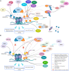
Pre-existing chromatin features and damage-induced histone modifications regulate the DNA repair pathways. The set of changes of HPTMs coordinately orchestrates the DDR around the damaged site, reorganizing chromatin structure in order to limit local transcription and to promote the DNA repair through the recruitment, the action and the retention of sensors, transducers and effectors of damage signaling cascade. In particular, methylation dynamics at different lysine residues, preferentially on H3 and H4 histone tails, regulate the response to DNA damage both temporally and spatially. Indeed, several histone methylation changes occur on H3K4, H3K9, H3K27, H3K36, H3K79, H4K16, H4K20, and H2AXK134 to aid in DNA damage repair. Lysine methylation is a highly dynamic histone modification owing to the interplay between KMTs (Lysine methyltransferases) and KDMs (Lysine demethylases). After the induction of genotoxic damage by ionizing radiation (IR), several KMTs and KDMs promote the formation of a chromatin landscape prone to DNA repair, acting in synergy or antagonistically to favor the HR and/or NHEJ pathway. Indeed, some demethylases and methyltransferases act at DSB sites, thus directly regulating the recruitment of DDR factors; the demethylase/methyltransferase activity of these epigenetic modifiers modulates the DDR not only through a direct role (non-dashed lines), but also indirectly (dashed lines) by regulating the expression of such DSB repair factors, acting at their promoter regions. The recruitment of some of these enzymes, including KDM5B, is specific and requires the presence of histone variant macroH2A1.1 and PARylation by PARP1. In other cases, such as KMT8, the recruitment arises thanks to the interaction with the histone variant macroH2A1.2 without PARylation; alternatively, the recruitment can take place thanks to reader domains which recognize specific histone marks (see also Figure 3). Created using BioRender.
FIGURE 2
FIGURE 2
KDM enzymes may play different roles in the DNA Damage Response (DDR) depending on the genotoxic agent type. Upon IR treatment (1), PARP1 activity is required to recruit both KDM5A and KDM5B at DSB sites, where they catalyze the local demethylation of H3K4me2/3, crucial for the recruitment of transcriptional repressive complex like ZMYND8/NuRD or DDR key proteins, including BRCA1 and Ku70. Conversely, in response to alkylation damage by MMS (2), Poly-SUMOylated KDM5B is Poly-ubiquitylated by the SUMO-targeted ubiquitin ligase RNF4 and degraded by the proteasome, while JARID1C is Mono-SUMOylated and recruited to the chromatin to demethylate histone H3K4me2/3. Created using BioRender.
FIGURE 3
FIGURE 3
Histone modifications, dynamically added or removed around the DNA damage site, constitute an integrated signal for the DSBs repair pathway choice. (A) 53BP1 is a master regulator of DNA double-strand break repair pathway choice toward NHEJ and its binding to chromatin is mainly regulated by specific histone modifications, comprising H2A ubiquitylation and H3 and H4 methylation. Indeed, specific HPTMs can generate a docking site for 53BP1, which binds the ubiquitinated H2A/H2A.X K13/15/119 through its UDR motif and H4K20me2, H4K16me1, and H3K79me2 through its tandem Tudor domain. Therefore, despite H4K20me2 being the main histone mark involved in 53BP1 recruitment, other marks may contribute to it. For example, the deacetylation of H4K16 by HDAC1/2 indirectly promotes the 53BP1 recruitment and the NHEJ over HR, because it makes possible the methylation of this lysine residue. Under normal conditions, the H4K20me2 mark is masked by various interacting proteins, including L3MBTL1 and KDM4A/JMJD2A. Upon DNA DSBs induction, the ATM phosphorylation cascade promotes the recruitment of the E3 ubiquitin ligase RNF8 to the DNA damage site, which, together with the E2 ubiquitin ligase UBC13, promotes the relocation of the E3 ubiquitin ligase RNF168 to the DNA break sites. LSD1 may favor the 53BP1 recruitment interacting with RNF168 and/or thanks to a possible epigenetic cross-talk between H3K4 demethylation and H2A ubiquitylation. RNF168 catalyzes in turn the H2A K13/15 mono-ubiquitination and the poly-ubiquitination of L3MBTL1 and KDM4A, leading to RNF8-dependent degradation of KDM4A and to VCP-mediated and ubiquitin-dependent removal of L3MBTL1 from chromatin, favoring the unmasking of H4K20me2 and the loading of 53BP1 around the damaged site. LSD1 can also disadvantage 53BP1 recruitment, negatively regulating the interaction between p53 and 53BP1 (not shown). After DSB induction by IR, the H3K36me2 markedly increases, improving the association of early NHEJ factors such as Ku70/Ku80 and NBS1 at the damage site. Once activated, ATM (not shown) may negatively regulate KDM2A chromatin-binding capacity, favoring its displacement, which favors an increase of H3K36 di-methylation mark near the DNA damage sites by SETMAR. H3K36me3 can favor the retention of the NHEJ-associated factor PHF1, which is recruited in a Ku70/Ku80 dependent way and which supports an open chromatin for the efficient DNA repair. In contrast to H3K36me2, H3K36me3 regulates both NHEJ and HR, probably depending on the relative abundance of its reader factors and/or by the chromatin context. In this regard, pre-existing transcription-associated and cell cycle-regulated histone modifications together with those damage-induced regulate the DNA repair pathways. (B) The pre-existing H3K36me3 mark at actively transcribed gene bodies favors DNA repair by HR pathway choice: in human cells, both LEDGF (Lens epithelium-derived growth factor), via its PWWP reader domain, and MRG15, via its chromodomain, can bind H3K36me3, promoting the recruitment of CtIP and PALB2, two crucial factors for the DNA end-resection and strand invasion, respectively. Moreover, mutually exclusive histone modifications can contribute to tip the balance between HR and NHEJ in DSB repair pathway choice: a switch between ubiquitylation and acetylation on H2AK15 and H4K16 can favor HR over NHEJ. The human NuA4/TIP60 acetyltransferase complex is able to inhibit the 53BP1 recruitment both through this mechanism and by targeting the same H4K20me2 mark, by binding H4K20me with the MBT (malignant brain tumor) domain of MBTD1, its stable subunit. Moreover, the human NuA4/TIP60 acetyltransferase complex promotes the repositioning of 53BP1 through other mechanisms, such as the acetylation of H2A/H2AX K13/15 that blocks the recruitment of RNF8/168 and through the acetylation of H4K16 that impedes the methylation of this lysin residue, limiting the recruitment of 53BP1. KDM4A, besides masking H3K20me2 and limiting the 53BP1 recruitment thus inhibiting the NHEJ pathway, negatively regulates the DDR also removing H3K36me3 mark thus inhibiting the HR pathway. Conversely, KMT3A favors the HR through the methylation of H3K36. KMT1E favors the HR though the methylation of H3K9. Indeed, the H3K9me2 mark is involved in the recruitment of NuA4/TIP60 acetyltransferase complex, and H3K9me3 is involved in the recruitment of BARD1/BRCA1 Ub ligase activity on H2AK127 favoring the DNA end-resection, the repositioning of 53BP1 and thus the HR. Created using BioRender.
FIGURE 4
FIGURE 4
Expression levels of the HMTs and HDMs in response to ionizing radiation. (A) Time course of the modulation of all the HMTs and HDMs which are reported on Supplementary Table 1 and of the DDR genes ATM, BRCA2, and CDKN1A after particle irradiation. Data for KDM5B are reported for particle irradiation (B) and for X-ray (C). Statistical analysis of experimental data was performed using analysis of variance (ANOVA) followed by multiple comparison Tukey’s test. The data was retrieved from the NCBI Gene Expression Omnibus (GEO) public repository from these datasets: GSE23901, GSE23903, GSE32091, GSE21059, (Ghandhi et al., 2011) GSE23807, GSE30240 (Rashi-Elkeles et al., 2011). All statistical analysis was carried out using native functions of R language version 3.6.3. Expression levels reported are the Log2(IR/control) values. **P < 0.01 and *P < 0.05.

Similar articles

Cited by

References

    1. Acs K., Luijsterburg M. S., Ackermann L., Salomons F. A., Hoppe T., Dantuma N. P. (2011). The AAA-ATPase VCP/p97 promotes 53BP1 recruitment by removing L3MBTL1 from DNA double-strand breaks. Nat. Struct. Mol. Biol. 18 1345–1350. 10.1038/nsmb.2188 - DOI - PubMed
    1. Agarwal P., Jackson S. P. (2016). G9a inhibition potentiates the anti-tumour activity of DNA double-strand break inducing agents by impairing DNA repair independent of p53 status. Cancer Lett. 380 467–475. 10.1016/j.canlet.2016.07.009 - DOI - PMC - PubMed
    1. Alagoz M., Katsuki Y., Ogiwara H., Ogi T., Shibata A., Kakarougkas A., et al. (2015). SETDB1, HP1 and SUV39 promote repositioning of 53BP1 to extend resection during homologous recombination in G2 cells. Nucleic Acids Res. 43 7931–7944. 10.1093/nar/gkv722 - DOI - PMC - PubMed
    1. Alonso-de Vega I., Paz-Cabrera M. C., Rother M. B., Wiegant W. W., Checa-Rodríguez C., Hernández-Fernaud J. R., et al. (2020). PHF2 regulates homology-directed DNA repair by controlling the resection of DNA double strand breaks. Nucleic Acids Res. 48 4915–4927. 10.1093/nar/gkaa196 - DOI - PMC - PubMed
    1. Amundson S. A., Bittner M., Meltzer P., Trent J., Fornace A. J. (2001). Induction of gene expression as a monitor of exposure to ionizing radiation. Radiat. Res. 156 657–661. - PubMed

LinkOut - more resources