. 2021 Jun;27(6):710-724.
doi: 10.1261/rna.078746.121. Epub 2021 Apr 14.

Ago2 protects Drosophila siRNAs and microRNAs from target-directed degradation, even in the absence of 2'- O-methylation

Affiliations

Ago2 protects Drosophila siRNAs and microRNAs from target-directed degradation, even in the absence of 2'- O-methylation

Elena R Kingston et al. RNA. 2021 Jun.

Erratum in

Abstract

Target-directed microRNA (miRNA) degradation (TDMD), which is mediated by the protein ZSWIM8, plays a widespread role in shaping miRNA abundances across bilateria. Some endogenous small interfering RNAs (siRNAs) of Drosophila cells have target sites resembling those that trigger TDMD, raising the question as to whether they too might undergo such regulation by Dora, the Drosophila ZSWIM8 homolog. Here, we find that some of these siRNAs are indeed sensitive to Dora when loaded into Ago1, the Argonaute paralog that preferentially associates with miRNAs. Despite this sensitivity when loaded into Ago1, these siRNAs are not detectably regulated by target-directed degradation because most molecules are loaded into Ago2, the Argonaute paralog that preferentially associates with siRNAs, and we find that siRNAs and miRNAs loaded into Ago2 are insensitive to Dora. One explanation for the protection of these small RNAs loaded into Ago2 is that these small RNAs are 2'-O-methylated at their 3' termini. However, 2'-O-methylation does not protect these RNAs from Dora-mediated target-directed degradation, which indicates that their protection is instead conferred by features of the Ago2 protein itself. Together, these observations clarify the requirements for regulation by target-directed degradation and expand our understanding of the role of 2'-O-methylation in small-RNA biology.

Keywords: RNA methylation; TDMD; endogenous siRNAs; posttranscriptional regulation; siRNA dynamics; target-directed degradation.


Figures

FIGURE 1.
FIGURE 1.
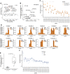
Methylated small RNAs are not susceptible to TDD. (A) Normalized abundance of the most highly expressed individual siRNAs in periodate-treated samples from wild-type (WT, dark green) and dora (light green) cells, as measured using sRNA-seq. Abundance is plotted as reads normalized to a cohort of abundant, Ago2-enriched, Dora-insensitive miRNAs (Supplemental Table S1). Shown are the results from three biological replicates for each genotype, each of which used a different clonal line. Significance was evaluated by a Welch two-sample t-test. (B) Changes in abundance of siRNAs mapping to annotated siRNA-generating loci observed upon loss of Dora in periodate-treated samples analyzed in A. Fold-changes in normalized abundance are plotted as a function of abundance observed in WT cells (RPKM, reads per kilobase per million mapped reads). Each point represents the mean from the three biological replicates, as determined by CuffDiff (Trapnell et al. 2012). The locus for which siRNAs significantly increased upon loss of Dora (CuffDiff adjusted P < 0.05) is indicated in red. (C) Changes in miRNA levels observed upon loss of Dora in periodate-treated samples analyzed A and B. Each point represents the mean from the three biological replicates, as determined by DESeq. Shown in red and labeled are results for Dora-sensitive miRNAs, identified as miRNAs that significantly increased (adjusted P < 2 × 10⁻⁷) upon loss of Dora in a different analysis that examined untreated, total-sRNA samples (Supplemental Fig. S1A; Shi et al. 2020). (D) Changes in miRNA levels observed upon loss of Dora, comparing results for total-sRNA (dark blue) and periodate-treated (dark green) samples for both Dora-sensitive and Dora-insensitive miRNAs. MicroRNAs were classified as Dora sensitive as in C. Significance was evaluated by a Welch two-sample t-test.
FIGURE 2.
FIGURE 2.
Small RNAs in Ago1 are susceptible to TDD. (A) Changes in levels of Ago1-associated miRNAs observed upon loss of Dora, as measured using sRNA-seq. Analysis was as in Figure 1C, but for miRNAs that copurified with Ago1. Results for miRNAs that significantly increased upon loss of Dora (adjusted P < 10−20) in the Ago1 samples are colored red and labeled, indicating with triangles those previously identified as Dora-sensitive in analyses of total-RNA samples (Supplemental Fig. S1A; Shi et al. 2020). (B) Relationship between the increased Dora sensitivity observed when examining Ago1-associated miRNAs and miRNA enrichment in Ago2. Results are shown for Ago1-associated miRNAs that most significantly increased upon loss of Dora (red points, A). Ago2 enrichment was inferred by dividing the average fraction of miRNA reads corresponding to that miRNA in the periodate-treated libraries by the average fraction of miRNA reads corresponding to that miRNA in the untreated libraries. The increased Dora sensitivity was computed by subtracting the log2-transformed fold-change observed upon loss of Dora in total-sRNA samples from the log2-transformed fold-change observed upon loss of Dora in the Ago1 samples. (C) Normalized abundance of the most highly expressed individual siRNAs in Ago1 samples from wild-type (WT, dark orange) and dora (orange) S2 cells, as measured using sRNA-seq. Significant differences are indicated (*) P > 0.05. Abundance is plotted as reads normalized to a cohort of abundant, Ago1-enriched, Dora-insensitive miRNAs (Supplemental Table S1); otherwise, this panel is as in Figure 1A. (D) Tailing and trimming of Dora-sensitive (top row) and Dora-insensitive (bottom row) siRNAs that passed the expression cutoff (an average of >40 reads across the wild-type samples) in Ago1 samples from wild-type (WT, dark orange) and dora (orange) S2 cells. Fractional abundance was quantified as the fraction of reads that the isoform contributed to the total reads for that siRNA and is shown for mature isoforms (defined as the most abundant isoform in wild-type cells), tailed isoforms with one to three additional nucleotides (N1, N2, N3), and trimmed isoforms with one to three fewer nucleotides (T1, T2, T3). Each of the two genotypes was represented by three clonal lines, and average fractional abundance and standard deviation (error bars) are shown for each isoform. (E) Changes in the fractions of trimmed isoforms observed upon loss of Dora for either the Dora-sensitive or Dora-insensitive siRNAs shown in D. Changes for each siRNA were quantified by summing the differences between the fractional abundances in dora and wild-type S2 cells for each trimmed isoform. Significance was evaluated by a Welch two-sample t-test. (F) Normalized abundance of the most highly expressed individual siRNAs in total-sRNA samples from wild-type (WT, dark blue) and dora (blue) S2 cells, as measured using sRNA-seq. Otherwise, this panel is as in C.
FIGURE 3.
FIGURE 3.
Loss of methylation does not alter sensitivity to TDD. (A) Tailing and trimming of Ago2-associated small RNAs from wild-type (WT, green) and hen1 (red) S2 cells. Ago2-associated small RNAs were enriched by either periodate treatment (WT) or co-IP with FLAG-Ago2 (hen1) (Supplemental Fig. S3B). Otherwise, this panel is as in Figure 2D. (B) Normalized abundance of individual Ago2-associated siRNAs from hen1 and hen1/dora cells, as measured using sRNA-seq. Ago2-associated siRNAs were isolated by IP of FLAG-Ago2. Otherwise, this panel is as in Figure 1A. (C) Changes in levels of Ago2-associated miRNAs observed upon loss of Dora in hen1 cells. Ago2-associated miRNAs were isolated by IP of FLAG-Ago2. Each point represents the mean from three biological replicates, as determined by DESeq. Results for miRNAs sensitive to loss of Dora in wild-type cells are labeled (Supplemental Fig. S1A; Shi et al. 2020), and results for miRNAs significantly up-regulated (P < 10−3) upon Dora loss in these Ago2 samples are colored in red. (D) Changes in abundance of siRNAs mapping to annotated siRNA-generating loci observed upon loss of Hen1. Fold-changes in normalized abundance are plotted as a function of abundance observed in WT cells (RPKM, reads per kilobase per million mapped reads), as determined by CuffDiff. Differential expression was determined by DESeq, after normalizing to a cohort of abundant, Ago1-enriched, Dora-insensitive miRNAs (Supplemental Table S1). Each point represents the mean from the three biological replicates. The loci for which siRNAs significantly changed upon loss of Hen1 (DESeq adjusted P < 0.05) are indicated in red. (E) Normalized abundance of individual siRNAs in total-sRNA wild-type (WT), hen1, and hen1/dora samples. Note that the WT data are those of Figure 2D, replotted here for comparison, and that the normalization was performed as in Figure 2D. Significance was evaluated by ANOVA and the Tukey test.
FIGURE 4.
FIGURE 4.
Methylation does not protect an Ago1-loaded miRNA from TDD. (A) Decay of methylated (2′-O-Me) or nonmethylated (2′-OH) miR-7 transfected into wild-type and dora cells. Decay was monitored on a northern blot probed both for miR-7 and endogenous miR-11, which served as a loading control. (B) As in A, but in the nibbler background. (C) Quantification of the results in B and those of one additional biological replicate, which used independent clonal cell lines (Supplemental Fig. S4E). The lines show the best least-squares fits of an exponential-decay model. Half-lives, indicated in parentheses, encapsulated the rates of both dilution and decay.

Similar articles

Cited by

References

    1. Agarwal V, Subtelny AO, Thiru P, Ulitsky I, Bartel DP. 2018. Predicting microRNA targeting efficacy in Drosophila. Genome Biol 19: 152. 10.1186/s13059-018-1504-3 - DOI - PMC - PubMed
    1. Ameres SL, Horwich MD, Hung J-H, Xu J, Ghildiyal M, Weng Z, Zamore PD. 2010. Target RNA–directed trimming and tailing of small silencing RNAs. Science 328: 1534–1539. 10.1126/science.1187058 - DOI - PMC - PubMed
    1. Ameres SL, Hung J-H, Xu J, Weng Z, Zamore PD. 2011. Target RNA-directed tailing and trimming purifies the sorting of endo-siRNAs between the two Drosophila Argonaute proteins. RNA 17: 54–63. 10.1261/rna.2498411 - DOI - PMC - PubMed
    1. Baccarini A, Chauhan H, Gardner TJ, Jayaprakash AD, Sachidanandam R, Brown BD. 2011. Kinetic analysis reveals the fate of a microRNA following target regulation in mammalian cells. Curr Biol 21: 369–376. 10.1016/j.cub.2011.01.067 - DOI - PMC - PubMed
    1. Bartel DP. 2009. MicroRNAs: target recognition and regulatory functions. Cell 136: 215–233. 10.1016/j.cell.2009.01.002 - DOI - PMC - PubMed

Publication types

LinkOut - more resources