

MiR-30a and miR-200c differentiate cholangiocarcinomas from gastrointestinal cancer liver metastases
- PMID: 33852640
- PMCID: PMC8046207
- DOI: 10.1371/journal.pone.0250083
MiR-30a and miR-200c differentiate cholangiocarcinomas from gastrointestinal cancer liver metastases
Abstract
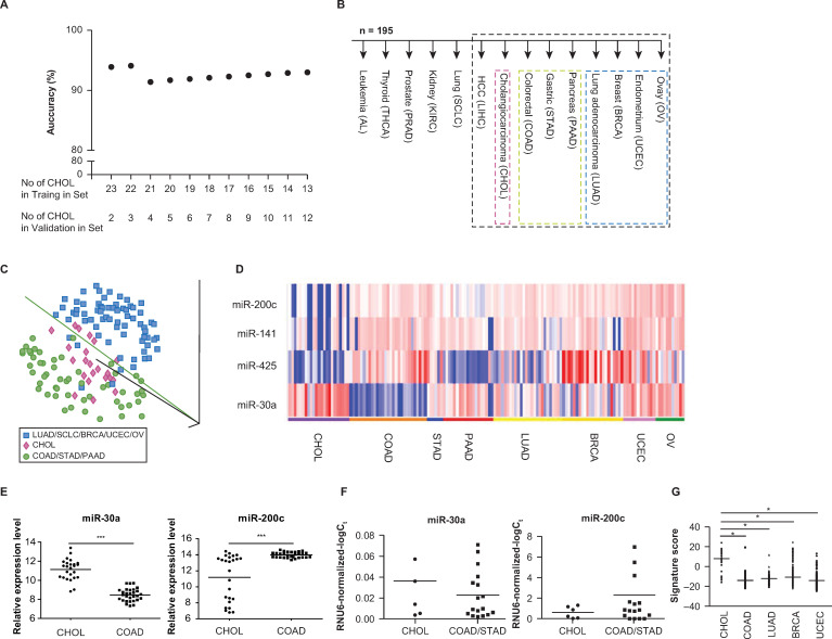
Prior studies have demonstrated the utility of microRNA assays for predicting some cancer tissue origins, but these assays need to be further optimized for predicting the tissue origins of adenocarcinomas of the liver. We performed microRNA profiling on 195 frozen primary tumor samples using 14 types of tumors that were either adenocarcinomas or differentiated from adenocarcinomas. The 1-nearest neighbor method predicted tissue-of-origin in 33 samples of a test set, with an accuracy of 93.9% at feature selection p values ranging from 10-4 to 10-10. According to binary decision tree analyses, the overexpression of miR-30a and the underexpression of miR-200 family members (miR-200c and miR-141) differentiated intrahepatic cholangiocarcinomas from extrahepatic adenocarcinomas. When binary decision tree analyses were performed using the test set, the prediction accuracy was 84.8%. The overexpression of miR-30a and the reduced expressions of miR-200c, miR-141, and miR-425 could distinguish intrahepatic cholangiocarcinomas from liver metastases from the gastrointestinal tract.
Conflict of interest statement
The authors have declared that no competing interests exist.
Figures
Similar articles
-
Transcriptomic profiling reveals hepatic stem-like gene signatures and interplay of miR-200c and epithelial-mesenchymal transition in intrahepatic cholangiocarcinoma.Hepatology. 2012 Nov;56(5):1792-803. doi: 10.1002/hep.25890. Epub 2012 Aug 22. Hepatology. 2012. PMID: 22707408 Free PMC article.
-
MicroRNA-21 regulates the invasion and metastasis in cholangiocarcinoma and may be a potential biomarker for cancer prognosis.Asian Pac J Cancer Prev. 2013;14(2):829-34. doi: 10.7314/apjcp.2013.14.2.829. Asian Pac J Cancer Prev. 2013. PMID: 23621247
-
Emerging Role of microRNA Dysregulation in Diagnosis and Prognosis of Extrahepatic Cholangiocarcinoma.Genes (Basel). 2022 Aug 19;13(8):1479. doi: 10.3390/genes13081479. Genes (Basel). 2022. PMID: 36011390 Free PMC article. Review.
-
Expression profile of microRNA-200 family in hepatocellular carcinoma with bile duct tumor thrombus.Ann Surg. 2014 Feb;259(2):346-54. doi: 10.1097/SLA.0000000000000223. Ann Surg. 2014. PMID: 24135722
-
[Gallbladder and bile duct carcinoma. Biology and pathology].Internist (Berl). 2004 Jan;45(1):33-41. doi: 10.1007/s00108-003-1110-6. Internist (Berl). 2004. PMID: 14735242 Review. German.
Cited by
-
Effect of miRNA-200b on the proliferation of liver cancer cells via targeting SMYD2/p53 signaling pathway.Zhong Nan Da Xue Xue Bao Yi Xue Ban. 2022 Oct 28;47(10):1303-1314. doi: 10.11817/j.issn.1672-7347.2022.210521. Zhong Nan Da Xue Xue Bao Yi Xue Ban. 2022. PMID: 36411681 Free PMC article.
-
Tumor Microenvironment Remodeling in Gastrointestinal Cancer: Role of miRNAs as Biomarkers of Tumor Invasion.Biomedicines. 2023 Jun 19;11(6):1761. doi: 10.3390/biomedicines11061761. Biomedicines. 2023. PMID: 37371856 Free PMC article. Review.
References
Publication types
MeSH terms
Substances
Grants and funding
LinkOut - more resources
Full Text Sources
Other Literature Sources
