. 2021 Apr 12;12(1):2185.
doi: 10.1038/s41467-021-22479-4.

Therapeutic B-cell depletion reverses progression of Alzheimer's disease

Affiliations

Therapeutic B-cell depletion reverses progression of Alzheimer's disease

Ki Kim et al. Nat Commun. .

Abstract

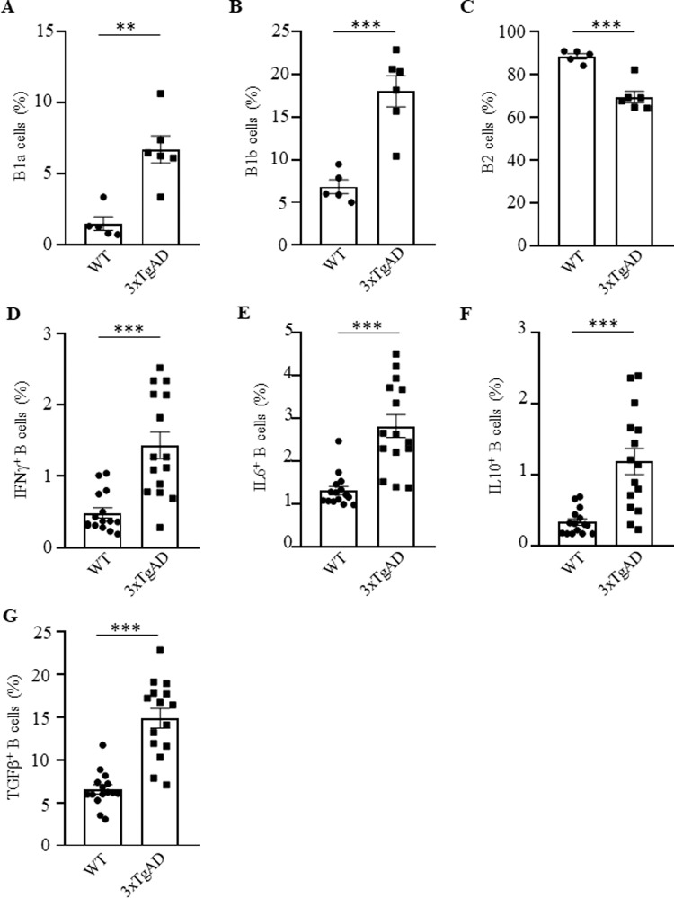
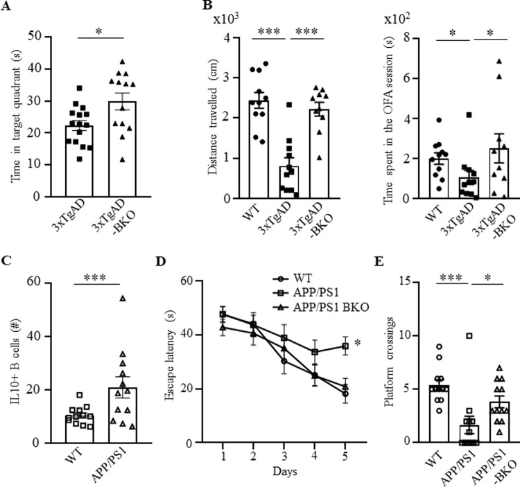
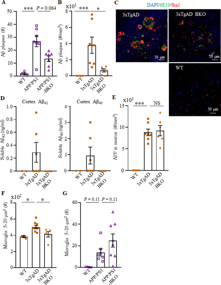
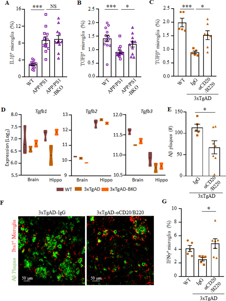
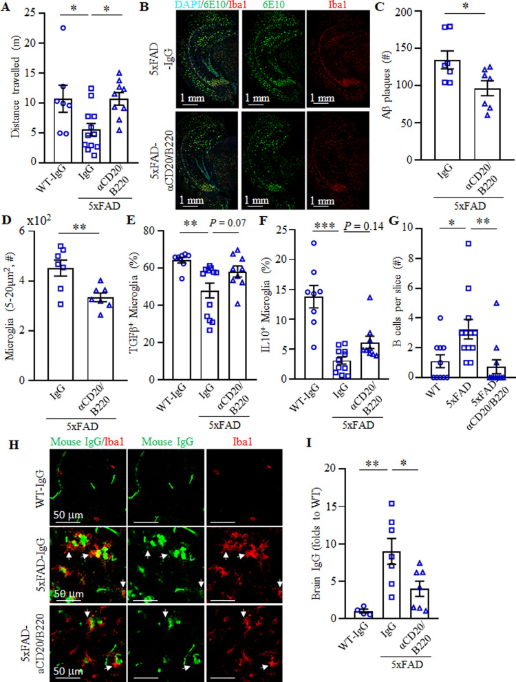
The function of B cells in Alzheimer's disease (AD) is not fully understood. While immunoglobulins that target amyloid beta (Aβ) may interfere with plaque formation and hence progression of the disease, B cells may contribute beyond merely producing immunoglobulins. Here we show that AD is associated with accumulation of activated B cells in circulation, and with infiltration of B cells into the brain parenchyma, resulting in immunoglobulin deposits around Aβ plaques. Using three different murine transgenic models, we provide counterintuitive evidence that the AD progression requires B cells. Despite expression of the AD-fostering transgenes, the loss of B cells alone is sufficient to reduce Aβ plaque burden and disease-associated microglia. It reverses behavioral and memory deficits and restores TGFβ+ microglia, respectively. Moreover, therapeutic depletion of B cells at the onset of the disease retards AD progression in mice, suggesting that targeting B cells may also benefit AD patients.


Conflict of interest statement

The authors declare no competing interests.

Figures

Fig. 1
Fig. 1. Activated B cells were increased in 3×TgAD mice.
Compared with congenic, age- and sex-matched WT mice, B1a (CD5+CD11b+CD19+, A) and B1b cells (CD5CD11b+CD19+, B) were increased, while B2 cells (CD5CD11bCD19+, C) were decreased in the cervical lymph nodes of 3×TgAD mice. AD also activated B cells, as they upregulated IFNγ (D), IL6 (E), IL10 (F), and TGFβ (G) in the peripheral blood of mice. Frequency (%) mean ± SEM is shown; each symbol is for a single mouse, n = 5–6 in AC, n = 15 in DG. Gating strategy is shown in Supplementary Fig. 9A. **p < 0.01; ***p < 0.001 in unpaired t test.
Fig. 2
Fig. 2. Progression of AD required B cells.
The impaired AD-associated cognitive (A, the MWM test) and noncognitive locomotion activity (B, total distance travelled, left panel, and time spent in center zone, right panel, in 30 min OFA test are shown) of 3×TgAD mice were improved in B-cell-deficient 3×TgAD-BKO mice (n = 10–15 female). Compared with WT littermate, APP/PS1 mice upregulated numbers of IL10+ B cells in the peripheral blood (C). The impaired cognitive ability of APP/PS1 mice (D, latency to reach the escape platform; E, platform crossing times in a probe test in MWM test) was reversed in APP/PS1-BKO mice. Mean ± SEM is shown; each symbol is for a single mouse. A, B and D, E were independently reproduced three and two times, respectively. Gating strategy is shown in Supplementary Fig. 9A. *p < 0.05; ***p < 0.001 in unpaired t test (A), Mann–Whitney test (C), one-way ANOVA (B, left), Kruskal–Wallis test (B, right and E) or two-way ANOVA (D).
Fig. 3
Fig. 3. B-cell deficiency reduced the Aβ plaque burden and microglial activation in the hippocampus of AD mice.
AC show the number of Aβ plaques in APP/PS1 (A) and 3×TgAD (B) mice (n = 4–7), and representative images of immune fluorescent staining of the subiculum of WT, 3×TgAD, and 3×TgAD-BKO mice (C, Aβ plaque (green), Iba1+ microglia (red), and DAPI (blue); scale, 50 μm). D The increase of soluble Aβ1–40 and Aβ1–42 peptides in the brain of 3×TgAD mice was reversed in 3×TgAD-BKO mice. The results of ELISA in the cortex of indicated mice (n = 2–6) are shown. The B-cell deficiency did not affect expression of the transgene, as both 3×TgAD and 3×TgAD-BKO highly expressed transgenic hAPP in hippocampal neurons (E, stained with 6E10 as in BC). The B-cell deficiency significantly decreased the number (#) of large-sized microglia (5–20 μm2) in 3×TgAD (n = 3–9, F), but not in APP/PS1 mice (n = 4–8, G). Mean ± SEM is shown; each symbol is for a single mouse. *p < 0.05; ***p < 0.001; NS not significant in Kruskal–Wallis test (A, B, G) or one-way ANOVA (E, F).
Fig. 4
Fig. 4. B-cell deficiency at least in part reversed the DAM phenotype.
The results of flow cytometric quantification of IL1β+ (A) and TGFβ+ (B, C) microglia (CD11b+CD45int) in the brains of indicated mice (n = 11–12) are shown. Genetic B-cell deficiency in APP/PS1 mice (APP/PS1-BKO, B) or transient depletion of circulating B cells (aCD20/B220, C) at the onset of AD (70–79 weeks of age 3×TgAD mice) reversed the AD-associated decrease of TGFβ+ microglia. mRNA microarray analyses of hippocampi and brains (without hippocampus) of 3×TgAD mice revealed that the B-cell deficiency (3×TgAD-BKO) upregulates expression of TGFβ1, but not TGFβ2 and TGFβ3 (D, n = 3). Therapeutic depletion of B cells (aCD20/B220) at the onset of AD ameliorated AD (EG), as it markedly decreased Aβ plaques in the subiculum (quantification and representative images are shown in E and F, respectively; Aβ plaque (green) and Iba1+ microglia (red, F), n = 6–8; independently reproduced twice). B-cell depletion reversed the reduction of IFNγ+ microglia in 3×TgAD mice (G, n = 5–7). Mean ± SEM is shown; each symbol is for a single mouse. Gating strategy is shown in Supplementary Fig. 9B. *p < 0.05; ***p < 0.001 in one-way ANOVA (AC, G) or unpaired t test (F).
Fig. 5
Fig. 5. Therapeutic depletion of B cells improves AD symptoms in 5×FAD mice.
OFA test revealed that B-cell depletion improves the retarded locomotion of 5×FAD mice (A, total distance travelled in first 5 min). B-cell depletion reduced Aβ plaque burden in the hippocampus (B, C). Representative immune fluorescent staining images for Aβ plaques (green), Iba1+ microglia (red), and DAPI (cyan) are shown in B. In C and D, the results of quantification of Aβ plaques and microglia are shown (D, large size, 5–20 μm2), respectively. AD Results were reproduced twice, n = 7–12. Flow cytometric evaluation of brain microglia (CD11b+CD45int) revealed that B-cell depletion increases the frequency (%) of TGFβ + (E) and IL10+ (F) microglia in 5×FAD mice (n = 10–12). As shown with immune fluorescent staining, B220+ B cells were markedly increased in the brain parenchyma of 5×FAD compared with WT controls, which was lost after transient B-cell depletion (G, n = 9–12). The brains of 5×FAD mice contained high levels of IgG as compared with WT controls, which was also reversed by transient depletion of B cells (H and I). In H, IgG is green and Iba1+ microglia is red. Brain IgG quantification is in I (n = 5–7). Mean ± SEM is shown; each symbol is for a single mouse. Gating strategy is shown in Supplementary Fig. 9B. *p < 0.05; **p < 0.01; ***p < 0.001 in one-way ANOVA (A, E, F, I), Kruskal–Wallis test (G), or unpaired t test (C, D).

Similar articles

Cited by

References

    1. Hardy J, Selkoe DJ. The amyloid hypothesis of Alzheimer’s disease: progress and problems on the road to therapeutics. Science. 2002;297:353–356. doi: 10.1126/science.1072994. - DOI - PubMed
    1. Ma D, et al. TGF-beta induced by interleukin-34-stimulated microglia regulates microglial proliferation and attenuates oligomeric amyloid beta neurotoxicity. Neurosci. Lett. 2012;529:86–91. doi: 10.1016/j.neulet.2012.08.071. - DOI - PubMed
    1. Tichauer JE, von Bernhardi R. Transforming growth factor-beta stimulates beta amyloid uptake by microglia through Smad3-dependent mechanisms. J. Neurosci. Res. 2012;90:1970–1980. doi: 10.1002/jnr.23082. - DOI - PubMed
    1. Keren-Shaul H, et al. A unique microglia type associated with restricting development of alzheimer’s disease. Cell. 2017;169:1276–1290 e1217. doi: 10.1016/j.cell.2017.05.018. - DOI - PubMed
    1. Brionne TC, Tesseur I, Masliah E, Wyss-Coray T. Loss of TGF-beta 1 leads to increased neuronal cell death and microgliosis in mouse brain. Neuron. 2003;40:1133–1145. doi: 10.1016/S0896-6273(03)00766-9. - DOI - PubMed

Publication types

Substances