. 2021 Apr 5;11(1):7486.
doi: 10.1038/s41598-021-86881-0.

Effects of fetuin-A-containing calciprotein particles on posttranslational modifications of fetuin-A in HepG2 cells

Affiliations

Effects of fetuin-A-containing calciprotein particles on posttranslational modifications of fetuin-A in HepG2 cells

Hideki Uedono et al. Sci Rep. .

Abstract

Fetuin-A is an inhibitor of ectopic calcification that is expressed mainly in hepatocytes and is secreted into the circulation after posttranslational processing, including glycosylation and phosphorylation. The molecular weight (MW) of fully modified fetuin-A (FM-fetuin-A) is approximately 60 kDa in an immunoblot, which is much higher than the estimated MW by amino acid sequence. Under conditions of calcification stress such as advanced stage chronic kidney disease, fetuin-A prevents calcification by forming colloidal complexes, which are referred to as calciprotein particles (CPP). Since the significance of CPP in this process is unclear, we investigated the effect of synthetic secondary CPP on the level of FM-fetuin-A in HepG2 cells. Secondary CPP increased the level of FM-fetuin-A in dose- and time-dependent manners, but did not affect expression of mRNA for fetuin-A. Treatment with O- and/or N-glycosidase caused a shift of the 60 kDa band of FM-fetuin-A to a lower MW. Preincubation with brefeldin A, an inhibitor of transport of newly synthesized proteins from the endoplasmic reticulum to the Golgi apparatus, completely blocked the secondary CPP-induced increase in FM-fetuin-A. Treatment with BAPTA-AM, an intracellular calcium chelating agent, also inhibited the CPP-induced increase in the FM-fetuin-A level. Secondary CPP accelerate posttranslational processing of fetuin-A in HepG2 cells.


Conflict of interest statement

The authors declare no competing interests.

Figures
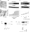
Figure 1
Figure 1
TEM imaging of synthetic CPP and effects of secondary CPP on FM-fetuin-A in HepG2 cells. (A1) After 1.5 h of incubation of CPP solution, small spherical-shaped CPP (primary CPP) were formed. (A2) At 24 h, larger crystalline-like CPP (secondary CPP) were observed. (B) HepG2 cells were treated with different concentrations (0, 1, 5, 10, 25, 50, 100, and 200 μg/mL) of synthetic secondary CPP for 24 h. Immunoblots of cell lysates were probed with fetuin-A (B, upper panel) and α-tubulin (B, middle panel) antibodies. FM-fetuin-A levels were quantified using α-tubulin as an endogenous reference (B, lower panel), with the fetuin-A level without CPP defined as 100% (control). (C) HepG2 cells were treated with 100 μg/mL secondary CPP for 0, 6, 12, 24 h. Immunoblots of cell lysates were probed with fetuin-A (C, upper panel) and α-tubulin (C, middle panel) antibodies. FM-fetuin-A levels were quantified as above (C, lower panel), with the fetuin-A level at 0 h defined as 100% (control). Data are shown as mean ± SD from at least three independent experiments. (D) Cell lysate from 100 μg/mL secondary CPP-treated HepG2 cells, 40 μg of secondary CPP as a protein level, and 40 μg of bovine fetuin-A were probed with human anti-fetuin-A antibody. (E) HepG2 cells were treated with or without 100 μg/mL secondary CPP for 24 h. Cell Lysate and the supernatant from cultured HepG2 cells were probed with an anti-fetuin-A antibody. *P < 0.05 vs. control. Full-length blots/gels (B, C, D, E) are presented in Supplementary Fig. X1 (full-size) (B, B′, C, D, D′, E, E′), respectively.
Figure 2
Figure 2
Effects of secondary CPP on expression of fetuin-A mRNA in HepG2 cells. (A) HepG2 cells were treated with different concentrations (0, 25, 50, and  100 μg/mL) of synthetic secondary CPP for 24 h. (B) HepG2 cells were treated with 100 μg/mL secondary CPP for 0, 6, 12, 24 h. Total RNA was isolated from cells and fetuin-A/ahsg mRNA expression was determined using quantitative real-time PCR. Levels of fetuin-A mRNA were calculated using the comparative cycle threshold method, with 18S ribosomal RNA as the endogenous reference. Fetuin-A mRNA levels without secondary CPP (A) or at 0 h (B) were defined as 100% (control). Data are shown as mean ± SD from at least three independent experiments. *P < 0.05 vs. control.
Figure 3
Figure 3
Localization of secondary CPP in HepG2 cells. HepG2 cells without incubation with secondary CPP (A) and 12 h (B) and 24 h (C) after addition of 100 μg/mL secondary CPP. TEM imaging showed extracellular crystalline-like structures (arrowhead) around treated HepG2 cells (B, C), whereas no such structure was present in control cells (A).
Figure 4
Figure 4
Deglycosylation of secondary CPP-induced FM-fetuin-A by O- and N-glycosidases. Secondary CPP-treated cell lysates were denatured at 100 °C for 10 min and subsequently incubated with O-glycosidase and/or N-glycosidase F (PNGase F) at 37 °C for 1–2 h. Immunoblots of enzyme-treated samples were probed with fetuin-A (upper panel) and α-tubulin (lower panel) antibodies. FM-fetuin-A levels were quantified using α-tubulin as an endogenous reference, with the fetuin-A level in the absence of CPP without O-glycosidase and N-glycosidase F defined as 100% (control). Data are shown as mean ± SD from at least three independent experiments. *P < 0.05 vs. control. Full-length blots/gels are presented in Supplementary Fig. X4 (full-size).
Figure 5
Figure 5
Effect of brefeldin A on secondary CPP-induced FM-fetuin-A. HepG2 cells were pre-treated for 30 min with or without 1 mg/mL brefeldin A and subsequently incubated in the absence or presence of the indicated concentrations of secondary CPP for 24 h. Immunoblots of cell lysates were probed with fetuin-A (upper panel) and α-tubulin (lower panel) antibodies. FM-fetuin-A levels were quantified using α-tubulin as an endogenous reference, with the fetuin-A level in the absence of CPP without brefeldin A defined as 100% (control). Data are shown as mean ± SD from at least three independent experiments. *P < 0.05 vs. control. Full-length blots/gels are presented in Supplementary Fig. X5 (full-size).
Figure 6
Figure 6
Effect of BAPTA-AM on secondary CPP-induced FM-fetuin-A. HepG2 cells were cultured with 100 μg/mL secondary CPP in the absence or presence of 500 μg/mL of BAPTA-AM for 24 h. Immunoblots of cell lysates were probed with fetuin-A (upper panel) and GAPDH (lower panel) antibodies. FM-fetuin-A levels were quantified using GAPDH as an endogenous reference, with the fetuin-A level in the absence of CPP without BAPTA-AM as 100% (control). Data are shown as mean ± SD from at least three independent experiments. *P < 0.05 vs. control. Full-length blots/gels are presented in Supplementary Fig. X6 (full size).

Similar articles

Cited by

References

    1. Jahnen-Dechent W, Heiss A, Schafer C, Ketteler M. Fetuin-A regulation of calcified matrix metabolism. Circ. Res. 2011;108:1494–1509. doi: 10.1161/CIRCRESAHA.110.234260. - DOI - PubMed
    1. Mori K, Emoto M, Inaba M. Fetuin-A: A multifunctional protein. Recent Pat. Endocr. Metab. Immune Drug. Discov. 2011;5:124–146. doi: 10.2174/187221411799015372. - DOI - PubMed
    1. Mori K, Emoto M, Inaba M. Fetuin-A and the cardiovascular system. Adv. Clin. Chem. 2012;56:175–195. doi: 10.1016/b978-0-12-394317-0.00010-8. - DOI - PubMed
    1. Jahnen-Dechent W, Smith ER. Nature's remedy to phosphate woes: Calciprotein particles regulate systemic mineral metabolism. Kidney Int. 2020;97:648–651. doi: 10.1016/j.kint.2019.12.018. - DOI - PubMed
    1. Kuro-o M. Klotho, phosphate and FGF-23 in ageing and disturbed mineral metabolism. Nat. Rev. Nephrol. 2013;9:650–660. doi: 10.1038/nrneph.2013.111. - DOI - PubMed

Publication types

LinkOut - more resources