. 2021 Mar 31;9(4):360.
doi: 10.3390/biomedicines9040360.

Probing Skin Barrier Recovery on Molecular Level Following Acute Wounds: An In Vivo/Ex Vivo Study on Pigs

Affiliations

Probing Skin Barrier Recovery on Molecular Level Following Acute Wounds: An In Vivo/Ex Vivo Study on Pigs

Enamul Haque Mojumdar et al. Biomedicines. .

Abstract

Proper skin barrier function is paramount for our survival, and, suffering injury, there is an acute need to restore the lost barrier and prevent development of a chronic wound. We hypothesize that rapid wound closure is more important than immediate perfection of the barrier, whereas specific treatment may facilitate perfection. The aim of the current project was therefore to evaluate the quality of restored tissue down to the molecular level. We used Göttingen minipigs with a multi-technique approach correlating wound healing progression in vivo over three weeks, monitored by classical methods (e.g., histology, trans-epidermal water loss (TEWL), pH) and subsequent physicochemical characterization of barrier recovery (i.e., small and wide-angle X-ray diffraction (SWAXD), polarization transfer solid-state NMR (PTssNMR), dynamic vapor sorption (DVS), Fourier transform infrared (FTIR)), providing a unique insight into molecular aspects of healing. We conclude that although acute wounds sealed within two weeks as expected, molecular investigation of stratum corneum (SC) revealed a poorly developed keratin organization and deviations in lipid lamellae formation. A higher lipid fluidity was also observed in regenerated tissue. This may have been due to incomplete lipid conversion during barrier recovery as glycosphingolipids, normally not present in SC, were indicated by infrared FTIR spectroscopy. Evidently, a molecular approach to skin barrier recovery could be a valuable tool in future development of products targeting wound healing.

Keywords: acute wound; histology; in vivo/ex vivo; lipid; pH; polarization transfer solid state NMR (PTssNMR); skin barrier; small and wide-angle X-ray diffraction (SWAXD); stratum corneum; trans-epidermal water loss (TEWL).


Conflict of interest statement

The authors declare no conflict of interest.

Figures

Figure 1
Figure 1
(A) A schematic of the wound healing study design. The study was performed on two non-naïve Göttingen minipigs. A total of 10 wounds were generated on each pig, five on the left side of the pigs back and five on the right side, as shown here with numbers from 6 to 10. The green filled circle inside the square indicates regions from where the control skin tissues were harvested at the end of the in vivo study. (B) A simplified drawing showing three major layers of the skin: the epidermis, the dermis, and the hypodermis. The wound incision area, which is approximately 2 × 2 cm2, is shown, along with the time scale for healing progression over 21 days. After the in vivo study, the recovered skin was harvested for further evaluation ex vivo. (C) A cartoon presenting the thin top layer of the epidermis, the stratum corneum, which can be illustrated with bricks and mortar. The bricks in the cartoon represent anucleated corneocytes, mainly composed of keratin filaments. The corneocytes are embedded in a lipid lamellar matrix constituting the mortar. These lipid lamellae mainly comprise ceramides, cholesterol, and free fatty acids.
Figure 2
Figure 2
(A) Representative wound images on pig 1 at various time points during the healing progression cycle. The arrow on top of the figure indicates ascending date for healing cycle and the readers are encouraged to follow the images in the arrow direction. The images presented origin from position 1 and were captured on the days when the dressing was changed. The name plates on top of the images indicate the in vivo wound study number, pig number, study days when the wound dressing was changed, and the wound position number. The ruler on the bottom of the images was used as a guide to set the scale when calculating the wound marked area, e.g., see marking on day 7. Calculated wound area was presented as a function of days during the healing cycle for pig 1 (B) and pig 2 (C). The labelling W1, W2 and so on in the figure legends indicate the various wound positions on each pig.
Figure 3
Figure 3
Variations in TEWL, skin temperature, and pH during healing. (A) Measured TEWL vs. time for control skin and healing skin for pig 1, position 1. On day 0, the readings were recorded before the wound incision. (B) TEWL values vs. time for individual wounds obtained from pig 1 when the wound dressings were changed. (C) Skin surface temperature vs. time measured on both pigs. (D) pH measurements on the wounds (position 1, pig 1, and position 5, pig 2) and corresponding control sites were plotted vs. time for both pigs. All the TEWL and pH values were recorded at ambient controlled conditions, i.e., approximately 50% relative humidity (RH) and 23 °C. Error bars depict standard deviation.
Figure 4
Figure 4
Classical hematoxylin and eosin (H&E) staining of control (A,B) and healed (C,D) pig skin. The skin surface is directed towards the top/left. The individual epidermal layers can be identified in the control skin labelled (A). Magnifications of the dermis part for control and healed skin are provided in (B,D).
Figure 5
Figure 5
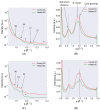
Small and wide-angle X-ray diffraction (SWAXD) spectra of control and healed stratum corneum (SC) excised from pig 1 and recorded at dry (A,B) and wet (97% RH) conditions (C,D). The SAXD spectra are provided on the left column whereas the WAXD spectra are on the right. In the SAXD region, several peaks attributed to lipid lamellar ordering and keratin packing are identified. The numbers with arrows indicate the d-spacing in Å for various peaks. Phase-separated crystalline Chol was also detected in some spectra and then indicated by an asterisk (*) sign. In the WAXD region, the shaded lines indicate peaks originating from the keratin interchain distance and show a change in peak position following hydration of the sample. The lipid acyl chain ordering indicated by dotted lines do not show a shift in peak position when comparing dry and hydrated conditions. The secondary β-sheet structure of keratin could also be detected and is marked in the plot. All SWAXD measurements were performed at 32 °C, which represents average physiological skin temperature.
Figure 6
Figure 6
(A) Water sorption measurements for control and healed SC at 32 °C, expressed as water content, (in wt % with respect to dry SC) plotted as a function of RH. (B) 1H NMR spectra recorded on control and healed SC at 97% RH. The peak close to 5 ppm in both spectra was due to water in the sample from hydration at 97% RH. Several sharp peaks attributed to lipid molecular segments (assignments in Figure 7E) were also detected, indicative of fluid lipids present in both samples. The arrow close to 0 ppm in the healed SC was due to the silicon present in the wound dressing.
Figure 7
Figure 7
Natural abundance 13C polarization transfer solid state NMR (PTssNMR) study on control (A,C) and healed (B,D) SC samples in dry (A,B) and wet (C,D) conditions. The individual direct polarization (DP) (grey), cross polarization (CP) (blue), and insensitive nuclei enhanced by polarization transfer (INEPT) (red) spectra were overlaid in all experiments for the purpose of comparison. The resonance lines originating from various SC lipid molecular segments along with Chol are labelled in the spectra. The labelling is provided in black for control and red in healed SC when changes in the INEPT signals were observed compared to control SC. The peak labeled with (*) in the healed SC at 97% RH might indicate the presence of glycosphingolipid. Assignments of various lipid molecular segments with their numberings are depicted with a representative ceramide (Cer) structure—Cer EOS linoleate (E), as well as for Chol (F).

Similar articles

Cited by

References

    1. Madison K.C. Barrier function of the skin: “La raison d’etre” of the epidermis. J. Investig. Dermytol. 2003;121:231–241. doi: 10.1046/j.1523-1747.2003.12359.x. - DOI - PubMed
    1. Proksch E., Brandner J.M., Jensen J.-M. The skin: An indispensable barrier. Exp. Dermatol. 2008;17:1063–1072. doi: 10.1111/j.1600-0625.2008.00786.x. - DOI - PubMed
    1. Blatteis C.M. Age-dependent changes in temperature regulation–A mini review. Gerontology. 2012;58:289–295. doi: 10.1159/000333148. - DOI - PubMed
    1. Schaefer H., Redelmeier T.E. Skin Barrier: Principles of Percutaneous Absorption. Karger; Basel, Switzerland: 1996.
    1. Scheuplein R.J. Permeability of the skin: A review of major concepts and some new developments. J. Investig. Dermatol. 1976;67:672–676. doi: 10.1111/1523-1747.ep12544513. - DOI

LinkOut - more resources