

Date Palm Pollen Extract Avert Doxorubicin-Induced Cardiomyopathy Fibrosis and Associated Oxidative/Nitrosative Stress, Inflammatory Cascade, and Apoptosis-Targeting Bax/Bcl-2 and Caspase-3 Signaling Pathways
- PMID: 33804672
- PMCID: PMC8003775
- DOI: 10.3390/ani11030886
Date Palm Pollen Extract Avert Doxorubicin-Induced Cardiomyopathy Fibrosis and Associated Oxidative/Nitrosative Stress, Inflammatory Cascade, and Apoptosis-Targeting Bax/Bcl-2 and Caspase-3 Signaling Pathways
Abstract
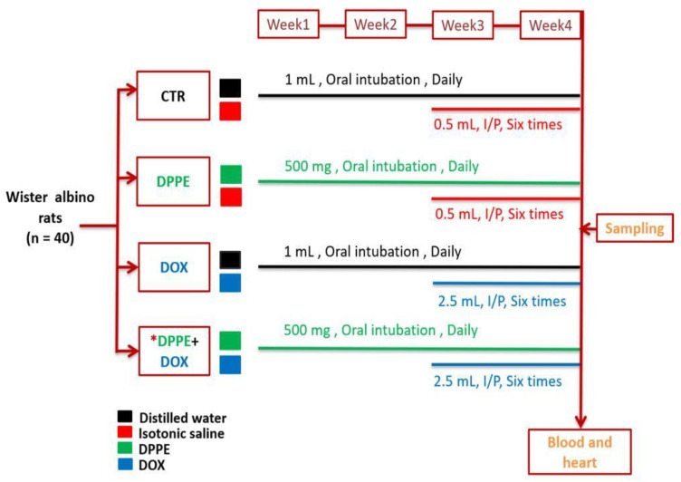
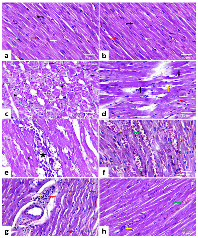
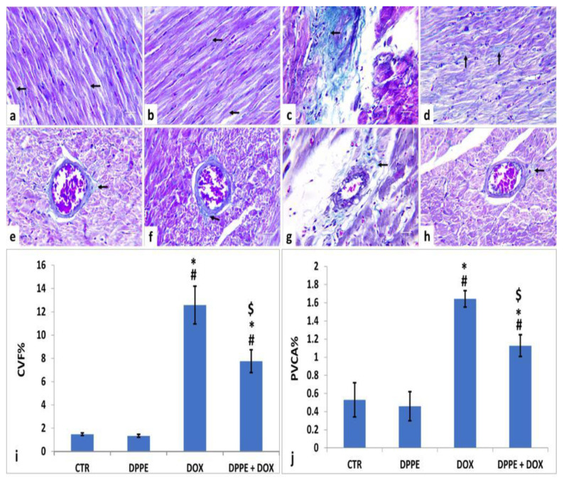
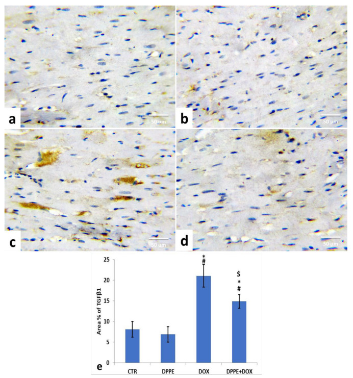
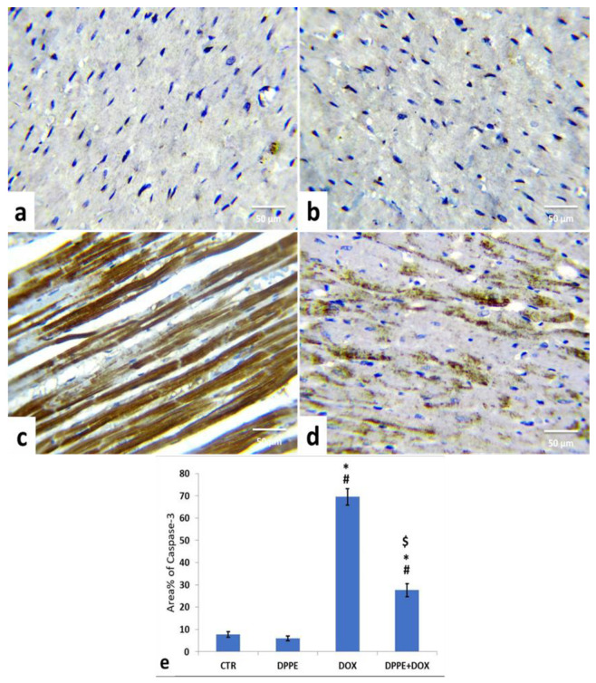
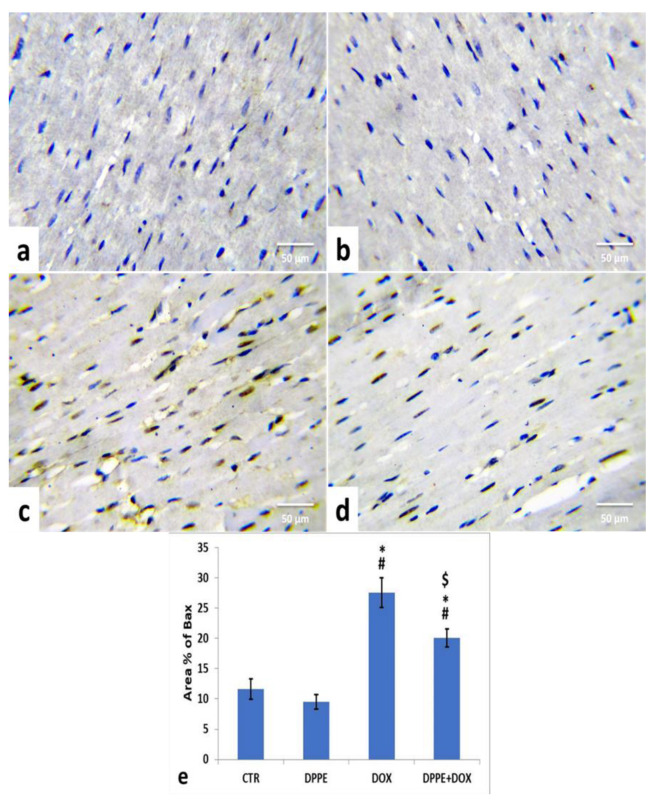
Doxorubicin (DOX) has a potent antineoplastic efficacy and is considered a cornerstone of chemotherapy. However, it causes several dose-dependent cardiotoxic results, which has substantially restricted its clinical application. This study was intended to explore the potential ameliorative effect of date palm pollen ethanolic extract (DPPE) against DOX-induced cardiotoxicity and the mechanisms underlying it. Forty male Wistar albino rats were equally allocated into Control (CTR), DPPE (500 mg/kg bw for 4 weeks), DOX (2.5 mg/kg bw, intraperitoneally six times over 2 weeks), and DPPE + DOX-treated groups. Pre-coadministration of DPPE with DOX partially ameliorated DOX-induced cardiotoxicity as DPPE improved DOX-induced body and heart weight changes and mitigated the elevated cardiac injury markers activities of serum aminotransferases, lactate dehydrogenase, creatine kinase, and creatine kinase-cardiac type isoenzyme. Additionally, the concentration of serum cardiac troponin I (cTnI), troponin T (cTnT), N-terminal pro-brain natriuretic peptide (NT-pro BNP), and cytosolic calcium (Ca+2) were amplified. DPPE also alleviated nitrosative status (nitric oxide) in DOX-treated animals, lipid peroxidation and antioxidant molecules as glutathione content, and glutathione peroxidase, catalase, and superoxide dismutase activities and inflammatory markers levels; NF-κB p65, TNF-α, IL-1β, and IL-6. As well, it ameliorated the severity of histopathological lesions, histomorphometric alteration and improved the immune-staining of the pro-fibrotic (TGF-β1), pro-apoptotic (caspase-3 and Bax), and anti-apoptotic (Bcl-2) proteins in cardiac tissues. Collectively, pre-coadministration of DPPE partially mitigated DOX-induced cardiac injuries via its antioxidant, anti-inflammatory, anti-fibrotic, and anti-apoptotic potential.
Keywords: Bax; Bcl-2; TGF-β1; cardiac injury markers; date palm (pollen extract); doxorubicin; histopathology; oxidative stress.
Conflict of interest statement
The authors declared no competing conflict of interest.
Figures

Similar articles
-
All-trans-retinoic acid ameliorates doxorubicin-induced cardiotoxicity: in vivo potential involvement of oxidative stress, inflammation, and apoptosis via caspase-3 and p53 down-expression.Naunyn Schmiedebergs Arch Pharmacol. 2018 Jan;391(1):59-70. doi: 10.1007/s00210-017-1437-5. Epub 2017 Oct 30. Naunyn Schmiedebergs Arch Pharmacol. 2018. PMID: 29085977
-
Irbesartan suppresses cardiac toxicity induced by doxorubicin via regulating the p38-MAPK/NF-κB and TGF-β1 pathways.Naunyn Schmiedebergs Arch Pharmacol. 2019 Jun;392(6):647-658. doi: 10.1007/s00210-019-01624-3. Epub 2019 Feb 7. Naunyn Schmiedebergs Arch Pharmacol. 2019. PMID: 30734091
-
Chrysin alleviates acute doxorubicin cardiotoxicity in rats via suppression of oxidative stress, inflammation and apoptosis.Eur J Pharmacol. 2014 Apr 5;728:107-18. doi: 10.1016/j.ejphar.2014.01.065. Epub 2014 Feb 6. Eur J Pharmacol. 2014. PMID: 24509133
-
The role of hesperidin as a cardioprotective strategy against doxorubicin-induced cardiotoxicity: The antioxidant, anti-inflammatory, antiapoptotic, and cytoprotective potentials.Open Vet J. 2023 Dec;13(12):1718-1728. doi: 10.5455/OVJ.2023.v13.i12.20. Epub 2023 Dec 31. Open Vet J. 2023. PMID: 38292716 Free PMC article.
-
Vitamin C mitigates oxidative/nitrosative stress and inflammation in doxorubicin-induced cardiomyopathy.Am J Physiol Heart Circ Physiol. 2017 Oct 1;313(4):H795-H809. doi: 10.1152/ajpheart.00253.2017. Epub 2017 Jul 14. Am J Physiol Heart Circ Physiol. 2017. PMID: 28710069
Cited by
-
Palm Fruit (Phoenix dactylifera L.) Pollen Extract Inhibits Cancer Cell and Enzyme Activities and DNA and Protein Damage.Nutrients. 2023 Jun 2;15(11):2614. doi: 10.3390/nu15112614. Nutrients. 2023. PMID: 37299576 Free PMC article.
-
Ang-1 and VEGF: central regulators of angiogenesis.Mol Cell Biochem. 2025 Feb;480(2):621-637. doi: 10.1007/s11010-024-05010-3. Epub 2024 Apr 23. Mol Cell Biochem. 2025. PMID: 38652215 Review.
-
Protective effect of limonin against doxorubicin-induced cardiotoxicity via activating nuclear factor - like 2 and Sirtuin 2 signaling pathways.Bioengineered. 2021 Dec;12(1):7975-7984. doi: 10.1080/21655979.2021.1985299. Bioengineered. 2021. PMID: 34565300 Free PMC article.
-
The cardioprotective properties of Persicaria maculosa and Citrus sinensis extracts against doxorubicin-induced cardiotoxicity in mice.Avicenna J Phytomed. 2024 Jul-Aug;14(4):455-469. doi: 10.22038/AJP.2024.24101. Avicenna J Phytomed. 2024. PMID: 38952773 Free PMC article.
-
Farnesol Protects against Cardiotoxicity Caused by Doxorubicin-Induced Stress, Inflammation, and Cell Death: An In Vivo Study in Wistar Rats.Molecules. 2022 Dec 6;27(23):8589. doi: 10.3390/molecules27238589. Molecules. 2022. PMID: 36500681 Free PMC article.
References
LinkOut - more resources
Full Text Sources
Other Literature Sources
Research Materials
