Review
. 2021 Mar 1;22(5):2482.
doi: 10.3390/ijms22052482.

The Role of TCOF1 Gene in Health and Disease: Beyond Treacher Collins Syndrome

Affiliations
Review

The Role of TCOF1 Gene in Health and Disease: Beyond Treacher Collins Syndrome

Małgorzata Grzanka et al. Int J Mol Sci. .

Abstract

The nucleoli are membrane-less nuclear substructures that govern ribosome biogenesis and participate in multiple other cellular processes such as cell cycle progression, stress sensing, and DNA damage response. The proper functioning of these organelles is ensured by specific proteins that maintain nucleolar structure and mediate key nucleolar activities. Among all nucleolar proteins, treacle encoded by TCOF1 gene emerges as one of the most crucial regulators of cellular processes. TCOF1 was initially discovered as a gene involved in the Treacher Collins syndrome, a rare genetic disorder characterized by severe craniofacial deformations. Later studies revealed that treacle regulates ribosome biogenesis, mitosis, proliferation, DNA damage response, and apoptosis. Importantly, several reports indicate that treacle is also involved in cancer development, progression, and response to therapies, and may contribute to other pathologies such as Hirschsprung disease. In this manuscript, we comprehensively review the structure, function, and the regulation of TCOF1/treacle in physiological and pathological processes.

Keywords: DDR; DNA damage response; TCOF1; Treacher Collins syndrome; cancer; nucleolus; ribosome biogenesis; treacle.


Conflict of interest statement

The authors declare no conflict of interest.

Figures

Figure 1
Figure 1
The structure of the major treacle isoform including amino acids encoded by exon 6A. The exon location was retrieved from Ensembl (https://www.ensembl.org), transcript ID: ENST00000377797.7, accessed on 11 May 2020. The N-terminus is encoded by six exons and contains nuclear export signal (NES) and potential nuclear localization signal (NLS) regions. The central domain encoded by exons 6A-16 consist of 11 repetitive motifs that are phosphorylated by CK2 and protein kinase C (PKC). The motif encoded by exon 6A has an additional NLS region. The C-terminus is encoded by exons 17–26 and contains several NLS regions in exons 23, 24 and 25 [12].
Figure 2
Figure 2
Ribosome biogenesis begins in the nucleolus and is continued during the transport via the nucleoplasm and the final maturation steps that occur in the cytoplasm. The treacle protein recruits the pre-initiation complex (UBF, SL-1, Pol I with associated TIF1A) to the rDNA promoter region [40]. The produced 47S pre-rRNA undergoes post-transcriptional processing (including methylation and possibly pseudouridylation), involving treacle, which binds ribonucleoprotein methylation complex composed of Nop56, fibrillarin, Nop58, and 15.5 kDa protein [32,41]. As a result of the post-transcriptional processing, 5.8S, 18S, and 28S RNAs are generated. The residual ribosome components are synthesized in the nucleoplasm and cytoplasm—Pol III transcribes a gene encoding 5S RNA, which is then transported to the nucleolus; Pol II transcribes genes encoding ribosomal proteins, resulting in mRNAs that are transported to the cytoplasm where translation of ribosomal proteins (RPs) takes place. RPs are then translocated to the nucleolus, where ribosome subunits are formed—the large one (60S), composed of 5S, 5.8S, 28S and 47RP, and the small one (40S), composed of 18S and 33RP. The ribosome subunits migrate to the cytoplasm where the mature ribosome is assembled [35].
Figure 3
Figure 3
The proposed role of treacle in response to rDNA damage. Treacle facilitates DNA damage response (DDR) mechanisms involving ATM and ATR kinases by recruiting their key adaptor proteins, NBS1 and TOPBP1, respectively. (1) Following rDNA damage, ATM and ATR kinases are initially activated at the sites of rDNA breaks (DSB) independently of treacle [56]. ATM- and CK2-catalyzed phosphorylation enables treacle to recruit NBS1, which mediates ATM interactions with MRN complex [21,27,55]. (2) ATM-mediated phosphorylation of treacle enables recruitment of TOPBP1. The efficient TOPBP1 recruitment requires the presence of NBS1 [56]. (3) NBS1 and TOPBP1 localized at the sites of rDNA damage enable further recruitment and activation of ATR kinase, needed for repression of Pol I-catalyzed transcription [56]. (4) Inhibition of rDNA transcription enables formation of the nucleolar caps. The localization of damaged rDNA in nucleolar periphery facilitates recruitment of repair factors from the nucleoplasm [58]. (5) The scheme of treacle with depicted phosphorylation sites involved in interactions with NBS1 and TOPBP1. NBS1 interacts with treacle via FHA and BRCT domains [21,56].
Figure 4
Figure 4
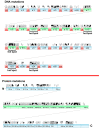
TCOF1 polymorphisms and mutations detected in patients with Treacher Collins syndrome (TCS) and described thus far in the literature. The exonic/intronic localization of mutations (upper panel) refers to data provided in Table S1. The localization of the corresponding amino acids (lower panel) is based on NCBI Reference Sequence: NP_001128715.1. Each pin represents one genetic change. Black pins indicate changes that result in a truncated protein. Detailed characteristics of the mutations are provided in Table S1.
Figure 5
Figure 5
The model of TCOF1 haploinsufficiency leading to TCS proposed by Jones et al. [82]. TCOF1 haploinsufficiency leads to disruption of ribosome biogenesis, impaired proliferation, and apoptotic loss of neuroepithelial and neural crest cells. This mechanism is further boosted by p53 tumor suppressor, which is stabilized in response to the nucleolar stress. p53 prevents the interaction between SL1 (promoter selectivity factor 1) and UBF, leading to inhibition of RNA polymerase I activity and attenuation of rRNA transcription [83]. This in turn leads to the reduced ribosome biogenesis and attenuated proliferation. On the other hand, p53 activates the transcription of cyclin G1, which leads to cell-cycle arrest in G1 phase, with the following induction of apoptosis. The apoptotic loss of neural crest precursors leads to craniofacial abnormalities and TCS development. Importantly, inhibition of p53 function in mouse TCS embryos reduces the activation of cyclin G1 and consequently attenuates apoptosis, preventing TCS in TCOF1+/− embryos. The above mechanism has been described by Jones et al. [82] as a mechanistic model describing the molecular background of TCS pathogenesis. Later studies revealed that treacle loss also results in dysfunction of Polo-like kinase 1 (Plk1), leading to mitotic arrest and cell cycle delay of developing brain neurons and contributing to the TCS neurodevelopmental disorders such as microcephaly [25]. Another postulated mechanism of TCOF1 haploinsufficiency resulting in TCS could be lowered rRNA production and global reprogramming of cellular translation program [32] (see text for details).
Figure 6
Figure 6
TCOF1 mutations in uterine carcinoma. (A) The frequency of TCOF1 alterations in 509 samples of uterine carcinomas, classified as uterine mixed endometrial carcinoma (n = 21), uterine endometrioid carcinoma (n = 383), and uterine serous carcinoma/uterine papillary serous carcinoma (n = 105). (B) Graphical representation of mutations identified in uterine carcinomas. In total, 55 TCOF1 mutations were found in 509 samples of uterine carcinomas. The figure shows results of bioinformatic analysis of the publicly available transcriptomic data of The Cancer Genome Atlas, retrieved and analyzed using cBio platform (http://www.cbioportal.org/, accessed on 11 May 2020).
Figure 7
Figure 7
The summary of the key treacle functions in the cell. The molecules interacting with specific amino acids of Table 140. and UBF. In response to DNA damage, treacle binds and recruits NBS1 to the nucleolus, enabling silencing of rRNA transcription. Interaction with TOPBP1 enables recruitment and activation of ATR kinase, a crucial regulator of DNA damage response. Treacle function in the antioxidative defense is tightly linked with its role in DNA damage repair. The ability of treacle to regulate mitosis and cell cycle progression relies on its interaction with Plk1.

Similar articles

Cited by

References

    1. Núñez Villacís L., Wong M.S., Ferguson L.L., Hein N., George A.J., Hannan K.M. New Roles for the Nucleolus in Health and Disease. Bioessays. 2018;40:e1700233. doi: 10.1002/bies.201700233. - DOI - PubMed
    1. Turi Z., Lacey M., Mistrik M., Moudry P. Impaired ribosome biogenesis: Mechanisms and relevance to cancer and aging. Aging (Albany NY) 2019;11:2512–2540. doi: 10.18632/aging.101922. - DOI - PMC - PubMed
    1. Correll C.C., Bartek J., Dundr M. The Nucleolus: A Multiphase Condensate Balancing Ribosome Synthesis and Translational Capacity in Health, Aging and Ribosomopathies. Cells. 2019;8:869. doi: 10.3390/cells8080869. - DOI - PMC - PubMed
    1. Sullivan G.J., Bridger J.M., Cuthbert A.P., Newbold R.F., Bickmore W.A., McStay B. Human acrocentric chromosomes with transcriptionally silent nucleolar organizer regions associate with nucleoli. EMBO J. 2001;20:2867–2874. doi: 10.1093/emboj/20.11.2867. - DOI - PMC - PubMed
    1. Hernandez-Verdun D. Assembly and disassembly of the nucleolus during the cell cycle. Nucleus. 2011;2:189–194. doi: 10.4161/nucl.2.3.16246. - DOI - PMC - PubMed

MeSH terms

LinkOut - more resources